Robot-assisted spleen preserving distal pancreatectomy: case report
←
→
Page content transcription
If your browser does not render page correctly, please read the page content below
Case Report
                                                                                                                                           Page 1 of 6

Robot-assisted spleen preserving distal pancreatectomy: case
report
Emanuele Federico Kauffmann, Niccolò Napoli, Francesca Menonna, Concetta Cacace, Valerio
Genovese, Fabio Vistoli, Ugo Boggi

Division of General and Transplant Surgery, University of Pisa, Pisa, Italy
Correspondence to: Emanuele Federico Kauffmann. Division of General and Transplant Surgery, University of Pisa, Via Paradisa 2, 56124 Pisa, Italy.
Email: emanuele.kauffmann@unipi.it.

                 Abstract: A 28-year-old female patient was incidentally diagnosed with a unilocular pancreatic cystic lesion
                 located in the body-tail of the pancreas. Findings at contrast-enhanced computed tomography scan and
                 magnetic resonance were both consistent with the diagnosis of mucinous cystadenoma. The patient was then
                 scheduled for a robot-assisted distal pancreatectomy with preservation of the spleen and the splenic vessels.
                 The procedure was completed safely in 255 minutes with minimal blood loss. The post-operative course was
                 uneventful and the patient was discharged on post-operative day 6. Final pathology confirmed the diagnosis
                 of mucinous cystadenoma. In the video presented herein we show the technique for robot-assisted distal
                 pancreatectomy with preservation of the spleen and the splenic vessels.

                 Keywords: Robotic; spleen; pancreas; distal pancreatectomy; case report

                 Received: 16 December 2019; Accepted: 19 March 2020; Published: 20 January 2021.
                 doi: 10.21037/ales.2020.03.14
                 View this article at: http://dx.doi.org/10.21037/ales.2020.03.14

Introduction                                                                  because of the enhanced dexterity offered by the robotic
                                                                              system (6).
According to the Miami international evidence-based
                                                                                 We herein describe a case of robot-assisted MIPD with
guidelines on minimally invasive pancreas resection,
                                                                              preservation of the spleen and the splenic vessels. The
minimally invasive distal pancreatectomy (MIPD) for
                                                                              attached video demonstrates the technical details of this
benign or low-grade malignant tumors is to be considered
                                                                              procedure as defined in a center with experience with over
over open distal pancreatectomy, since it is associated with                  380 robotic pancreatic resections.
a shorter hospital stay, reduced blood loss, and equivalent                      We present the following case in accordance with the
complication rates (1). Additionally, whenever oncologically                  CARE reporting checklist (available at http://dx.doi.
indicated, the spleen should be preserved because of                          org/10.21037/ales.2020.03.14).
hematological and immunological advantages (2). During
MIPD the spleen can be spared along with the splenic
vessels (3) or despite the sacrifice of the splenic vessels (so               Case presentation
called procedure) (4). In the Warshaw procedure, spleen                       A 28-year-old female patient was incidentally diagnosed
supply is maintained through the left gastro-epiploic arcade                  with a cystic lesion located in the body-tail of the
and the short gastric vessels. This collateral circulation,                   pancreas during routine abdominal ultrasonography. She
however, may not be sufficient to ensure spleen viability                     was otherwise completely healthy (American Society of
in all patients, while sometimes the lack of optimal venous                   Anesthesiologists score class 1).
outflow results in sinistral portal hypertension with the                       The patient was further investigated by assay of
development of gastric varices (5). Robotic assistance                        tumor markers (Ca19.9, Ca15.3 and Ca125), contrast-
facilitates spleen preservation during MIPD, possibly                         enhanced computed tomography (CT) scan and magnetic

© Annals of Laparoscopic and Endoscopic Surgery. All rights reserved.     Ann Laparosc Endosc Surg 2021;6:13 | http://dx.doi.org/10.21037/ales.2020.03.14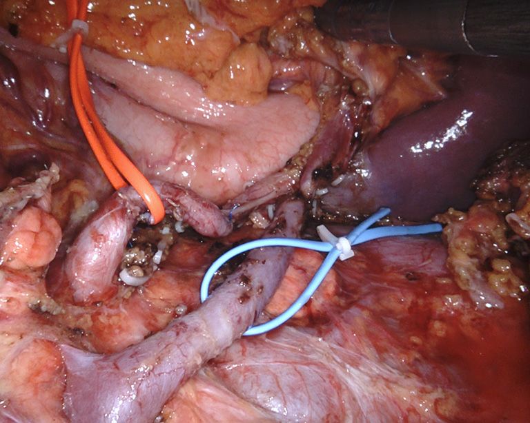
Page 2 of 6                                                                               Annals of Laparoscopic and Endoscopic Surgery, 2021

resonance imaging (MRI). Tumor markers were negative.                           Operative procedure
CT confirmed the presence of a pancreatic cystic lesion,
                                                                                The patient was placed supine with the legs parted on an
measuring 40 mm with internal septae (Figure 1). MRI
                                                                                operating table equipped with a thermic blanket. She was
demonstrated also the presence of two mural nodules
                                                                                secured to the table with wide bandings and pneumatic
showing restricted diffusion (Figure 2). The case was
                                                                                intermittent cuffs were placed around the legs to reduce the
discussed at a multidisciplinary tumor board with the
                                                                                risk of venous thrombosis (Figure 3). The table was tilted
recommendation for robot-assisted distal pancreatectomy
                                                                                on the patient’s right side (5–8°) and adjusted in reverse
with preservation of the spleen and the splenic vessels.
Endoscopic ultrasonography, possibly associated with fine                       Trendelenburg position (15–20°).
needle aspiration of the cystic fluid, was not recommended                         Using a Verres needle, pneumoperitoneum was created
because the results of this additional test were not deemed                     and maintained at 10 mmHg. A total of five ports were used,
to possibly change the indication for surgery. After standard                   including four 8 mm robotic ports and one 12 mm assistant
preoperative work-up the patient was then scheduled for                         port (Figure 4). After docking of a da Vinci Xi robotic
surgery.                                                                        system (Intuitive Surgical, Inc., Sunnyvale, CA, USA), the
                                                                                liver was suspended by hanging the round ligament to the
                                                                                abdominal wall using a transparietal suture and suturing the
                                                                                anterior margin of liver segment three to the diaphragmatic
                                                                                dome (Figure 5).
                                                                                   The operation began by opening the lesser sac dividing
                                                                                the reflection of colon and omentum. Next the inferior
                                                                                margin of the pancreas was identified and dissection
                                                                                proceeded to the left until the left colonic flexure was fully
                                                                                mobilized. Further mobilization of the pancreatic body-
                                                                                tail allowed visualization of landmark structures, such as
                                                                                the inferior mesenteric vein, the left renal vein and the
                                                                                left adrenal gland. The posteriorly located cystic tumor
Figure 1 Preoperative contrast-enhanced CT scan showing the                     could also be visualized. After identification of the left
pancreatic cystic lesion with internal septae.                                  gastric artery at the superior margin of the pancreatic body,

                     A                                                  C

                     B

Figure 2 Magnetic resonance imaging. (A) The cyst demonstrates two nodules with restricted diffusion. In the T2 (B) and the
cholangiographic (C) sequences the lesion appears dishomogeneous with septae and nodules.

© Annals of Laparoscopic and Endoscopic Surgery. All rights reserved.       Ann Laparosc Endosc Surg 2021;6:13 | http://dx.doi.org/10.21037/ales.2020.03.14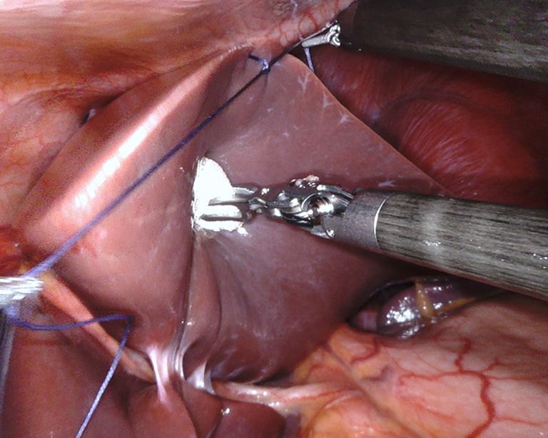
Annals of Laparoscopic and Endoscopic Surgery, 2021 Page 3 of 6
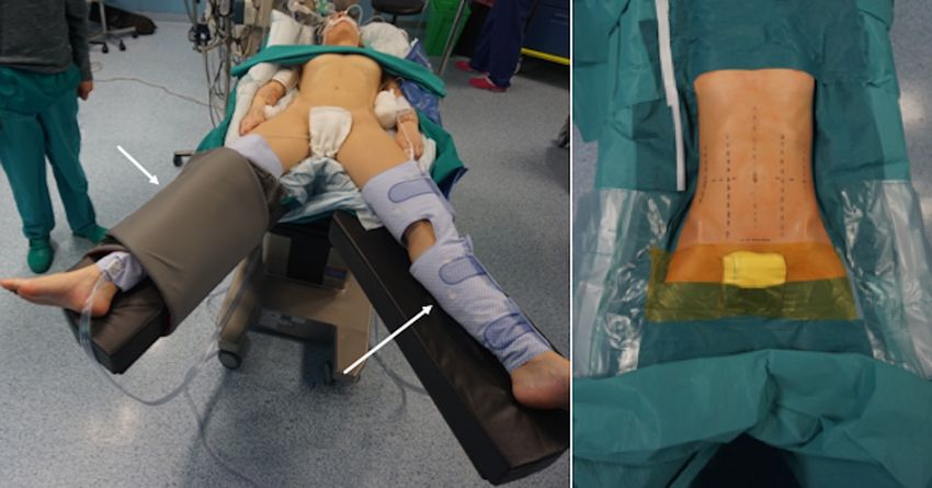
A B Operative field
Bandings
Intermittent
pneumatic cuffs
Figure 3 Patient’s position. (A) The patient is placed on the operative table in a French position and secured using wide bandings; (B) the
patient is widely prepped.
Figure 5 Liver segment number 3 being anchored to the
diaphragmatic dome.
dissection was further carried out posteriorly by detaching
the splenic vein from the pancreas. Dissection employed
Figure 4 Port position with respect to anatomical lines (1 right bipolar Maryland forceps and electrified scissors. The
anterior axillary line; 2 right pararectal line; 3 mid line; 4 left splenic artery was then identified at the superior border of
pararectal line; 5 left anterior axillary line; 6 transverse umbilical the pancreas and dissected off likewise with selective ligatures
line; 7 suprapubic extraction site). All ports are placed along the of pancreatic arteries using linen sutures (Figure 6). Both
transverse umbilical line. The optic port is placed at the umbilicus splenic vessels were encircled with vessel loops (Figure 7).
(R2). The remaining ports are placed to the right and to the Once a tunnel was developed behind the pancreatic neck,
left along the right (R1) and left (R4) axillary lines and the left the gland was looped and a robotized stapler, using a
pararectal line (R3). reinforced stapler armed with a purple cartridge. After
© Annals of Laparoscopic and Endoscopic Surgery. All rights reserved. Ann Laparosc Endosc Surg 2021;6:13 | http://dx.doi.org/10.21037/ales.2020.03.14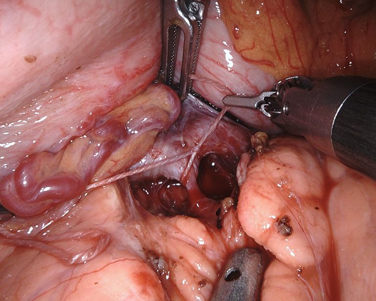
Page 4 of 6                                                                           Annals of Laparoscopic and Endoscopic Surgery, 2021

                                                                            division of the pancreas, dissection was carried out along
                                                                            the splenic vessels until the splenic hilum. Lymph nodes
                                                                            around the pancreatic artery were also removed. Spurting
                                                                            bleeders were fixed using 5/0 polypropylene sutures. At the
                                                                            end of the procedure both splenic vessels were completely
                                                                            skeletonized (Figure 8). The specimen was placed in an
                                                                            endoscopic bag and retrieved from a small suprapubic
                                                                            transverse incision. The round ligament of the liver was
                                                                            mobilized and placed around the pancreatic stump. A 14 Fr
                                                                            pig-tail drain was placed close to the pancreatic stump and
                                                                            left to drain by gravity.

Figure 6 Selective ligature of a pancreatic artery using linen
                                                                            Post-operative course
sutures.
                                                                            The post-operative course was uneventful, and the patient
                                                                            was discharged on postoperative day 6.

                                                                            Pathology

                                                                            Histology demonstrated a mucinous cystadenoma with low
                                                                            grade dysplasia measuring 42 mm.

                                                                            Discussion

                                                                            The first robotic distal pancreatectomy was reported in
                                                                            2003 (7). Several studies have shown that robotic distal
                                                                            pancreatectomy is not just feasible but also safe with
                                                                            respect to competing surgical approaches (8). While all
Figure 7 The splenic vessels and the pancreas are encircled with            types of minimally-invasive pancreatic resections have
vessel loops.                                                               been performed using laparoscopic technique, the use of
                                                                            robotic assistance is believed to be rewarding when fine
                                                                            dissections and intracorporeal sutures are needed (9).
                                                                            Distal pancreatectomy with preservation of both spleen
                                                                            and splenic vessels is certainly a procedure that requires
                                                                            fine intracorporeal dissections. Indeed the use of robotic
                                                                            assistance, when compared to laparoscopy in the setting of
                                                                            robotic spleen-preserving distal pancreatectomy, was shown
                                                                            to increase the rate of spleen preservation (10-12), to reduce
                                                                            the rate of conversion to open surgery, and to shorten
                                                                            the length of hospital stay (12). Additionally, laparoscopic
                                                                            spleen-preserving distal pancreatectomy has been associated
                                                                            with high rates of post-operative thrombosis of the splenic
                                                                            vessels leading to the formation of gastric varices. Although
                                                                            no direct comparison is available, the robotic technique was
Figure 8 Final intraoperative view of the spared splenic vessels.           associated with high rates of long-term patency of splenic

© Annals of Laparoscopic and Endoscopic Surgery. All rights reserved.   Ann Laparosc Endosc Surg 2021;6:13 | http://dx.doi.org/10.21037/ales.2020.03.14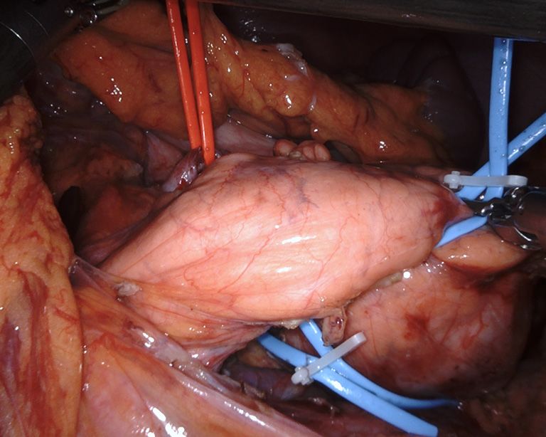
Annals of Laparoscopic and Endoscopic Surgery, 2021                                                                                      Page 5 of 6

vessels (13). The main argument against the use of robotic                  and any accompanying images. A copy of the written
assistance, especially in procedures that can be performed                  consent is available for review by the Editor-in-Chief of this
laparoscopically, is costs (14). Current estimates, however,                journal.
mostly take into account a quite rough evaluation of costs
associated to the use of the robot. More sophisticated                      Open Access Statement: This is an Open Access article
studies are clearly needed to define the economic                           distributed in accordance with the Creative Commons
sustainability of robotic assistance, as currently known, for               Attribution-NonCommercial-NoDerivs 4.0 International
procedures that can also per performed by conventional                      License (CC BY-NC-ND 4.0), which permits the non-
laparoscopic techniques (15).                                               commercial replication and distribution of the article with
   As specifically regards the technique shown in this video,               the strict proviso that no changes or edits are made and the
we have decided to remove the lymph nodes along the                         original work is properly cited (including links to both the
splenic artery. This is not standard and may not be required                formal publication through the relevant DOI and the license).
even in the context of mucinous cystadenocarcinoma due                      See: https://creativecommons.org/licenses/by-nc-nd/4.0/.
to the low rate of lymph node metastasis associated with
this tumor type (16). We prefer to remove the lymph nodes
                                                                            References
along the splenic vessels in case of a different diagnosis at
final pathology to have a complete staging of the tumor.                    1.  Asbun HJ, Moekotte AL, Vissers FL, et al. The Miami
   In conclusion in this video, we have demonstrated                            International Evidence-based Guidelines on Minimally
the technique for robotic spleen-preserving distal                              Invasive Pancreas Resection. Ann Surg 2020;271:1-14.
pancreatectomy, developed at a center having performed                      2. Shoup M, Brennan MF, McWhite K, et al. The value of
380 robotic pancreatic resections.                                              splenic preservation with distal pancreatectomy. Arch Surg
                                                                                2002;137:164-8.
                                                                            3. Kimura W, Inoue T, Futakawa N, et al. Spleen-preserving
Acknowledgments
                                                                                distal pancreatectomy with conservation of the splenic
Funding: None.                                                                  artery and vein. Surgery 1996;120:885-90.
                                                                            4. Warshaw AL. Conservation of the spleen with distal
                                                                                pancreatectomy. Arch Surg 1988;123:550-3.
Footnote
                                                                            5. Elabbasy F, Gadde R, Hanna MM, et al. Minimally
Reporting Checklist: The authors have completed the CARE                        invasive spleen-preserving distal pancreatectomy: Does
reporting checklist. Available at http://dx.doi.org/10.21037/                   splenic vessel preservation have better postoperative
ales.2020.03.14                                                                 outcomes? A systematic review and meta-analysis.
                                                                                Hepatobiliary Pancreat Dis Int 2015;14:346-53.
Conflicts of Interest: All authors have completed the ICMJE                 6. Chen S, Zhan Q, Chen JZ, et al. Robotic approach
uniform disclosure form (available at http://dx.doi.                            improves spleen-preserving rate and shortens postoperative
org/10.21037/ales.2020.03.14). The authors have no                              hospital stay of laparoscopic distal pancreatectomy: a
conflicts of interest to declare.                                               matched cohort study. Surg Endosc 2015;29:3507-18.
                                                                            7. Melvin WS, Needleman BJ, Krause KR, et al. Robotic
Ethical Statement: The authors are accountable for all                          resection of pancreatic neuroendocrine tumor. J
aspects of the work in ensuring that questions related                          Laparoendosc Adv Surg Tech A 2003;13:33-6.
to the accuracy or integrity of any part of the work are                    8. Cirocchi R, Partelli S, Coratti A, et al. Current status of
appropriately investigated and resolved. All procedures                         robotic distal pancreatectomy: a systematic review. Surg
performed in studies involving human participants were in                       Oncol 2013;22:201-7.
accordance with the ethical standards of the institutional                  9. Napoli N, Kauffmann EF, Menonna F, et al. Indications,
and/or national research committee(s) and with the Helsinki                     technique, and results of robotic pancreatoduodenectomy.
Declaration (as revised in 2013). Written informed consent                      Updates Surg 2016;68:295-305.
was obtained from the patient for publication of this study                 10. Kang CM, Kim DH, Lee WJ, et al. Conventional

© Annals of Laparoscopic and Endoscopic Surgery. All rights reserved.   Ann Laparosc Endosc Surg 2021;6:13 | http://dx.doi.org/10.21037/ales.2020.03.14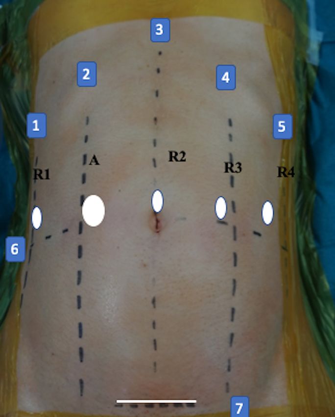
Page 6 of 6                                                                           Annals of Laparoscopic and Endoscopic Surgery, 2021

    laparoscopic and robot-assisted spleen-preserving                           Endosc 2013;27:774-81.
    pancreatectomy: does da Vinci have clinical advantages?                 14. Souche R, Herrero A, Bourel G, et al. Robotic versus
    Surg Endosc 2011;25:2004-9.                                                 laparoscopic distal pancreatectomy: a French prospective
11. Zhou JY, Xin C, Mou YP, et al. Robotic versus                               single-center experience and cost-effectiveness analysis.
    Laparoscopic Distal Pancreatectomy: A Meta-Analysis of                      Surg Endosc 2018;32:3562-9.
    Short-Term Outcomes. PLoS One 2016;11:e0151189.                         15. Patti JC, Ore AS, Barrows C, et al. Value-based assessment
12. Guerrini GP, Lauretta A, Belluco C, et al. Robotic versus                   of robotic pancreas and liver surgery. Hepatobiliary Surg
    laparoscopic distal pancreatectomy: an up-to-date meta-                     Nutr 2017;6:246-57.
    analysis. BMC Surg 2017;17:105.                                         16. Doulamis IP, Mylonas KS, Kalfountzos CE, et al.
13. Hwang HK, Kang CM, Chung YE, et al. Robot-assisted                          Pancreatic mucinous cystadenocarcinoma: Epidemiology
    spleen-preserving distal pancreatectomy: a single surgeon's                 and outcomes. Int J Surg 2016;35:76-82.
    experiences and proposal of clinical application. Surg

 doi: 10.21037/ales.2020.03.14
 Cite this article as: Kauffmann EF, Napoli N, Menonna F,
 Cacace C, Genovese V, Vistoli F, Boggi U. Robot-assisted
 spleen preserving distal pancreatectomy: case report. Ann
 Laparosc Endosc Surg 2021;6:13.

© Annals of Laparoscopic and Endoscopic Surgery. All rights reserved.   Ann Laparosc Endosc Surg 2021;6:13 | http://dx.doi.org/10.21037/ales.2020.03.14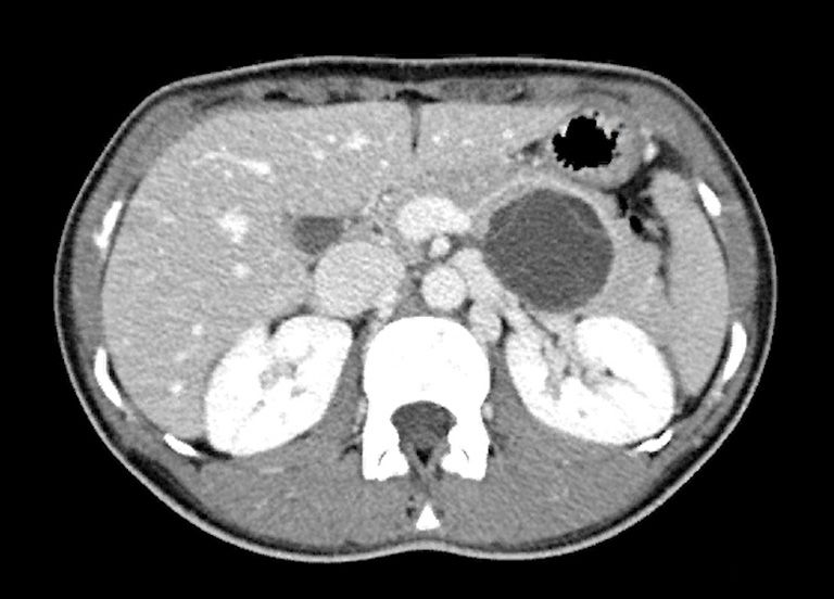
You can also read