Combined associating liver partition and portal vein ligation for staged hepatectomy (ALPPS) followed by left trisectionectomy and Whipple ...
←
→
Page content transcription
If your browser does not render page correctly, please read the page content below
Case Report

Combined associating liver partition and portal vein ligation for
staged hepatectomy (ALPPS) followed by left trisectionectomy
and Whipple operation for PNET
Ren Ji1, Shi Zuo1, Siyuan Qiu1, Ping Li2, Albert Chan1, William Sharr1, Chung Mau Lo1
1
    Division of Hepatobiliary and Pancreatic Surgery, Department of Surgery, 2Division of Pathology, University of Hong Kong-Shenzhen Hospital,
Shenzhen 518053, China
Correspondence to: Ren Ji, MS. Division of Hepatobiliary and Pancreatic Surgery, Department of Surgery, University of Hong Kong-Shenzhen
Hospital, Shenzhen 518053, China. Email: jir@hku-szh.org.

                   Abstract: Pancreatic neuroendocrine tumor (PNET) is slow-growing, and account only for 2% of all
                   pancreatic primary tumors. Surgical resection is still the only curative treatment for PNET patients.
                   Unfortunately, most of PNETs was found with unresectable multiple liver metastases and extrahepatic
                   metastasis as their characteristics of non-functional and asymptomatic. With advances in liver surgery in
                   these years, especially combined associating liver partition and portal vein ligation for staged hepatectomy
                   (ALPPS), provide a new curative surgical treatment for PNET with liver metastases patient. Here we report
                   a PNET with multiple liver metastases case underwent ALPPS (followed by left trisectionectomy) and
                   Whipple operation within one-stage.

                   Keywords: Neuroendocrine tumor; liver metastases; surgical resection

                   Submitted Sep 26, 2017. Accepted for publication Nov 20, 2017.
                   doi: 10.21037/gs.2017.11.15
                   View this article at: http://dx.doi.org/10.21037/gs.2017.11.15

Introduction                                                                   Case presentation

Pancreatic neuroendocrine tumor (PNET) is slow-                                We report on a 37-year-old male patient (176 cm, 68 kg,
growing (1), and account only for 2% of all pancreatic                         good past medical history) who presented uncharacteristic
primary tumors. Surgical resection is still the only                           symptoms (i.e., epigastric pain, diarrhea, weight lost)
curative treatment for PNET patients. Unfortunately,                           2 months prior to admittance, which led to the initiation of
most of PNETs was found with unresectable multiple                             diagnostic procedures by a gastroenterologist and included
liver metastases and extrahepatic metastasis as their                          an esophagogastroscopy, abdominal computed tomography
characteristics of non-functional and asymptomatic.                            (CT) (Figure 1) and PET-CT. The esophagogastroscopy
With advances in liver surgery in these years, especially                      revealed multiple ulcers of the gastric antrum and
combined associating liver partition and portal vein                           duodenum (Figure 2). Based on the CT imaging studies
ligation for staged hepatectomy (ALPPS), provide a new                         bilobular hepatocellular carcinoma (HCC) was diagnosed,
curative surgical treatment for PNET with liver metastases                     although the HBVs Ag is negative and the serum tumor
patient. Here we reported a case of a 36-year-old man who                      marker alpha fetoprotein is normal. Other suspicious for
was diagnosed PNET with liver metastases and underwent                         metastatic disease (i.e., pancreas, prostate, colon), were
ALPPS (followed by left trisectionectomy) and Whipple                          excluded by CT and PET-CT. In addition, the serum
operation within one-stage.                                                    tumor marker CEA, CA19-9, and prostate-specific antigen

© Gland Surgery. All rights reserved.                                gs.amegroups.com                                Gland Surg 2018;7(1):47-53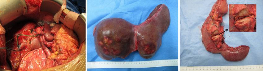
48                                                                                                             Ji et al. Combined ALPPS

                    A                                                 B

                    C                                                 D

Figure 1 Upper abdominal computed tomography. (A) Plain phase; (B) arterial phase; (C) portal vein phase; (D) portal vein phase: no
suspicious of pancreatic head.

                     A                                               B

Figure 2 esophagogastroscopy photos. (A) Multiple ulcers of the gastric antrum; (B) multiple ulcers of duodenum.

were within normal reference range. Besides the patient                  ultrasound (IOUS) in order to evaluate resectability of the
was feeling fine with no clinical symptoms of diabetes,                  hepatic lesions. The hepatic lesions identified by IOUS
hypoglycemia, hypopituitarism, hypoparathyroidism.                       were resected by multiple sub-segmental resections (n=6,
                                                                         SI/II/III/IVa/IVb/V/VIII). As calculating the future liver
                                                                         remnant (FLR)/estimated standard liver volume (ESLV)
Clinical course—hepatic surgery (ALPPS stage 1)
                                                                         ratio just 24%, significant lower than our standard of 30%
After routine exploration for abdominal surgery, we                      (no cirrosis). ALPPS stage 1 was performed, during the
fully mobilized the liver and performed intraoperative                   operation we ligated the left portal vein and partition the

© Gland Surgery. All rights reserved.                         gs.amegroups.com                                     Gland Surg 2018;7(1):47-53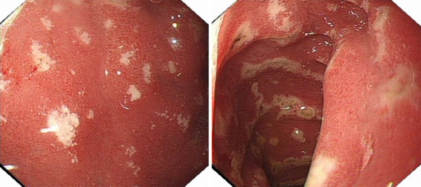
Gland Surgery, Vol 7, No1 February 2018 49
A B C
Figure 3 Intraoperative photos. (A) Tumor; (B) anatomy of right pedicle; (C) CUSA partition the SVI/VII from SV/VIII. CUSA, Cavitron
Ultrasonic Surgical Aspirator.
A B
C D
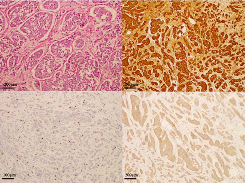
Figure 4 Microscopically, pancreatic tumor consists of relative uniform cells forming solid nests. (A) HE staining showed small sized tumor
cells with bland feature (×20); (B) immunohistochemically, the tumor cells were positive for chromogranin (×20); (C) Ki-67 staining (proliferation)
showed a lower proliferation rate with index about 5% (×10); (D) immunohistochemically, the tumor cells were positive for synaptophysin (×20).
SVI/VII from SV/VIII by CUSA (Cavitron Ultrasonic leakage and liver failure. On day 10, the histopathological
Surgical Aspirator, Valleylab, Boulder, CO, USA). One examinations of the resected liver specimen and the
additional lesion (2 cm) in SIVb and some enlarged lymph lymph nodes in ligamentum hepatoduodenal revealed
nodes in ligamentum hepatoduodenal was resected for metastases of a NET. Microscopically, both the specimen
pathologic diagnosis (Figure 3A-C). showed small-sized ovoid cells arranged in nests separated
by broad fibrous bands and glandular duct formation.
They displayed poorly differentiated with large nuclei.
Clinical course—diagnosis correcting
Immunohistochemically, the tumors were positive for
The ALPPS stage 1 hepatic surgery postoperative chromogranin A, synaptophysin, Ki-67 (with index about
course was uneventful, with no evidence of bleeding, bile 5%), but negative for CD56 (Figure 4).
© Gland Surgery. All rights reserved. gs.amegroups.com Gland Surg 2018;7(1):47-53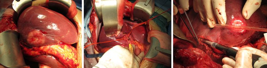
50                                                                                                              Ji et al. Combined ALPPS

                                                                           As the primary liver NET is rare, and most primary
                                                                        NET come from pancreas. We performed an emergency
                                                                        MRI examination which identified a small size lesion located
                                                                        at the pancreatic head (Figure 5). In general, corresponded
                                                                        to the resected metastatic liver NET, the lymph nodes
                                                                        in ligamentum hepatoduodenal and the MRI image, we
                                                                        corrected the diagnosis with PNET with liver metastases.

                                                                        Clinical course—stage 2 surgery (ALPPS stage 2
                                                                        combine Whipple procedure)

                                                                        As the patient is young, and recover very smoothly from
                                                                        the stage 1 operation, the SVI/VII volumetry (FLR/ESLV)
                                                                        from 24% increase to 38% (Figure 6), we performed ALPPS
Figure 5 MRI revealed a lesion (size 1.6 cm × 2.3 cm) in pancreatic     stage 2 (left trisectionectomy) combine Whipple procedure
head. The arrow indicates the tumor of pancreatic head.                 at the same time (Figure 7A). During operation pancreatic
                                                                        head tumor identified by IOUS and frozen biopsy section.
                                                                        The histopathological examinations of the resected liver
                                                                        specimen (Figure 7B) revealed metastases of a NET, that, in
                                                                        general, corresponded to the resected primary lesion in the
                                                                        pancreatic head (Figure 7C). Both the primary pancreatic
                                                                        tumor and the liver metastases showed the same typical
                                                                        neuroendocrine tumor.

                                                                        Follow up

                                                                        Four years passed, the patient did not accept chemotherapy
Figure 6 Hypertrophy of right posterior lobe (from 328 to               as patient’s personal reasons. There was no evidence for
504 cm3).                                                               NET recurrence within the liver or pancreas by CT or MRI

            A                                     B                                         C

Figure 7 Intraoperative photo of stage 2 operation and the specimens. (A) specimen removed during stage 2 operation; (B) bilobed multiple
solid tumor; (C) Pancreatic head solid tumor, size about 2.0 cm ×1.8 cm. The arrow indicates the tumor of pancreatic head.

© Gland Surgery. All rights reserved.                          gs.amegroups.com                                  Gland Surg 2018;7(1):47-53
Gland Surgery, Vol 7, No1 February 2018 51
bowel, prostate) (4). Just like this case we report, NETs
are often present in an advanced stage with an unknown
primary or initial diagnosed with metastasis (5,6). For the
PNET liver metastasis patients, besides the resection of
liver metastatic lesions, it is more important to find the
primary site of pancreas, because it has great impact on the
patient’s outcome by resection of the primary pancreatic
tumor (4). We should be aware of, primary liver-NET, is an
extremely rare occurrence, and until 2015, there were only
less than 150 reported cases in the English literature (7). So,
once we detected PNET in liver, ought to try to identify
the primary for an optimal therapy. Over the last few years
the detection rate of primary NET in the pancreas by MRI
diffusion weighted imaging (DWI) has been improved (8).
Recently, somatostatin receptor PET/CT scan technique
was one of topics of most concern, as studies reported it
can detects about 59% unknown NET primaries. However,
which means, even by using such a sensitive technique,
more than 1/3 of patients’ primaries could not be found (9).
Liver transplantation (LT) for unresectable metastases
has essentially been abandoned as its high rate of tumor
recurrence (10). Without exception, several reports showed
poor results on LT apply to PNET patients with liver
Figure 8 Recently CT scan: no evidence for tumor recurrence
metastases between 1960 and the 1980s. In the present
within the liver and pancreas.
critical situation of donor shortage, LT apply to PNET
patients with liver metastases should be strictly controlled.
It has been wildly accepted that only highly selected PNET
(once per 6 months) (Figure 8), the patient general condition
patients with liver metastases may be candidates for LT
is good, diarrhea symptom also improved obviously.
(11-13). The only prospective study recommended strict
selection criteria (i.e., low grade, removal of primary tumor,
Discussion liver involvement52 Ji et al. Combined ALPPS
mentioning is the NET patients’ liver is normal, which with other neuroendocrine tumors. Cancer Treat Rev
always contribute a rapid increase of FLR hypertrophy, are 2011;37:358-65.
good cases for this procedure. 7. Mousavi SR, Ahadi M. Primary Neuroendocrine Tumor
Only very few literatures compare the outcome of one- of Liver (Rare Tumor of Liver). Iran J Cancer Prev
stage or two-stage hepatectomy for metastatic liver tumor 2015;8:e3144.
in patients undergoing Whipple operation, a dual center 8. Brenner R, Metens T, Bali M, et al. Pancreatic
analysis showed the advantage of less abscess incidence (7%) neuroendocrine tumor: added value of fusion of T2-
in one-stage operation group (22). To our knowledge, by weighted imaging and high b-value diffusion-weighted
retrieving literatures, we find no literature on the ALPPS imaging for tumor detection. Eur J Radiol 2012;81:e746-9.
combine Whipple operation for PNET within one-stage. 9. Prasad V, Ambrosini V, Hommann M, et al. Detection of
In conclusion, combined ALPPS and Whipple operation unknown primary neuroendocrine tumours (CUP-NET)
within one-stage appears to be a valuable option for patients using (68)Ga-DOTA-NOC receptor PET/CT. Eur J Nucl
with advanced pancreatic NETs metastatic to the liver, thus Med Mol Imaging 2010;37:67-77.
facilitating curative liver surgery. 10. Chapman WC. Liver transplantation for unresectable
metastases to the liver: a new era in transplantation or a
time for caution? Ann Surg 2013;257:816-7.
Acknowledgements
11. Gottwald T, Koveker G, Busing M, et al. Diagnosis and
None. management of metastatic gastrinoma by multimodality
treatment including liver transplantation: report of a case.
Surg Today 1998;28:551-8.
Footnote
12. Le Treut YP, Gregoire E, Klempnauer J, et al. Liver
Conflicts of Interest: The authors have no conflicts of interest transplantation for neuroendocrine tumors in Europe-
to declare. results and trends in patient selection: a 213-case European
liver transplant registry study. Ann Surg 2013;257:807-15.
Informed Consent: Written informed consent was obtained 13. Mazzaferro V, Pulvirenti A, Coppa J. Neuroendocrine
from the patient for publication of this case report and any tumors metastatic to the liver: how to select patients for
accompanying images. liver transplantation? J Hepatol 2007;47:460-6.
14. Schnitzbauer AA, Lang SA, Goessmann H, et al. Right
portal vein ligation combined with in situ splitting induces
References
rapid left lateral liver lobe hypertrophy enabling 2-staged
1. Kimura W, Tezuka K, Hirai I. Surgical management extended right hepatic resection in small-for-size settings.
of pancreatic neuroendocrine tumors. Surg Today Ann Surg 2012;255:405-14.
2011;41:1332-43. 15. Donati M, Stavrou GA, Oldhafer KJ. Current position
2. Frilling A, Modlin IM, Kidd M, et al. Recommendations of ALPPS in the surgical landscape of CRLM treatment
for management of patients with neuroendocrine liver proposals. World J Gastroenterol 2013;19:6548-54.
metastases. Lancet Oncol 2014;15:e8-21. 16. Aloia TA, Vauthey JN. Associating liver partition and
3. Berardi R, Rinaldi S, Torniai M, et al. Gastrointestinal portal vein ligation for staged hepatectomy (ALPPS): what
neuroendocrine tumors: Searching the optimal treatment is gained and what is lost? Ann Surg 2012;256:e9; author
strategy--A literature review. Crit Rev Oncol Hematol reply e16-9.
2016;98:264-74. 17. Yaprak O, Guler N, Altaca G, et al. Ratio of remnant to
4. Bergsland EK, Nakakura EK. Neuroendocrine tumors of total liver volume or remnant to body weight: which one
unknown primary: is the primary site really not known? is more predictive on donor outcomes? HPB (Oxford)
JAMA Surg 2014;149:889-90. 2012;14:476-82.
5. Morris GJ, Greco FA, Hainsworth JD, et al. Cancer of 18. Torres OJ, Fernandes Ede S, et al. Associating liver
unknown primary site. Semin Oncol 2010;37:71-9. partition and portal vein ligation for staged hepatectomy
6. Stoyianni A, Pentheroudakis G, Pavlidis N. (ALPPS): the Brazilian experience. Arq Bras Cir Dig
Neuroendocrine carcinoma of unknown primary: a 2013;26:40-3.
systematic review of the literature and a comparative study 19. de Santibañes E, Clavien PA. Playing Play-Doh to prevent
© Gland Surgery. All rights reserved. gs.amegroups.com Gland Surg 2018;7(1):47-53Gland Surgery, Vol 7, No1 February 2018 53
postoperative liver failure: the "ALPPS" approach. Ann carcinoma with major vascular invasion. World J Surg
Surg 2012;255:415-7. 2014;38:1498-503.
20. Andriani OC. Long-term results with associating liver 22. De Jong MC, Farnell MB, et al. Liver-directed
partition and portal vein ligation for staged hepatectomy therapy for hepatic metastases in patients undergoing
(ALPPS). Ann Surg 2012;256:e5; author reply e16-9. pancreaticoduodenectomy: a dual-center analysis. Ann
21. Vennarecci G, Laurenzi A, Santoro R, et al. The Surg 2010;252:142-8.
ALPPS procedure: a surgical option for hepatocellular
Cite this article as: Ji R, Zuo S, Qiu S, Li P, Chan A, Sharr
W, Lo CM. Combined associating liver partition and portal
vein ligation for staged hepatectomy (ALPPS) followed by left
trisectionectomy and Whipple operation for PNET. Gland
Surg 2018;7(1):47-53. doi: 10.21037/gs.2017.11.15
© Gland Surgery. All rights reserved. gs.amegroups.com Gland Surg 2018;7(1):47-53You can also read