Clear Cell Sarcoma of the Kidney: Correlational Study
←
→
Page content transcription
If your browser does not render page correctly, please read the page content below
JMSCR Vol||08||Issue||07||Page 77-82||July                                                  2020
                                                                        http://jmscr.igmpublication.org/home/
                                                                   ISSN (e)-2347-176x ISSN (p) 2455-0450
                                                               DOI: https://dx.doi.org/10.18535/jmscr/v8i7.17

               Clear Cell Sarcoma of the Kidney: Correlational Study
                                                  Authors
                           Teena Sharma , Hema Udawat2, Ranjana Solanki3,
                                        1*

                                  Deepika Hemrajani4, Arpita Jindal5
                    1 rd
                   3 year Resident, Department of Pathology, SMS Medical College, Jaipur
                2
                  Associate Professor, Department of Pathology SMS medical College, Jaipur
                  3
                    Senior Professor, Department of Pathology, SMS Medical College, Jaipur
                4
                  Associate Professor, Department of Pathology, SMS Medical College, Jaipur
                  5
                    Senior Professor, Department of Pathology, SMS Medical College, Jaipur
                                            *Corresponding Author
                                               Teena Sharma
                         Department of Pathology, SMS Medical College, Jaipur, India
  Abstract
  Clear cell sarcoma of kidney is a rare malignant renal neoplasm of childhood, known for its
  aggressiveness, its tendency for recurrence and metastasis to bone. Three cases of clear cell sarcoma are
  being reported.
  Case 1: A two year old male child having complaint off pain in right lumbar region.USG abdomen
  revealed right hypoechoic lesion. CECT abdomen revealed 13x11x1ocm heterogenous mass involving
  right kidney. Microscopy revealed clear cell sarcoma of kidney (classic pattern). Immunohistochemistry
  showed strong vimentin positivity and negative for cytokeratin and WT-1.
  Case 2: A thirty months old male child having complaint of pain in left abdominal region since 8 months.
  There was history of fever and increased micturition. There was no history of weight loss. His vitals
  stable. Biochemical and hematological profiles were normal. USG revealed left heterogenous mass lesion
  ms 6x4x2cm involving left kidney. CT revealed hypoechoic heterogenous mass ms 6x4x2cm involving left
  kidney. Microscopy revealed clear cell sarcoma.
  Case 3: A 2 year old male child presented with right abdominal pain since 1 year. There was no history of
  fever, weight loss, hematuria. On examination patient had pallor only. Hematological finding normal.
  Urine examination normal. Chest X-ray normal. USG revealed right hypoechoic heterogenous mass
  ms13x10x10cm involving right kidney. CT revealed hypoechoic heterogenous mass ms 13x10x10cm
  involving right kidney.
  The purpose of this paper is to review the published series and case reports of CCSK and to create
  an up-to-date overview of clinical and histological features and IHC.
  Keywords: Tumor, Malignant, Bone metastasizing, child.

 Introduction-                                            metastasis to bone1,2. Its peak incidence is in 3-5
 Clear cell sarcoma of kidney (CCSK) is a rare            years with slight male preponderance3. In this
 malignant neoplasm of childhood, known for its           study, we report three cases of clear cell sarcoma
 aggressiveness, its tendency for recurrence and

 Teena Sharma et al JMSCR Volume 08 Issue 07 July 2020                                              Page 77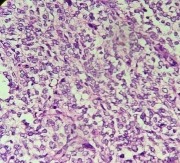
JMSCR Vol||08||Issue||07||Page 77-82||July 2020
kidney, which were diagnosed in Department of lesion ms 6x4x2cm involving left kidney. CT
Pathology, SMS medical college, Jaipur. revealed hypoechoic heterogenous mass ms
6x4x2cm involving left kidney. Patient underwent
Case 1 surgery and specimen was sent for
A two year old male child presented with histopathological examination. Gross examination
complaint of pain in right lumbar region since 6 showed unoriented grey white soft tissue mass ms
months. There was no history of hematuria, 6x4x2cm. External surface was grey white. Cut
vomiting, loss of weight. On examination patient surface fleshy grey white. Microscopically
had pallor, Vitals were stable. Investigations showed round to oval cells having vesicular
revealed: Hemoglobin of 11gm%, normal total nuclei, inconspicuous nucleoli, clear to
and differential cell count, ESR 15mm/hr. Urine eosinophilic cytoplasm arranged diffusely and in
examination was normal.USG whole abdomen nest, surrounded by delicate arborizing
revealed a 13x10x10cm hypoechoic heterogenous vasculature. Occasional nuclear grooving also
mass uniformly involving right kidney. CECT noted. Mitosis are brisk. Feature favoured
abdomen revealed 13x8x6cm heterogenous mass diagnosis of clear cell sarcoma kidney (Classic
involving right kidney. Provisional diagnosis of pattern).
Wilms tumor was made on radiology. Right
nephrectomy was done and specimen was sent for Case 3
histopathological examination. On gross A 2 year old male child presented with right
examination nephrectomy specimen measured abdominal pain since 1 year. There was no history
15x12x12cm. External surface grey brown of fever, weight loss, hematuria. On examination
bosselated, covered by Gerotas fascia with no patient had pallor only. Hematological profile was
capsular breach. On cutting tumor ms normal. Urine examination normal. Chest X-ray
13x10x10cm, and grey white, glistening and normal. USG revealed right hypoechoic
showed focal whorled appearance. At one end heterogenous mass ms13x10x10cm involving
normal kidney parenchyma identified (Fig 1). right kidney. CT revealed hypoechoic
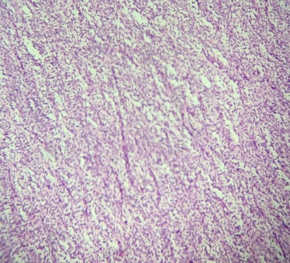
Microscopically revealed presence of heterogenous mass ms 13x10x10cm involving
monomorphic cells arranged in sheets and nests right kidney. Right nephrectomy was done and
separated by delicate arborizing fibro vascular specimen was sent for histopathological
network. The cells were polygonal to spindle examination. On gross examination specimen
shaped with indistinct cell borders and vesicular measured 15x12x10cm. External surface
nucleus showing grooves at many places and bosselated in appearance. No breaching of capsule
variable mitosis. Diagnosis of clear cell sarcoma seen. Cut surface showed a tumor ms
of kidney (classic pattern) was made (Fig 2a and 13x10x10cm and grey white, showed whorling
2b). Immunohisto chemistry showed strong appearance. At one end normal kidney
positivity for vimentin and negativity for parenchyma identified. Microscopically revealed
cytokeratin and WT-1 (Fig 3a, b, c). presence of monomorphic tumor cells arranged in
cords, sheets, nests and alveolar pattern separated
Case 2 by thin capillary network. The cells were spindle
An thirty months old male child presented with shaped to oval with distinct cell borders and
complaint of pain in left abdominal region since 8 vesicular nucleus showing nuclear grooves at
months. There was past history of fever and many places and variable mitosis surrounded by
increased micturition. His vitals were stable. compressed normal renal tissue. Overall
Biochemical and Hematological profiles were morphology favoured diagnosis of clear cell
normal. USG revealed left heterogenous mass sarcoma of kidney (Classic pattern).
Teena Sharma et al JMSCR Volume 08 Issue 07 July 2020 Page 78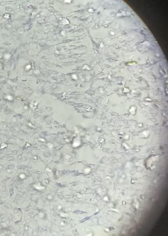
JMSCR Vol||08||Issue||07||Page 77-82||July                                          2020
                                                   3a

 Fig 1.Right nephrectomy specimen of clear cell
 sarcoma kidney. Cut surface grey white,           3b
 transluscent.

 2a

                                                   3c

 2b

                                                   Fig 3a). Tumor cells show vimentin positivity.
                                                   b). Cytokeratin negative. c) WT1 negative.

                                                   Clinical Features
 Fig 2. Microscopic view a).Tumor cells arranged   The mean age of presentation in my study is about
 in nest pattern. (10x) b). Tumor cells having     24 months. Argani et al found that 50% of their
 vacuolated cytoplasm and surrounds by delicate    351 cases were diagnosed between 2 and 3 years
 vasculature.(40x)                                 of age4-6. CCSK is extremely rare in the first 6
                                                   months of life and in adults.7-20 The youngest

 Teena Sharma et al JMSCR Volume 08 Issue 07 July 2020                                      Page 79
JMSCR Vol||08||Issue||07||Page 77-82||July 2020
CCSK patient reported in literature was a foetus CCSK from other renal tumors, it is important that
(at 31week gestation) and the oldest patient was suspected CCSK cases are reviewed by
58 years old.8,16 pathologists who were experts in paediatric renal
Contrary to WT’s slight propensity to the female tumors.
gender, a male predominance has been noted in all
large CCSK series (average male to female ratio Immunohistochemistry
of about 2:1).4,6 In my study all three cases male Immunohistochemistry can help to distinguish
predominace noted. CCSK from other renal tumors of childhood. IHC
All three patients in my study series show done in one of the 3 cases shows non specific
common clinical presentation of abdominal pain. vimentin positivity and negativity for WT-1 and
One case has fever and increased micturition also cytokeratin.
noted. Other symptoms that have been described
are hematuria, vomiting, decreased oral intake, Discussion and Conclusion
constipation and hypertension21-25 in other studies Clear cell sarcoma also known as “Bone
which were absent in my study series. Some study metastasizing tumor of Childhood” comprise 4%
suggested a predilection for involvement of the of all primary childhood renal tumors30.
right kidney, in my study case 2 out 3 showed Histological variants of CCSK include spindle
right predilection. cell, sclerosing, epithelioid, myxoid, pallisading,
storiform and anaplastic clear cell sarcoma31.
Pathology Accurate and early diagnosis of clear cell sarcoma
All CCSK typically presents as a large, unicentric, as a separate entity from wilms tumor is of
well circumscribed and sharply demarcated mass. extreme importance. Careful study of
The tumor arises from the central region and morphology, the characteristic delicate arborizing
replaces normal kidney tissue or is located in the vascular septae, monomorphous population and
medulla of the kidney.26 The diameter of the invasive property will help to distinguish it from
tumor ranges in my study from 6 to 13cm. CCSK more common and less aggressive counterparts of
is soft, tan grey in color. External surface childhood renal neoplasm like Wilms tumor32.
bosselated and cut surface glistened due to Points helpful in this matter are: Foci of blastema
mucinous material in all cases. are not seen in clear cell sarcoma; nonrenal
Microscopically classic pattern predominant on elements such as cartilage or muscle are not found
other histologic patterns. In other studies also in clear cell sarcomas. Clear cell sarcomas are
classic pattern is more common. The classic unilateral and unicentric, and sclerotic stroma is
subtype of CCSK is characterized by round /oval uncommon in wilms tumor before therapy. The
cells with clear cytoplasm, fairly uniform round/ vascular pattern typical of clear cell sarcoma is
oval and often vesicular bland nuclei with finely often often helpful in distinguishing it from Wilms
dispersed chromatin, inconspicuous nucleoli and tumor. The border with the kidney is usually
infrequent mitotic figures.26,27 In the classic infiltrative in CCSK, whereas the border of Wilms
pattern, tumor cells are arranged in nests or cords, tumor is typically pushing33.
separated by fibrovascular septa.28 Other pattern Immunohistochemistry for Clear cell sarcoma
that have been described include myxoid, which is positive for vimentin negative for WT1,
sclerosing, cellular, epitheloid, pallisading, and wilms tumor positive for WT 1. Treatment of
spindle cell, storiform and anaplastic.29 CCSK generaly involve surgical intervention
The histological heterogeneity emphasizes the coupled with radiation and chemotheraphy with
importance of discriminating the disease from cyclophosphamide, etoposide and vincristin and
other entities. Considering the low incidence of doxorubicin for 24 weeks It is of considerable
Teena Sharma et al JMSCR Volume 08 Issue 07 July 2020 Page 80JMSCR Vol||08||Issue||07||Page 77-82||July 2020
therapeutic importance that CCSK be not only 2005;446:566–8.
correctly diagnosed but early too. 9. Kural AR, Onal B, Ozkara H, Cakarir C,
Ayan I, Agaoglu FY. Clear cell sarcoma
References of the kidney: a case report. BMC Urol
1. Sharma SC, Menon PA.Clear cell sarcoma 2006;6:11.
of the kidney. J Postgrade Med 2001; 10. Rosso D, Ghignone GP, Bernardi D, et
47:206-7. al. Clear cell sarcoma of the kidney with
2. Namaoui RY, Castex MP, Vial J , Galinier invasion of the inferior vena cava. Urol Int
P, Rubie H, Laprie Mazieres A, et al.Clear 2003;70:251–2.
cell sarcoma of the kidney: About a 11. Amin MB, Peralta-Venturina MN, Rossi
pediatric case.Prog Urol 2010;20:465-8. JY, et al. Clear cell sarcoma of the
3. Argani P, Perlman EJ, Breslow NE, kidney in an adolescent and in young
Browning NG, Green DM, D Angio et adults. A report of four cases with
al.Clear cell srcoma kidney: A review of ultrastructural, immunohistochemical
351 cases from the National Wilms Tumor and DNA flow cytometric analysis. Am
Study Group Pathology Center.Am J Surg J Surg Pathol 1999;23:1455–63.
Pathol 2000;24:4-18. 12. Bhayani SB, Liapsis H, Kibel AS. Adult
4. Seibel NL, Sierra LI, Breslow NE, clear cell sarcoma of the kidney with
Beckwith JB, Green DM. Eff ect of atrial tumor thrombus. J Urol
duration of treatment on treatment 2001;165:896–7.
outcome for patients with clear-cell 13. Oda H, Shiga J, Machinami R. Clear cell
sarcoma of the kidney: a report from the sarcoma of the kidney: two cases in
National Wilms’ Study Group. J Clin adults. Cancer 1993;71:2286–91.
Oncol 2004;22:468–73. 14. Mishra VK, Krishani N, Bandari M.
5. Green DM, Breslow NE, Beckwith JB, Clear cell sarcoma of the kidney in an
Moksness J, Finklestein JZ, D’Angio adult. Br J Urol 1993;72:118.
GJ. Treatment of children with clear-cell 15. Toyoda Y, Yamashiota C, Sugimoto T,
sarcoma of the kidney: a report from the Yoshida M, Okada M. Clear cell
National Wilms’ Tumor Study Group. J sarcoma of the kidney with tumor
Clin Oncol 1994;12:2132–7. extension into the right atrium. J
6. Seibel N, Sun J, Andersen JR. Outcome Cardiovasc Surg (Torino) 1998;39:489–
of clear cell sarcoma of the kidney 91.
(CCSK) treated on the National Wilms’ 16. Benchekroun A, Ghadouane M,
Tumor Study-5 (NWTS). 24,5022006 Zannoud M, Alami M, Amhajji R, Faik
Ref Type Conf Proc Ref ID 2006:6076. M. Clear cell sarcoma of the kidney in
7. Van den Heuvel-Eibrink MM, Grundy an adult. Ann Urol (Paris) 2002;36:33–
P, Graf N, et al. Charac- teristics and 5.
survival of 750 children diagnosed with 17. Adnani A, Latib R, Bouklata S, Ajana A,
a renal tumor in the first seven months Hammani L, Imani F. Sarcome à cellules
of life: a collaborative study by the claires du rein chez l’adulte: à propos
SIOP/ GPOH/SFOP, NWTSG, and d’un cas. J Radiol 2006;87:136–8.
UKCCSG Wilms’ tumor study groups. 18. Suzuki H, Honzumi M, Itoh Y,
Pediatr Blood Cancer 2008;50:1130–4. Umehara N, Moriyama S, FunadaM.
8. Hung N. Congenital “clear cell sarcoma of Clear cell sarcoma of the kidney seen in
the kidney”. Virchows Arch a 3-day old newborn.Z Kinderchir
Teena Sharma et al JMSCR Volume 08 Issue 07 July 2020 Page 81JMSCR Vol||08||Issue||07||Page 77-82||July 2020
1983;38:422–4. the kidney. JKMS 1997;12:473–76.
19. Newbould MJ, Kelsey AM. Clear cell 30. Bo Xie, Jiajun Ling.Clear cell sarcoma of
sarcoma of the kidney in a 4-month-old the kidney-A case report. Chinese J Clin
infant: a case report. Med Pediatr Oncol Onco. 2006; 2:151-152.
1993;21:525–8. 31. Murphy WM, Beckwith JB, Farrow
20. Mazzoleni S, Vecchiato L, Alaggio R, GM.Tumors of the kidney. In: Murphy
Cecchetto G, Zorzi C, Carli M. Clear WM, editor. Tumors of the Kidney,
cell sarcoma of the kidney in a newborn. Bladder and Related Urinary Structures.
Med Pediatr Oncol 2003;41:153–5. Washington D.C, The Armed Forces
21. Wood DP, Kay R, Norris D. Renal Institute of Pathology; 1993. p.67-81.
sarcomas of childhood. Urology 32. Sandstedt BE, Delemarre JF, Harms D,
1990;36:73–8. Tournade MF. Sarcomatous Wilms tumor
22. Yumura-Yagi K, Inoue M, Wakabayashi with clear cells and hyalinization. A study
R, et al. Successful double autografts for of 38 tumors in children from the SIOP
patients with relapsed clear cell sarcoma nephroblastoma file. Histopathology
of the kidney. Bone Marrow Transplant 1987;11:273-85.
1998;22:381–3. 33. Murphy WM, Beckwith JB, Farrow GM.
23. Parikh SH, Chintagumpala M, Hicks J, Tumors of the kidney, bladder and related
et al. Clear cell sarcoma of the kidney: uriary structures. Washington D.C. The
an unusual presentation and review of Armed Forces Institute of Pathology;
the literature. J Pediatr Hematol Oncol 1993. p.67-81.
1998;20:165–8.
24. Sharma SC, Menon PA. Clear cell
sarcoma of the kidney. J Postgrad Med
2001;47:206–7.
25. Kagan AR, Steckel RJ, Sleight G, Lock
MM. Clear cell sarcoma of the kidney: a
renal tumor of childhood that
metastasize to bone. AJR 1986;146:64–
6.
26. Balarezo FS, Joshi VV. Clear cell
sarcoma of the pediatric kidney: detailed
description and analysis of variant
histologic patterns of a tumor with many
faces. Adv Anat Pathol 2001;8:98–108.
27. Watts KE, Hansel DE, MacLennan GT.
Clear cell sarcoma of the kidney. J Urol
2011;185:279–80.
28. Boo YJ, Fisher JC, Haley MJ, Cowles
RA, Kandel JJ, Yamashiro DJ. Vascular
characterization of clear cell sarcoma of
the kidney in a child: a case report and
review. J Pediatr Surg 2009;44:2031–6.
29. Park DY KY, Chi JG. Intracranial
metastasis from clear cell sarcoma of
Teena Sharma et al JMSCR Volume 08 Issue 07 July 2020 Page 82You can also read