The Coronal Pulp Cavity Index an aid in age determination -A Cone Beam Computed Tomography Study
←
→
Page content transcription
If your browser does not render page correctly, please read the page content below
896     Indian Journal of Forensic Medicine & Toxicology, January-March 2021, Vol. 15, No. 1

The Coronal Pulp Cavity Index an aid in age determination -A
        Cone Beam Computed Tomography Study

                  Ceena Denny E1, Bastian TS2, Srikant Natarajan3, Nithin Thilak4, Almas Binnal5
1Associate   Professor, 2Professor and Head, 3Professor and Head, 4Post Graduate, Associate Professor, Department
      of Oral and Maxillofacial Pathology, Manipal College of Dental Sciences, Mangalore, Manipal Academy of
                                     Higher Education, Manipal, Karnataka.India

                                                              Abstract
      Background: The objective of the present study was to assess the accuracy of age estimation from TCI of
      mandibular molars (except wisdom teeth) of both sides using CBCT images of known age and sex, so that
      we can apply this method to estimate age in both living individuals and skeletal material of unknown age.

      Methods: A cross sectional retrospective study was conducted on a total of 100 CBCT images of subjects
      who were referred to the department for varied diagnostic purposes of known age and gender. All CBCT
      images with a fully visible pulp cavity were selected. Two measurements were taken at two different levels.
      Height of the crown (CH) and height of the coronal pulp cavity (CPCH). Then tooth – coronal index (TCI)
      for each tooth was calculated as follows: TCI = CPCH X 100 / CH.

      Results: Comparison of TCI value between male and female showed no significant difference between the
      gender. Among the four mandibular molars (lower left and right1stand 2nd molars) 46 and 47 TCI correlates
      well with age. TCI of 46 has significant prediction of age in female with an R value of 0.426. Inter observer
      measurements showed a moderate to good agreement of the measures.

      Conclusions: TCI method of mandibular teeth was found to be a reliable method for age estimation and not
      gender determination. CBCT can be used in age determination for forensic purposes as it is non- invasive
      and also makes it possible to reconstruct the images in different planes showing the anatomical and imaged
      structures at different planes.

      Key words: Coronal Pulp Cavity Index, Age Estimation, Cone Beam Computed Tomography, Forensic
      Odontology.

                        Introduction                                   vacuolization, reticular atrophy, fibrosis of pulp, hyaline
                                                                       and mucoid degeneration and diffuse calcification.
    Saunders, in 1837 was the first to publish an article
                                                                       Reparative dentine formation also results in decrease of
‘‘Teeth A Test of Age’’ implying the importance
                                                                       the pulpal volume due to wasting disease, trauma and
of teeth in age estimation.1 In forensic dentistry,
                                                                       restoration.3
age determination using teeth plays a vital role in
identification of the victim during mass disaster, criminal                 Age estimation using Tooth coronal index (TCI)
cases or social issues. Teeth being the hardest structure              has been done using 2 dimensional (2D) radiographs
also is least resistant to decomposition. Various age                  like intra oral periapical (IOPA) using the paralleling
estimation methods require extracted tooth by sectioning               technique and orthopantomograph (OPG). It was found
which is not feasible in a living individual.2                         to be simple and cost effective than histological methods
                                                                       and could be used in both living and un identified
     The size of the canal and the pulp chamber is
                                                                       dead for age estimation.4 Even though, conventional
inversely proportional to human age. The various age
                                                                       radiographs have been widely as a non-destructive
related changes seen associated are odontoblastic
                                                                       method in the measurement of the pulp chamber. The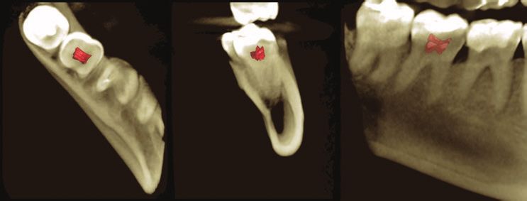
Indian Journal of Forensic Medicine & Toxicology, January-March 2021, Vol. 15, No. 1   897

disadvantages of these 2D radiographs is that it could          coronal section. All CBCT images which with a fully
have projection errors and the tooth could not be assessed      visible pulp cavity were selected. Two measurements
in all directions from a single radiograph. Cone Beam           were taken at two different levels. Height of the crown
Computed Tomography(CBCT) scans has overcome                    (CH) and height of the coronal pulp cavity (CPCH).
these disadvantages of 2D imaging modalities as it is           Then tooth – coronal index (TCI) for each tooth was
also non-invasive. A 3 dimensional (3D) reconstructed           calculated as follows: TCI = CPCH X 100 / CH.6
image can be obtained which allows us to visualise the
                                                                     All measurements were taken using the same
morphology of a tooth from all angles without any image
                                                                machine. To ensure the accuracy of the technique used
distortion.5
                                                                for measuring TCI detailed reference points were used:
    The present study was done to assess the accuracy           Cervical line that connect two landmarks to be measured;
of age estimation from TCI of mandibular molars                 the mesial and distal cemento-enamel junction points;
(except wisdom teeth) of both sides using CBCT images           and divides the tooth into crown and root. Crown height
of known age and sex, so that we can apply this method          is the maximum perpendicular distance from the cervical
to estimate age in both living individuals and skeletal         line to the tip of the highest cusp of teeth. While pulp
material of unknown age.                                        height is the distance from cervical line to the coronal tip
                                                                of the pulp chamber as shown in Figure 1.
                        Method
                                                                    After the procedures of image acquisition and
     A cross sectional retrospective study was conducted
                                                                measurement of the height of the crown and the pulp
on a total of 100 CBCT images of subjects who were
                                                                cavity as described above, the volume of pulp chamber
referred to the department of Oral Medicine and
                                                                was calculated using the region growing tool using the
Radiology for varied diagnostic purposes of known
                                                                Romexis software as shown in Figure 2.
age and gender. The study protocol was approved
by the Institutional Ethics committee. The inclusion                The measurements were taken using the software–
criteria for selecting the images were those images of          based calibrated measurement tool. All measurements
patients with healthy teeth without any periapical or           were carried out twice by two observers and the mean
periodontal pathologies. The second requirement was             was recorded to minimize intra and inter - observer
that the scanned images were of good diagnostic quality         errors.
without any artefacts. The images excluded were of
those patients with history of trauma or pathology to the                          Statistical Analysis
teeth and in which the pulp that could not be identified.           The statistical analysis of data was done by using
Images of patients with syndromes or any congenital             excel program for figures and SPSS (SPSS, Inc,
disorders were excluded. The study images were taken            Chicago, IL) program statistical package for social
using Promax 3D, Mid version (Planmeca Oy., Helsinki,           science version 20. Independent t-test was used for
Finland) CBCT unit. CBCT images were chosen over                comparison of gender. Linear regression analysis was
panoramic images as the measurements were more                  used for prediction of age using TCI of 36,37,46 and
accurate in terms of magnification and better individual        47. Interclass correlation coefficient for interobserver
detail could be obtained as there were no superimposition       variability between 2 observers for the calculated
of other structures.                                            parameters of TCI.
     The study images were of 200 subjects (56 females
                                                                                           Results
and 44 males). Four mandibular molars (lower left and
right,1stand 2ndmolars) excluding the 3rd molars were               Interobserver variability was carried out using
assessed. The individual tooth was assessed in the axial        Interaclass correlation coefficient test. The ICC values
section and was aligned in the coronal section so that          were 0.730, 0.592, 0.679 for male, female and total
the long axis of the tooth was perpendicular to the lower       population respectively for measurement of TCI (P value
border of mandible. The measurements were taken in the          of
898 Indian Journal of Forensic Medicine & Toxicology, January-March 2021, Vol. 15, No. 1
Independent students t test was used to compare 47. TCI values, with an R value of 0.333 and 0.241
TCI value between male and female and showed no respectively.
significant difference between the gender (Table1)
Independent t test was done to compare the volume
Linear regression analysis was performed to predict of the pulp among both the sexes and it showed a
the age of the individual using the TCI of the first and statistical significant difference between the gender in
second molars. Significant association was seen with the mandibular left 1st molar and both molars on the
the TCI values of the tooth 46. Among the four molar right side with p value ofIndian Journal of Forensic Medicine & Toxicology, January-March 2021, Vol. 15, No. 1 899
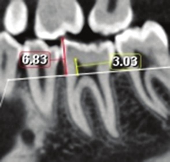
Fig1: Cone beam computed tomography images of the mandibular left molar obtained in the sagittal section
shows the measurement of CH (red line) and CPCH (yellow line).
Fig II: Cone beam computed tomography images showing the pulp volume of mandibular left molar
obtained in 3D rendered view (axial, coronal, and sagittal views)
Table I: Independent T Test for comparison of the TCI in male and female
FEMALE(n=56) MALE(n=44)
t df P VALUE
Mean Sd Mean sd
TCI 36 31.999 10.840 34.191 9.049 -1.078 98 0.284
TCI 37 27.966 9.096 26.881 7.479 0.639 98 0.524
TCI 46 31.946 12.357 34.775 10.026 -1.232 98 0.221
TCI 47 25.479 8.818 26.829 8.614 -0.768 98 0.444
Fig1: Cone beam computed tomography images of the mandibular left molar obtained in the sagittal section
shows the measurement of CH (red line) and CPCH (yellow line).900 Indian Journal of Forensic Medicine & Toxicology, January-March 2021, Vol. 15, No. 1
Fig II: Cone beam computed tomography images showing the pulp volume of mandibular left molar
obtained in 3D rendered view (axial, coronal, and sagittal views)
Discussion images of tooth without magnification, distortion, no
superimpositions and images can be viewed in multiple
Tooth coronal index aids in age estimation in people
sections at multiple levels. Due to the spatial resolution
whose age is unknown for a variety of reasons like,
of CBCT, it can be used to visualise the pulp chamber
minor children are made to work for various reasons,
and anatomic variations. Radiation dose is relatively less
among people who migrate for various reasons, in natural
when compared to other 3 D imaging modalities.
disasters and also from archaeological specimens.
In our study we measured both coronal and pulp
Secondary dentine is formed due to wasting
height in sagittal section as it was the ideal section for
diseases (erosion, abrasions and attrition), ageing,
measuring the maximal height of both tooth and pulp
caries etc. which gradually results in the decrease in
chamber when compared to other sections.
the pulp volume. Secondary dentine is considered as
an age predictor. As age advances the pulpal volume Comparing the TCI value between the genders
decreases due to apposition of secondary dentine along showed no significant results which was similar to
the dentinal walls.7 The second reason being that it can the study done by Drusini AG; Khattab et al.2017
withstand damages caused due environmental factors in and Nawaya and Burhan 2016; 10,11,12 but was not
human remains as it is encased in a harder tissue.2 This in accordance with the studies done by Igbigbi P and
deposition of secondary dentine is not uniform and can Nyirenda SK 200513, they found that gender had
vary in different parts in the pulp chamber.7 significance influence on age using TCI. Few studies
support this theory explaining that it could be due to the
Pulp cavity size decrease as age advances due to
influence of estrogen in secondary dentine deposition.6
secondary dentine getting deposited along the roof
and floor thereby reducing the height rather than the In our study using images taken in CBCT we
width. Pulpal changes were also seen after orthodontic found that the mandibular right molars (46,47) the TCI
treatment.8 This decrease in pulpal height acts as a values corelated well with age and it was found that
biomarker in age estimation. Sectioning of tooth and the lower right first molar (46) showed a significant
radiographs can be used for measuring secondary prediction of age in females. This was the best predictor
dentine deposition.9 in the equation, which was in accordance to a study by
Agematsu H et al and EL Morsi DA 201514,6. As R value
CBCT, a three dimensional imaging modality
is positive it shows a positive correlation indicating that
has advantages over 2 D imaging as it providesIndian Journal of Forensic Medicine & Toxicology, January-March 2021, Vol. 15, No. 1 901
as age increases the index increases and also it was SA, Saddiwal RS, Nisa SU. Age estimation in
more in females when compared to the males which was Indian adults by the coronal pulp cavity index. J
similar to the study done by EL Morsi DA 2015 which Forensic Dent Sci. 2017;9(3):177.
could be related to the hormonal influence, which could 5. Elmoazen R, Salaheldin MS, Abdalkhalek W. Age
lead to increase in the sequential obliteration of the pulp Estimation in an Egyptian Population Using Pulp/
with growth.6 Tooth Volume Ratio Obtained from Cone Beam
Computed Tomography. Egyptian dental journal
Pulp volume measurement was not statistically 63; (2), 635-43.
significant; it could be because the measurements were 6. El Morsi DA, Rezk HM, Aziza A, El–Sherbiny
taken from images of patients with an age range of 15-30 M. Tooth Coronal Pulp Index as a Tool for Age
years. But in our study there was a significant change in Estimation in Egyptian Population. J Forensic Sci
the volume of pulp when compared among both sexes. Criminol.2015; 3(2): 201. doi: 10.15744/2348-
The volume of the pulp chamber was stronger for the 9804.2.501
males in the present study which was in accordance to a 7. Star H, Thevissen P, Jacobs R, Fieuws S, Solheim
study done by Agematsu H et al. 14. T, Willems G. Human dental age estimation by
calculation of pulp-tooth volume ratios yielded
Conclusion on clinically acquired cone beam computed
CBCT plays an important in age as it gives a more tomography images of monoradicular teeth. J
accurate picture regarding the dimensions in all planes Forensic Sci. 2011;56 Suppl 1:S77-S82.
without with less radiation dose. CBCT could prove 8. Venkatesh S, Ajmera S, Ganeshkar SV. Volumetric
useful in forensic odontology as important diagnostic pulp changes after orthodontic treatment determined
tool in age and sex determination in living and also by cone-beam computed tomography. J Endod.
aid in diagnosing the age and gender of victims in 2014;40(11):1758-1763.
mass disasters. In present study TCI was useful in in 9. Singaraju S, Sharada P. Age estimation using pulp/
age estimation this method was not useful in gender tooth area ratio: A digital image analysis. J Forensic
determination. Dent Sci 2009;1:37-41
10. Drusini AG. The coronal pulp cavity index: A
Ethical Clearance- Taken from, Institutional forensic tool for age determination in human adults.
Ethics Committee Manipal College of Dental Sciences, Cuad Med Forense 2008;14:235‑49.
Mangaluru IEC Protocol Ref No.18070 11. Khattab NA, Marzouk HM, Abdel Wahab TM.
Application of tooth coronal index for age
Source of Funding- Nil
estimation among adult Egyptians. Schoolary Res.
Conflict of Interest - Nil 2013;1-15.
12. Nawaya FR, Burhan AS. Use of the Tooth Coronal
References Pulp Index for Recognition of the Pubertal Growth
1. Panchbhai AS. Dental radiographic indicators, a Period. The Journal of Contemporary Dental
key to age estimation. Dentomaxillofac Radiol. Practice. 2016 Nov;17(11):884-889. DOI: 10.5005/
2011;40(4):199-212. jp-journals-10024-1948.
2. Rai A, Acharya AB, Naikmasur VG. Age estimation 13. Igbigbi PS, Nyirenda SK. Age estimation of
by pulp-to-tooth area ratio using cone-beam Malawian adults from dental radiographs. West Afr
computed tomography: A preliminary analysis. J J Med. 2005; 24:329–33.
Forensic Dent Sci. 2016;8(3):150-154. 14. Agematsu H, Someda H, Hashimoto M, Matsunaga
3. Von Böhl M, Ren Y, Kuijpers-Jagtman AM, Fudalej S, Abe S, Kim HJ, et al. Three-dimensional
PS, Maltha JC. Age-related changes of dental pulp observation of decrease in pulp cavity volume
tissue after experimental tooth movement in rats. using micro-CT: Age-related change. Bull Tokyo
PeerJ 2016;4:e1625. Dent Coll. 2010; 51:1–6.
4. Koranne VV, Mhapuskar AA, Marathe SP, JoshiYou can also read