Epithelioid inflammatory myofibroblastic sarcoma: the youngest case reported - Autopsy ...
←
→
Page content transcription
If your browser does not render page correctly, please read the page content below

Clinical Case Report
Epithelioid inflammatory myofibroblastic sarcoma: the youngest case
reported
Sajida Batool1 Arvind Ahuja1 , Devender Singh Chauhan1,
Minakshi Bhardwaj1, Atul Kumar Meena2
How to cite: Batool S, Ahuja A, Chauhan DS, Bhardwaj M, Meena AK. Epithelioid inflammatory myofibroblastic sarcoma: the
youngest case reported. Autops Case Rep [Internet]. 2021;11:e2021288. https://doi.org/10.4322/acr.2021.288
ABSTRACT
Epithelioid inflammatory myofibroblastic sarcoma (EIMS) is a rare variant of the inflammatory myofibroblastic tumor. It
has an aggressive clinical course and a high rate of recurrence. EIMS primarily affects children and young adults. Hereby,
we report this entity in a 4-month-old infant who presented with an abdominal mass. Imaging studies revealed a large
hypodense mesentery-based lesion involving the right half and mid-region of the abdomen. The mass with an attached
segment of the small bowel was excised in toto. Grossly, a large encapsulated tumor was identified arising from the
mesentery of the small bowel. The histological examination showed a tumor consisting of epithelioid to spindle cells
loosely arranged in a myxoid background with numerous blood vessels and lymphoplasmacytic inflammatory infiltrate.
On immunohistochemistry, the tumor cells showed positivity for ALK1 (nuclear), desmin, SMA, CD68, and focal positivity
for CD30. A final diagnosis of EIMS of the small intestine was rendered. To the best of our knowledge, this case is the
youngest reported case in literature.
Keywords
Sarcoma; Epithelioid Cells; Intestine, Small; Mesentery; Anaplastic Lymphoma Kinase.
INTRODUCTION
Inflammatory myofibroblastic tumor (IMT) is a It is often intra-abdominal and clinicopathologically
mesenchymal neoplasm with borderline malignant different from conventional IMT in that it is more
potential. It often affects children and young adults aggressive with a higher rate of recurrence and poorer
with the mean age at diagnosis of 10 years. It mostly prognosis.3 It is mainly composed of plump round to
arises in the mesentery, omentum, and abdominal
epithelioid or histiocytoid tumor cells with a minor
soft tissue, followed by lung, mediastinum, and head
spindle cell component. Cells are arranged in clusters
and neck.1 Histologically, it is a distinctive neoplasm
or non-cohesive sheets within a stroma, which is
composed of spindled myofibroblastic cells, and
inflammatory infiltrate in an edematous myxoid frequently myxoid. Necrosis is uncommon, and mitosis
or hyalinized stroma with abundant blood vessels. is variable. Most EIMS express ALK and desmin with
Epithelioid inflammatory myofibroblastic sarcoma variable smooth muscle actin and CD30 positivity. Focal
(EIMS) is a rare and recently defined variant of IMT.2 CD68 positivity is seen in histiocytic appearing cells.1
1
Atal Bihari Vajpayee Institute of Medical Sciences, Dr Ram Manohar Lohia Hospital, Department of Pathology, New Delhi, India
2
Atal Bihari Vajpayee Institute of Medical Sciences, Dr Ram Manohar Lohia Hospital, Department of Pediatric Surgery, New Delhi, India
Copyright: © 2021 The Authors. This is an Open Access article distributed under the terms of the Creative
Commons Attribution License, which permits unrestricted use, distribution, and reproduction in any medium,
provided the original work is properly cited.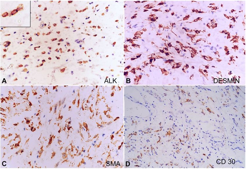
Epithelioid inflammatory myofibroblastic sarcoma: the youngest case reported

We report this rare entity in a four-month-old infant,                  also noted in many areas with few binucleated cells.
which is the youngest reported case in literature.                      Variable mitosis (4-5/10hpf) was noted. Necrosis was
                                                                        not seen. There was a small amount of spindle cell
                                                                        component comprising about 15-20% of the tumor
CASE REPORT
                                                                        and was mostly scattered all around the tumor.
     A 4-month-old female patient presented with a                      There was mild to moderate chronic inflammatory
rapidly growing abdominal lump. On the abdominal                        infiltrate within the tumor, mainly comprising mature
examination, a large non-tender mass with ill-defined                   lymphocytes with scattered plasma cells. A large
borders and side-to-side mobility was palpable in the                   panel of IHC was applied, and the tumor cells showed
right lumbar and umbilical region. The abdominal                        positivity for ALK1 (nuclear membrane), desmin, SMA,
contrast-enhanced computed tomography (CECT)                            CD68, and focal positivity for CD30 (Table 1) (Figure 3).
revealed a large hypodense abdominal lesion                                  Based on the morphology and
(11.4x11x8cm) involving the right half and mid-                         immunohistochemistry results, a final diagnosis of
region, displacing the bowel loops posterolaterally.                    epithelioid inflammatory myofibroblastic sarcoma
However, tumor invasion was not seen. No evidence                       of the small intestine was made. Both gross and
of septations or solid enhancement was seen within                      microscopic examinations from the small bowel’s
the lesion. Clinically, the diagnosis of mesenteric cyst                attached segment were unremarkable and not involved
or teratoma was considered. The infant was operated                     by the tumor. Few lymph nodes were dissected out
on, and an encapsulated irregular mass attached to a                    which showed reactive lymphoid hyperplasia. The
small bowel segment was excised in toto, along with                     immediate postoperative period was uneventful. The
mesenteric lymph nodes. On gross examination, a large                   patient was discharged after ileo-ileal anastomosis, in
encapsulated tumor (12x11x7cm) was identified arising                   a stable condition and normal passage of stool. On
from the mesentery. The external surface of the mass                    follow-up after 6 months, the general condition of the
was encapsulated and lobulated. The cut section was                     patient was fair, and she did not have any complaint.
greyish-white and glistening with a myxoid appearance
and soft to firm in consistency (Figure 1).
                                                                        DISCUSSION
     On the histopathological examination, the tumor
consisted of epithelioid to spindle cells loosely arranged
                                                                             EIMS occurs mostly in children and adolescents and
in a myxoid background with numerous blood vessels
                                                                        has a male predilection, contrary to the conventional
(Figure 2).The tumor cells had moderate eosinophilic
                                                                        IMT, which has a slight female predominance. The
cytoplasm, oval to spindle nuclei, and conspicuous
                                                                        exact frequency of EIMS remains underestimated, as
nucleoli. Epithelioid to round cell morphology was
                                                                        it has not been widely documented. Only 43 cases
                                                                        have been reported in the literature.4-7 The age of the
                                                                        patients ranged from 7 months to 63 years.4 Most
                                                                        of the tumors were intra-abdominal, of which 12

                                                                        Table 1. Antibodies used for immunohistochemical
                                                                        (IHC) staining and their interpretation
                                                                           Result                   IHC markers
                                                                          Positive   ALK, SMA, CD30, CD68, and Desmin
                                                                                     Pan CK, EMA, CD34, CD117, S100, CD99,
                                                                          Negative   BCL2
                                                                                     MyoD1, Myogenin
                                                                        ALK, anaplastic lymphoma kinase; BCL, B-cell lymphoma; CD,
Figure 1. Gross examination of the tumor showing a                      cluster of differentiation; CK, cytokeratin; EMA, epithelial
mass with attached ileal segment and its cut surface                    membrane antigen; MyoD1, myoblast determination
                                                                        protein 1; SMA, smooth muscle actin; S100, solubility in
(scale bar = 9 cm).                                                     100%ammonium sulphate.

2-6                                                                                      Autops Case Rep (São Paulo). 2021;11:e2021288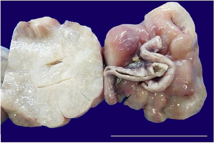
Batool S, Ahuja A, Chauhan DS, Bhardwaj M, Meena AK

Figure 2. Histopathological examination of the tumor. A – Moderately cellular tumor with intervening small blood
vessel (H&E, x100); B – Tumor cells with moderate eosinophilic cytoplasm and oval to spindle nuclei (H&E, x200);
C – Myxoid area along with mild lymphoplasmacytic inflammatory infiltrate (H&E, x200); D – Epithelioid to round
cell morphology of tumor cells with conspicuous nucleoli and few binucleated forms (H&E, x400).

Figure 3. Immunohistochemistry. A – Tumor cells showing ALK nuclear membranous positivity (inset) (x400);
B – Diffuse desmin positivity (x400); C – SMA positivity (x400); D – Focal CD30 positivity (x200).

Autops Case Rep (São Paulo). 2021;11:e2021288                                                                      3-6
Epithelioid inflammatory myofibroblastic sarcoma: the youngest case reported

Table 2. Main differential diagnoses of EIMS
              Tumor                                                      Immunohistochemistry
               EIMS                                 ALK +, SMA +, EMA +/-, CK +/-, CD30 +, CD34 -, and Desmin +
               GIST                                             CD117 +, DOG1 +, CD34 +, and Desmin -
               ALCL                                           ALK +, SMA +, EMA +, CD30 +, and Desmin -
               ELMS                                                 ALK -, SMA +, EMA + and Desmin +
              LGFMS                                     EMA +, MUC4 +, CD99, BCL2, SMA +/-, and Desmin +/-
               RMS                                 ALK+/-, Myogenin +, MyoD1 +, Desmin +, SMA +, EMA -, CD30 -
                MM                                              CK5 +, Calretinin +, EMA +/-, and Desmin -
             Our case                         ALK +, SMA +, CD30 +, CK-, EMA -, CD34 -, and Desmin +
ALCL, anaplastic large cell lymphoma; ALK, anaplastic lymphoma kinase ; BCL, B cell lymphoma; CD, cluster of
differentiation; CK, cytokeratin; DOG1, discovered on GIST 1 ; EIMS, epithelioid inflammatory myofibroblastic sarcoma;
EMA, epithelial membrane antigen; ELMS, epithelioid leiomyosarcoma, ; GIST, gastrointestinal stromal tumor; LGFMS,
low grade fibromyxoid sarcoma; MM, malignant mesothelioma; MUC, mucin; MyoD1, myoblast determination protein 1;
RMS, rhabdomyosarcoma; SMA, smooth muscle actin.

involved the mesentery, and out of these 12 cases,                      sarcoma (LGFMS) and malignant mesothelioma (MM).
9 involved the mesentery of the small bowel. Most                       ALCL can be difficult to distinguish from EIMS, as the
cases were unifocal, with a tumor size ranging from                     rare sarcomatoid variant of ALCL can exhibit spindle
5 to 26 cm in its longest axis. All cases were ALK-                     cell morphology and overlapping immunostaining,
positive on immunohistochemistry. Clinically, these                     including reactivity for CD30, ALK, and SMA and
patients presented with abdominal pain or masses                        non-reactivity for EMA. However, strong expression
and sometimes with ascites. Our case is the 10th case                   of desmin and the distinctive nuclear membrane
of small bowel mesentery and the youngest among all                     pattern of ALK staining are not observed in ALCL. The
the reported cases.                                                     RANBP2-ALK has never been reported in ALCL either.
     EIMS often displays the following common                           Epithelioid GIST is positive for CD117, DOG1, CD34
histomorphological and immunohistochemical                              while negative for ALK staining. Mutations of c-Kit
characteristics: i) round to epithelioid tumor cells with               and platelet-derived growth factor-α are also present
scattered inflammatory infiltrates, ii) abundant myxoid                 in GIST. ELMS usually displays higher cellularity and
stroma, iii) expression of ALK (nuclear membrane),                      pleomorphism. Also, it generally lacks an extensive
and desmin (cytoplasmic, diffuse, and strong).8 In                      myxoid background, inflammatory infiltrates, and
addition, it can also display a variable expression of                  ALK expression. The solid variant of alveolar RMS is
SMA, CD30, CD68, and cytokeratin.4 EIMS may show                        frequently ALK-positive; however, it lacks fibrovascular
nuclear membrane staining pattern or cytoplasmic                        stroma and forms sheets of round cells with variable
positivity with focal perinuclear accentuation pattern                  rhabdomyoblastic differentiation. Myogenin and MyoD
of ALK.9 There is aberrant expression of anaplastic                     are highly specific and sensitive for its diagnosis. LGFMS
lymphoma kinase (ALK) protein in 50-60% of the                          shows bland spindle cells with a whorling growth
cases due to clonal rearrangements of the ALK gene                      pattern and arcades of curvilinear blood vessels in an
located on chromosome 2p23.10 Several ALK fusion                        admixture of collagenous and myxoid stroma. LGFMS
proteins, including RNA binding protein 2 (RANBP2)-                     is positive for MUC4 and EMA, focal SMA, and desmin
ALK, tropomyosin 3 (TPM3)-ALK, and ribosome binding                     positivity while not expressing ALK. CK5 and calretinin
protein 1 (RRBP1)-ALK are identified in EIMS and are                    are positive in MM, but ALK and desmin are absent.1,8
associated with an aggressive clinical course.11-13                     Panel of IHC commonly used for differential diagnosis
     Microscopic diagnosis of EIMS can be challenging                   is depicted in Table 2.
due to its unique epithelioid to round cell morphology                       It is important to recognize EIMS as a distinct
and atypical nuclear features, which resembles                          variant of IMT as patients with ALK-rearranged EIMS
many other tumors like a gastrointestinal stromal                       may benefit from targeted therapy.3The mainstay of
tumor (GIST), anaplastic large cell lymphoma                            treatment for IMT/EIMS is complete surgical resection
(ALCL), and epithelioid leiomyosarcoma (ELMS),                          when possible. However, recurrence is common. A
rhabdomyosarcoma (RMS), low-grade fibromyxoid                           few case reports described the combined therapy

4-6                                                                                      Autops Case Rep (São Paulo). 2021;11:e2021288
Batool S, Ahuja A, Chauhan DS, Bhardwaj M, Meena AK
of surgery and systemic therapy with ALK inhibitors 5. Xu P, Shen P, Jin Y, Wang L, Wu W. Epithelioid
when an ALK mutation is present.14 The effectiveness inflammatory myofibroblastic sarcoma of stomach:
diagnostic pitfalls and clinical characteristics. Int J Clin
of alternative treatment modalities like radiotherapy
Exp Pathol. 2019;12(5):1738-44. PMid:31933992.
and chemotherapy is uncertain.
6. Garg R, Kaul S, Arora D, Kashyap V. Posttransplant
epithelioid inflammatory myofibroblastic sarcoma: a
CONCLUSION case report. Indian J Pathol Microbiol. 2019;62(2):303.
http://dx.doi.org/10.4103/IJPM.IJPM_284_17. PMID:
30971562.
Epithelioid inflammatory myofibroblastic sarcoma is
7. Zhang S, Wang Z. A case report on epithelioid
a rare and distinct variant of IMT, which mainly consists
inflammatory myofibroblastic sarcoma in the abdominal
of cells with an epithelioid or round cell morphology, cavity. Int J Clin Exp Pathol. 2019;12(10):3934-9.
with a high potential of recurrence and a poorer PMid:31933785.
prognosis. Only 43 cases have been reported in the 8. John R, Goldblum SW, Weiss ALF. Enzinger and weiss’s
literature, of which only 9 were located in the mesentery soft tissue tumors. 6th ed. Philadelphia, USA: Elsevier
of the small bowel. This is the 10th case reported in the Saunders; 2014. P. 304-10.
mesentery of small bowel and youngest among all the 9. Cook JR, Dehner LP, Collins MH, et al. Anaplastic lymphoma
reported cases to the best of our knowledge kinase (ALK) expression in the inflammatory myofibroblastic
tumor: a comparative immunohistochemical study.
Am J Surg Pathol. 2001;25(11):1364-71. http://
dx.doi.org/10.1097/00000478-200111000-00003.
REFERENCES PMid:11684952.
1. Fletcher CDM, Bridge JA, Hogendoorn PCW, Mertens F. 10. Griffin CA, Hawkins AL, Dvorak C, Henkle C, Ellingham
World Health Organization Classification of Tumors of T, Perlman EJ. Recurrent involvement of 2p23 in
Soft Tissue and Bone. 4th ed. Lyon, France: IARC Press; inflammatory myofibroblastic tumors. Cancer Res.
2013. p. 83-4. 1999;59(12):2776-80. PMID: 10383129.
2. Marino-Enriquez A, Wang W-L, Roy A, Lopez-Terrada D, 11. Hallin M, Thway K. Epithelioid inflammatory
Lazar AJF, Fletcher CDM, et al. Epithelioid inflammatory myofibroblastic sarcoma. Int J Surg Pathol. 2019;27(1):69-
myofibroblastic sarcoma: an aggressive intra-abdominal 71. http://dx.doi.org/10.1177/1066896918767557.
PMid:29623737.
variant of inflammatory myofibroblastic tumor with
nuclear membrane or perinuclear ALK. Am J Surg 12. Lee J-C, Li C-F, Huang H-Y, et al. ALK oncoproteins
Pathol. 2011;35(1):135-44. http://dx.doi.org/10.1097/ in atypical inflammatory myofibroblastic tumours:
PAS.0b013e318200cfd5. PMID: 21164297. novel RRBP1-ALK fusions in epithelioid inflammatory
myofibroblastic sarcoma. J Pathol. 2017;241(3):316-23.
3. Du X, Gao Y, Zhao H, Li B, Xue W, Wang D.
http://dx.doi.org/10.1002/path.4836. PMid:27874193.
Clinicopathological analysis of epithelioid inflammatory
myofibroblastic sarcoma. Oncology Letters. 13. Li J, Yin W, Takeuchi K, Guan H, Huang Y, Chan JKC.
2018;15(6):9317-26. http://dx.doi.org/10.3892/ Inflammatory myofibroblastic tumor with RANBP2 and
ol.2018.8530. PMID: 29805657. ALK gene rearrangement: a report of two cases and
literature review. Diagn Pathol. 2013;8(1):147. http://
4. Yu L, Liu J, Lao IW, Luo Z, Wang J. Epithelioid inflammatory
dx.doi.org/10.1186/1746-1596-8-147. PMid:24034896.
myofibroblastic sarcoma: a clinicopathological,
immunohistochemical and molecular cytogenetic analysis 14. Fujiya M, Kohgo Y. ALK inhibition for the treatment
of five additional cases and review of the literature. of refractory epithelioid inflammatory myofibroblastic
Diagn Pathol. 2016;11(1):67. http://dx.doi.org/10.1186/ sarcoma. Intern Med. 2014;53(19):2177-8. http://dx.doi.
s13000-016-0517-z. PMid:27460384. org/10.2169/internalmedicine.53.3038. PMid:25274227.
This study was carried out at Atal Bihari Vajpayee Institute of Medical Sciences, Dr RML Hospital, New Delhi.
Authors’ contributions: All authors have equally contributed for the study and manuscript’s conception. All
authors did proof reading and approved the final version for publication.
Ethics statement: The authors retain informed consent signed by the patient’s guardian and the manuscript
was approved by the Institutional Ethics Committee.
Conflict of interest: Nil.
Autops Case Rep (São Paulo). 2021;11:e2021288 5-6Epithelioid inflammatory myofibroblastic sarcoma: the youngest case reported Financial support: Nil. Submitted on: January 10th, 2021 Accepted on: April 11th, 2021 Correspondence Arvind Ahuja Atal Bihari Vajpayee Institute of Medical Sciences, Dr. Ram Manohar Lohia Hospital, Department of Pathology Baba Kharak Singh Marg, New Delhi-110001, India Phone: +91 (011) 23404344 drarvindahuja@gmail.com 6-6 Autops Case Rep (São Paulo). 2021;11:e2021288
You can also read