Giant Calculus with More than 100 Small Calculi in Choledochal Cysts
←
→
Page content transcription
If your browser does not render page correctly, please read the page content below
Case Rep Gastroenterol 2021;15:244–252
                                DOI: 10.1159/000513145                           © 2021 The Author(s)
                                Published online: February 26, 2021              Published by S. Karger AG, Basel
                                                                                 www.karger.com/crg

                                This article is licensed under the Creative Commons Attribution-NonCommercial 4.0
                                International License (CC BY-NC) (http://www.karger.com/Services/OpenAccessLicense).
                                Usage and distribution for commercial purposes requires written permission.

Case and Review

Giant Calculus with More than 100
Small Calculi in Choledochal Cysts
Akshay Bahadura Vijay Thakur b               Lovenish Bains c            Prerna Arora d
Yanshul Rathia Ashish Shukla a
a Departmentof Surgery, Dr. Hedgewar Arogya Sansthan, Delhi, India; bDepartment of
General Surgery, Lal Bahadur Shastri Hospital, Delhi, India; cDepartment of Surgery,
Maulana Azad Medical College, New Delhi, India; dDepartment of Pathology, Maulana
Azad Medical College, New Delhi, India

Keywords
Common bile duct · Choledochal cyst · Giant calculus · Heterotopic pancreas

Abstract
Giant biliary calculus in the common bile duct (CBD) is rare. Giant calculus of choledochal cyst
(CC) is even rarer, and no case of giant calculus of CC with more than 100 calculi has been
reported in the indexed literature. We present the case of a 8.0 × 4.5 × 4.0 cm sized giant
calculus with >100 small calculi in type IVa CCs with heterotopic pancreas in a 45-year-old
male, which is a surprisingly rare occurrence. Magnetic resonance cholangiopancreatography
showed multifocal irregular dilatation of intrahepatic biliary radicles with multiple filling de-
fects with a giant calculus in CC with cholelithiasis. The case was successfully managed with
open cholecystectomy and choledochotomy with retrieval of 1 giant and more than 100 small
calculi with excision of CC with Roux-en-Y hepaticojejunostomy. Histopathological examina-
tion (HPE) showed inflamed CC identified with focal areas of surface ulceration with increased
fibrosis areas in the wall and few pancreatic acini. A bile duct calculus is defined as “giant”
when the size is 5 cm or more. Stone formation within is the most frequent complication of
CC. Most intracystic calculi have been described as soft, earthy, and pigmented in appearance,

                                 Akshay Bahadur
                                 Department of Surgery
                                 Dr. Hedgewar Arogya Sansthan
                                 Delhi 110032 (India)
                                 drakshay@live.com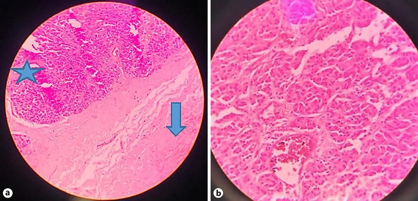
Case Rep Gastroenterol 2021;15:244–252                                                   245
                                   DOI: 10.1159/000513145          © 2021 The Author(s). Published by S. Karger AG, Basel
                                                                   www.karger.com/crg

                                   Bahadur et al.: Giant Calculus with More than 100 Small Calculi in Choledochal Cysts

supporting bile stasis as a primary etiologic factor. The only treatment for giant calculus of
CBD or CC is surgical. Endoscopic treatment is mostly unsuccessful and open surgery is the
treatment of choice due to giant size, increased load of calculus, and presence of calculi in the
left and right hepatic ducts.                                          © 2021 The Author(s)
                                                                                        Published by S. Karger AG, Basel

    Introduction

     Cystolithiasis and cholecystolithiasis are the most frequent conditions occurring in 70%
of adults with choledochal cysts (CCs) [1]. A bile duct calculus is defined as “large” if it is more
than 1.5 cm in size and as “giant” when it is 5 cm or more in size [2–4]. Though giant calculus
of the gallbladder is common, giant calculus in the common bile duct (CBD) is rare [5]. Only
few cases of giant biliary calculus in the CBD measuring 5 cm or more have been published [2,
3]. Only 1 case of more than 100 biliary calculi in the bile duct has been reported [6]. We report
probably the largest giant calculus of CC with more than 100 biliary calculi in CCs with pan-
creatic acini in the wall of CCs.

    Case Report

     Our patient, a 45-year-old male, came to Outpatient Department (OPD) with a history of
pain in the right upper quadrant, which was colicky in nature, mild to moderate in severity,
and recurrent for the past 15–20 days. The patient also gave a history of discomfort in the
same area with nausea off and on, especially after taking heavy meals for the past 4 years. The
patient was well nourished, nonicteric, afebrile with a pulse of 80/min and blood pressure of
110/70 mm Hg. Clinical examination revealed mild tenderness in the right hypochondrium
on deep palpation with no rebound tenderness. The remaining examination of the abdomen
was unremarkable.
     Blood investigations revealed hemoglobin of 15 g/dL, white cell count of 11,100/cumm
with 64% polymorphs, 30% lymphocytes, 4% monocytes, and 2% eosinophils. Liver function
test showed total serum bilirubin of 0.64 mg/dL, AST 18 U/L, and ALT 17 U/L. Renal function
test, serum electrolyte, serum glucose, and urine analysis were all normal.
     Ultrasonography (USG) revealed a partially distended gall bladder containing multiple
calculi in the CBD and CCs with CC of 5.0 × 8.0 cm in size with a giant calculus. Magnetic reso-
nance cholangiopancreatography (MRCP) showed gross fusiform dilatation of the CBD
throughout its length measuring approximately 4.8 cm, possibly representing CC. A large sig-
nal void measuring approximately 4.0 × 7.6 cm is seen within the CBD, which is likely a calcu-
lus. There was gross dilatation of bilobar intrahepatic biliary radicles (CCs) loaded with mul-
tiple calculi within. The gallbladder was distended with multiple calculi in the lumen (Fig. 1).
     On exploration, the gall bladder was distended, containing multiple calculi. There was a
large CC arising 1 cm distal to the confluence of the right and left hepatic duct, extending just
proximal to the opening of the pancreatic duct, containing a hard calculus and multiple calculi.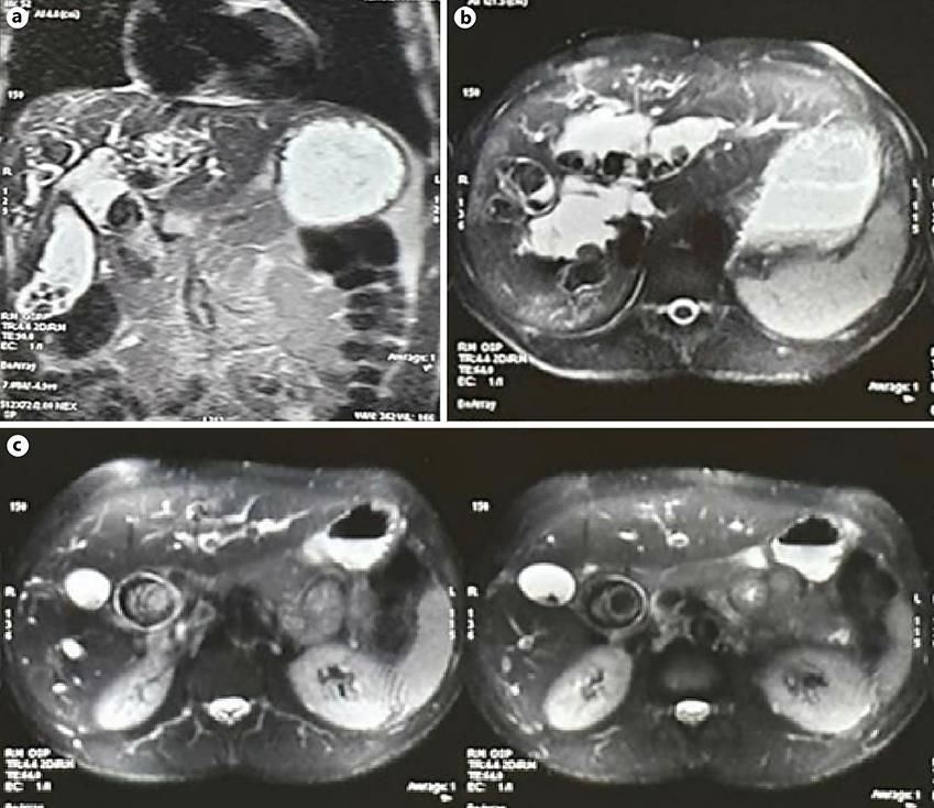
Case Rep Gastroenterol 2021;15:244–252 246
DOI: 10.1159/000513145 © 2021 The Author(s). Published by S. Karger AG, Basel
www.karger.com/crg
Bahadur et al.: Giant Calculus with More than 100 Small Calculi in Choledochal Cysts
The omentum and duodenum were adherent to the CC. With meticulous and gradual dissec-
tion, the surrounding structures were separated; a choledochotomy of approximately 1.5 cm
was made on anterior wall of the CC. The choledochotomy was enlarged due to the giant cal-
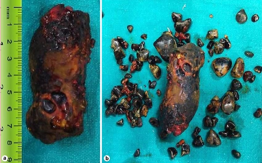
culus, which had completely occluded the cyst. The calculus of the size of 8.0 × 4.5 × 4.0 cm
was retrieved as a single piece. More than 100 small calculi were also retrieved from extrahe-
patic and intrahepatic CCs (Fig. 2). Cholecystectomy with excision of extrahepatic CC with
Roux-en-Y hepaticojejunostomy was performed. No liver biopsy was performed.
From the 4th postoperative day (POD), clear fluid through the right subhepatic drain was
noticed, which was 200–300 mL for the first 2 days and increased to 1,000 mL/day from day
6. Biochemical analysis of the fluid showed more than 20,000 U/L amylase. The patient was
kept on total parental nutrition along with octreotide injection 100 μg intravenously every
8 hours. Gradually, the daily drain output started decreasing. On the 19th POD, the daily drain
output was reduced to less than 50 mL. The patient was again started on entral nutrition. On
the 25th POD, the patient was discharged in a satisfactory condition. Histopathology showed
chronic cholecystitis with inflamed CC identified with focal areas of surface ulceration with
the underlying wall showing presence of areas of increased fibrosis and few pancreatic acini
(Fig. 3). The patient is presently doing well at 3 years of follow-up.
Discussion
A bile duct calculus is defined as giant if it is 5 cm or more in size [2–4]. Though giant
calculus of the gallbladder is common, giant calculus in the CBD is rare [5]. Only few cases of
giant biliary calculus in the CBD measuring 5 cm or more have been published (Table 1) [2–5,
7–15]. The presence of such a giant calculus without an associated jaundice is rare [14, 15].
Out of a total of 12 cases of giant biliary calculus in the CBD of 5 cm or larger found in the
literature, 6 cases had associated jaundice, 5 cases, including our case, had normal bilirubin,
and status of jaundice in 2 cases was not available.
It is reported that in most cases of choledocholithiasis, a solitary calculus is found in the
CBD [16]. Walter and Snell [17] reported a solitary calculus in two-thirds of their cases. Rob-
son and Dobson [18] once counted 88 calculi. There is only 1 case, as reported by Judd and
Marshall [6], in which more than 100 biliary calculi were found in the bile duct. To our
knowledge, no case of giant biliary calculus in confirmed CC nor any case of more than 100
biliary calculi in CC has been reported in the indexed literature to date. We came across such
a rare entity of a giant calculus with more than 100 small biliary calculi in CCs.
Stone formation within the CC (cystolithiasis) is the most frequent complication of CC.
The prevalence of intracystic calculi ranges from 2 to 72% in adults [19–21] and is increasing
in incidence with age [22]. Most intracystic calculi have been described as soft, earthy, and
pigmented in appearance, supporting bile stasis as a primary etiologic factor [21].
Although secondary calculi are the most commonly observed CBD calculi, particularly in
Europe and North America, primary calculi are encountered more commonly in Asia [23]. In
the West, the majority of CBD calculi are composed of cholesterol calculi that originated from
the gallbladder. Less than 10% of CBD calculi are formed de novo within the CBD. On the otherCase Rep Gastroenterol 2021;15:244–252 247
DOI: 10.1159/000513145 © 2021 The Author(s). Published by S. Karger AG, Basel
www.karger.com/crg
Bahadur et al.: Giant Calculus with More than 100 Small Calculi in Choledochal Cysts
hand, in the East, because of a higher incidence of chronic biliary tree infection and infestation,
the occurrence of pigmented calculi is much more common. It is characterized by recurrent
cholangitis due to the presence of multiple pigment calculi formed inside the intrahepatic
ducts [24].
Heterotopic pancreatic tissue present on the wall of CCs is a very rare entity. Heterotopic
pancreas is defined as pancreatic tissue that is not found in direct continuity with the main
pancreas [25]. It is difficult to explain embryologically the association of heterotopic pancre-
atic tissue on the wall of CCs. A possible hypothesis is that there is fetal migration of pancreatic
cells into the biliary tree, followed by release of pancreatic enzymes from the heterotopic rest
present on the wall, which may result in damage and dissolution of the wall leading to dilata-
tion [25].
MRCP has proved to be an accurate noninvasive imaging method for choledocholithiasis
[24], CC, and their associated anomalies [26]. Endoscopic treatment for giant calculus of the
CBD is often unsuccessful because of the calculus size, and the treatment is always surgical
[4]. Treatment of CC is complete excision of the affected biliary tract with reconstruction by a
bilioenteric anastomosis in order to reduce potential long-term complications [27]. Biliary
continuity may be established either by a Roux-en-Y hepaticojejunostomy or hepaticoduode-
nostomy [27]. In our case, open cholecystectomy with excision of extrahepatic CC with Roux-
en-Y hepaticojejunostomy was performed.
In conclusion, giant calculus of the CBD is uncommon and giant calculus in CC is rare. En-
doscopy has no role in the treatment of giant calculus. The treatment is always surgical, usu-
ally by open technique due to giant calculus, increased calculus load, and calculi in proximal
ducts. Roux-en-Y bilioenteric anastomosis may be considered as a first line of treatment in
cases of giant calculus of CBD/CC, multiple CBD calculi, or recurrent CBD calculi.
Statement of Ethics
Approval for case reports by the institutional ethics committee is not required. Written
informed consent was obtained from the patient regarding publication of this case.
Conflict of Interest Statement
There are no conflicts of interest to be reported.
Funding Sources
There was no source of funding.Case Rep Gastroenterol 2021;15:244–252 248
DOI: 10.1159/000513145 © 2021 The Author(s). Published by S. Karger AG, Basel
www.karger.com/crg
Bahadur et al.: Giant Calculus with More than 100 Small Calculi in Choledochal Cysts
Author Contributions
A.B. conceptualized the manuscript. A.B., V.T., and L.B. reviewed the literature and ana-
lyzed the data. A.B. wrote the manuscript with the help of L.B., Y.R., and A.S. A.B. and V.T. per-
formed the clinical examination, surgical treatment, and clinical follow-up. P.A. provided the
histopathological examination. A.B., V.T., L.B., P.A., Y.R., and A.S. performed the final review
and editing of the manuscript. All authors have read and approved the final version of the
manuscript.
References
1 Weyant MJ, Maluccio MA, Bertagnolli MM, Daly JM. Choledochal cysts in adults: a report of two cases and
review of the literature. Am J Gastroenterol. 1998 Dec;93(12):2580–3.
2 Bahuleyan CK. Giant common bile duct calculus. Indian J Surg. 1975;37:82.
3 Jayant M, Dalal AK, Attri AK, Sachdev A. Giant staghorn stone in common bile duct. Indian J Gastroenterol.
2010 Sep;29(5):212.
4 Hajong R, Topno N, Baruah AJ, Khongwar D. Giant staghorn common bile duct calculus. Indian J
Gastroenterol. 2012 Dec;31(6):357.
5 Bhattarai SR, Bhattarai A, Tamrakar KK. Giant staghorn common bile duct calculus: a case report. J Chitwan
Med Coll. 2019;9(4):72–4.
6 Judd ES, Marshall JM. Gallstones in the common bile duct. Arch Surg. 1931 Aug;23(2):175–81.
7 Okano A, Takakuwa H, Nishio A. Giant stone in the common bile duct. Endoscopy. 2001 Oct;33(10):907.
8 Sharma M, Gupta A, Singal R, Jain R, Khatri A, Sharda P, et al. A rare presentation of common bile duct stone.
Oncol Gastroenterol Hepatol Reports. 2016;5(1):29.
9 Ahmed MN, Bhat DP, Zargar HU, Kh an M. Giant common bile duct stone (A case report). J Postgrad Med.
1982;28(4):233–4.
10 Bhat J. Giant Staghorn Common Bile Duct Calculus (A Case Report). J Gastrointest Dig Syst. 2017;07(05):4–5.
11 Bektas H, Duzkoylu Y, Cakar E, Buyukasık K, Colak S. Giant choledochal calculosis: surgical treatment. N Am J
Med Sci. 2014 Oct;6(10):536–9.
12 Jarrar MS, Ben Hadj Khalifa MH, Ghrissi R, Ben Mansour I, Hamila F, Elghali A, et al. Giant staghorn common
bile duct calculus. Tunis Med. 2016 Jul;94(7):401–3.
13 Hamid R, Bhat NA, Ahmad M, Singh B. Choledochal Cyst (CDC). In: Neri V, editor. Gastrointestinal Surgery.
Rijeka: IntechOpen; 2018. https://doi.org/10.5772/intechopen.72938.
14 Hussain A, Jeelani G, Zargar HU, Bhan B. Giant common bile duct calculus. Int Surg. 1977 Sep;62(9):476.
15 Han HJ, Lee JS, Song TJ. Giant common bile duct stone. Cent Eur J Med. 2012;7(1):59–62.
16 Arid I. A Companion in Surgical Studies. 2nd ed. Edinburgh and London; 1949.
17 Walters W. Diseases of the gallbladder and bile ducts. South Med J. 1940;33(4):446.
18 Robson AW, Dobson JF. Diseases of the gall-bladder and bile-ducts, including gall-stones. New York: W
Wood; 1904.
19 Nagorney DM, McIlrath DC, Adson MA. Choledochal cysts in adults: clinical management. Surgery. 1984
Oct;96(4):656–63.
20 Chijiiwa K, Koga A. Surgical management and long-term follow-up of patients with choledochal cysts. Am J
Surg. 1993 Feb;165(2):238–42.
21 Khandelwal C, Anand U, Kumar B, Priyadarshi RN. Diagnosis and management of choledochal cysts. Indian J
Surg. 2012 Feb;74(1):29–34.
22 Matsumoto Y, Uchida K, Nakase A, Honjo I. Congenital cystic dilatation of the common bile duct as a cause of
primary bile duct stone. Am J Surg. 1977 Sep;134(3):346–52.
23 Shah KN, Clary BM. Chapter 36A – Stones in the bile duct: Clinical features and open surgical approaches and
techniques. In: Jarnagin W, editor. Blumgart's Surgery of the Liver, Biliary Tract and Pancreas. 2-vol set. 6th
ed. Philadelphia: Elsevier; 2016. p. 585–603.e3.
24 Lee YT, Sung J. Chapter 33: Choledocholithiasis. In: Baron TH, Kozarek R, Carr-Locke DL, editors. ERCP.
Edinburgh: W.B. Saunders; 2008. p. 357–66.Case Rep Gastroenterol 2021;15:244–252 249
DOI: 10.1159/000513145 © 2021 The Author(s). Published by S. Karger AG, Basel
www.karger.com/crg
Bahadur et al.: Giant Calculus with More than 100 Small Calculi in Choledochal Cysts
25 Suzuki K, Uchida T, Nakayama H, Ugajin W, Inaniwa Y, Sugitani M, et al. Heterotopic pancreatic tissue
associated with intra- and extrahepatic choledochal cysts. Pathol Int. 1999 Aug;49(8):759–62.
26 Sacher VY, Davis JS, Sleeman D, Casillas J. Role of magnetic resonance cholangiopancreatography in
diagnosing choledochal cysts: case series and review. World J Radiol. 2013 Aug;5(8):304–12.
27 Machado NO, Chopra PJ, Al-Zadjali A, Younas S. Choledochal Cyst in Adults: Etiopathogenesis, Presentation,
Management, and Outcome-Case Series and Review. Gastroenterol Res Pract. 2015;2015:602591.
Fig. 1. Magnetic resonance cholangiopancreatography. a Coronal view showing giant calculus within cho-
ledochal cyst with cholelithiasis. b Axial image demonstrates intrahepatic choledochal cysts with multiple
filling defects within calculi. c Axial view shows giant calculus within choledochal cyst with prominent bi-
lateral intrahepatic biliary radicals (choledochal cysts).Case Rep Gastroenterol 2021;15:244–252 250
DOI: 10.1159/000513145 © 2021 The Author(s). Published by S. Karger AG, Basel
www.karger.com/crg
Bahadur et al.: Giant Calculus with More than 100 Small Calculi in Choledochal Cysts
Fig. 2. Calculi retrieved from choledochal cysts. a 8.0 × 4.5 × 4.0 cm sized giant calculus of a extrahepatic
choledochal cyst. b Giant calculus with around 100 small calculi of extra- and intrahepatic choledochal
cysts.Case Rep Gastroenterol 2021;15:244–252 251
DOI: 10.1159/000513145 © 2021 The Author(s). Published by S. Karger AG, Basel
www.karger.com/crg
Bahadur et al.: Giant Calculus with More than 100 Small Calculi in Choledochal Cysts
Fig. 3. Histopathological examination of choledochal cyst showing areas of fibrosis (arrow) with pancreatic
acini (star) in the wall of a choledochal cyst (×200) (a) and pancreatic acini in the wall of a choledochal
cyst (×400) (b).Case Rep Gastroenterol 2021;15:244–252 252
DOI: 10.1159/000513145 © 2021 The Author(s). Published by S. Karger AG, Basel
www.karger.com/crg
Bahadur et al.: Giant Calculus with More than 100 Small Calculi in Choledochal Cysts
Table 1. Details of giant biliary calculus of 5 cm or more of the CBD/CC reported to date
No. Authors [ref.], year Age, Sex Number and size of calculus Jaundice Location of Treatment
years calculus
1 Bhat [10], 2017 38 F 11.5×4 cm Present In CBD Open cholecystectomy,
Single giant staghorn calculus choledochotomy, and T-tube
drainage
2 Bektas et al. [11], 2014 59 F 11×4.1 cm Absent In CBD Open cholecystectomy, T-tube
Single giant calculus choledochostomy, and
choledochodudenostomy
3 Jarrar et al. [12], 2016 65 9×4.5 cm Present In CBD Open cholecystectomy,
Single giant calculus choledochotomy
4 Jayant et al. [3], 2010 65 M 9×4 cm Absent In CBD Open cholecystectomy,
Calculus in two pieces from CBD choledochotomy, and
choledochodudenostomy
5 Bhattarai et al. [5], 2019 47 M 9×3 cm Present In CBD and both Open cholecystectomy,
Single large calculus hepatic ducts choledochotomy, and T-tube
drainage
6 Bahadur et al. (present case), 45 M 8×4.5×4 cm Absent In CC Open cholecystectomy,
2021 One giant calculus with excision of CC, and Roux-en-Y
>100 small calculi hepaticojejunostomy
7 Ahmed et al. [9], 1982 70 M 8.5×3.5 cm Present In CBD Open cholecystectomy,
Single giant calculus choledochotomy, and T-tube
drainage
8 Hussain et al. [14], 1977 NA NA 8×2 cm Present In CBD Not mentioned
Single giant calculus Open*
9 Hajong et al. [4], 2012 48 F 8×6 cm Present In CBD and both Open cholecystectomy,
Single giant calculus hepatic ducts choledochotomy, and
hepaticojejunostomy
10 Han et al. [15], 2012 72 F 7.5×4.0×4.0 cm Absent In CBD Open cholecystectomy,
Single giant calculus choledochotomy, CBD partially
excised, and T tube
11 Bahuleyan [2], 1975 38 F 6.5×3 cm NA In CBD Not mentioned
Single giant calculus Open*
12 Okano et al. [7], 2001 NA NA 5×5 cm NA In CBD Open laparotomy
Single giant calculus
13 Sharma et al. [8], 2016 55 F 5×3×4 cm Absent In CBD Open cholecystectomy,
Single giant calculus choledochotomy, and T-tube
drainage
CBD, common bile duct; CC, choledochal cyst. * Laparoscopic era started later.You can also read