Learning to be EXACT - BVM Workshop 2021
←
→
Page content transcription
If your browser does not render page correctly, please read the page content below
A3020
                      Learning to be EXACT
Cell Detection for Asthma on Partially Annotated Whole
                      Slide Images

  Christian Marzahl1,2 , Christof A. Bertram3 , Frauke Wilm1 , Jörn Voigt2 ,
Ann K. Barton4 , Robert Klopfleisch3 , Katharina Breininger1 , Andreas Maier1 ,
                              Marc Aubreville5
           1
              Pattern Recognition Lab, Department of Computer Science,
       Friedrich-Alexander-Universität Erlangen-Nürnberg (FAU), Germany
       2
         R & D Projects, EUROIMMUN Medizinische Labordiagnostika AG
       3
         Institute of Veterinary Pathology, Freie Universität Berlin, Germany
               4
                 Equine Clinic, Freie Universität Berlin, Berlin, Germany
                5
                  Technische Hochschule Ingolstadt, Ingolstadt, Germany
                                c.marzahl@euroimmun.de

      Abstract. Asthma is a chronic inflammatory disorder of the lower res-
      piratory tract and naturally occurs in humans and animals including
      horses. The annotation of an asthma microscopy whole slide image (WSI)
      is an extremely labour-intensive task due to the hundreds of thousands
      of cells per WSI. To overcome the limitation of annotating WSI incom-
      pletely, we developed a training pipeline which can train a deep learning-
      based object detection model with partially annotated WSIs and com-
      pensate class imbalances on the fly. With this approach we can freely
      sample from annotated WSIs areas and are not restricted to fully anno-
      tated extracted sub-images of the WSI as with classical approaches. We
      evaluated our pipeline in a cross-validation setup with a fixed training set
      using a dataset of six equine WSIs of which four are partially annotated
      and used for training, and two fully annotated WSI are used for valida-
      tion and testing. Our WSI-based training approach outperformed classi-
      cal sub-image-based training methods by up to 15% mAP and yielded
      human-like performance when compared to the annotations of ten trained
      pathologists.

1   Introduction
Asthma is a chronic inflammatory disorder of the lower respiratory tract and can
occur in multiple species. While asthma can affect humans, horses can also suffer
from asthma and are often used as models for human disease [1] due to their sim-
ilar symptoms and pathogenesis. The gold standard for diagnosis of equine and
human asthma is to collect bronchoalveolar lavage fluid (BALF) and to examine
the sample under a microscope or on digitised whole slide images (WSIs). Asthma
and other pulmonary disorders are diagnosed based on the relative proportion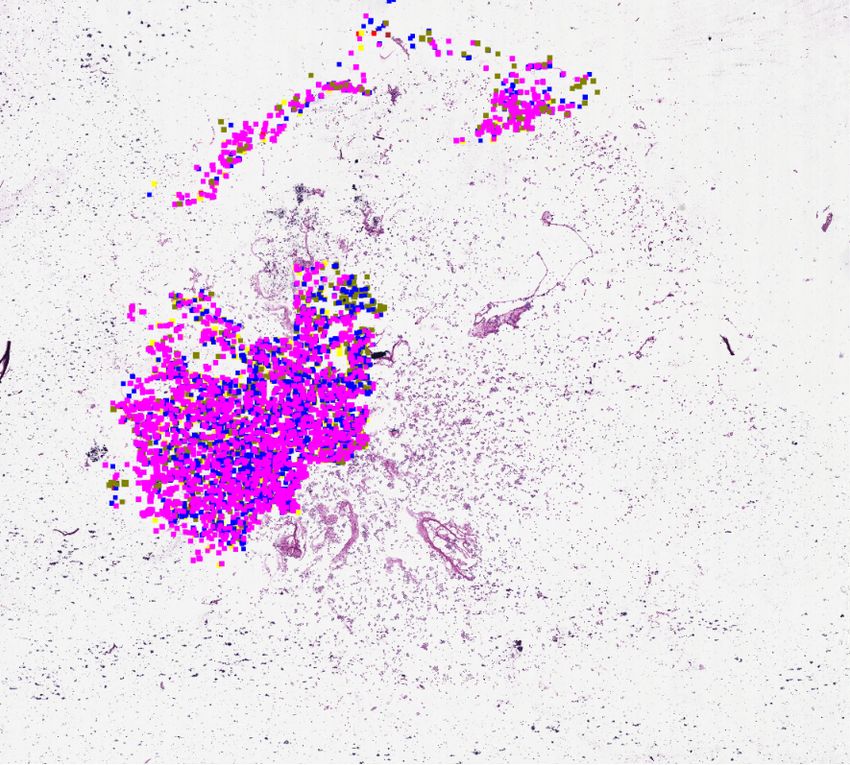
2                                Marzahl et al.

of different cell types including eosinophils, mast cells, neutrophils, macrophages
and lymphocytes. This typically requires manual counting of 300-500 cells and
is therefore time-consuming and strenuous for the pathologist [2]. Therefore, au-
tomatic solutions that support this task are of high interest. Although asthma
is a common disease in horses and humans, to the author’s knowledge there is
no trained model or method published analysing asthma on WSI automatically.
For the development of machine learning algorithms, huge annotated datasets are
generally required. A particular challenge for the annotation process of asthma is
the large number of cells per WSI, which can easily reach hundreds of thousands.
This makes the annotation process very labour-intensive and time-consuming.
In order to increase the efficiency of the annotation process, expert-algorithm
collaboration can be used where experts enhance pre-computed annotations of
a trained model. Marzahl et al.[2] showed this for mitotic figures or pulmonary
haemorrhage. An alternative option is to annotate multiple WSIs only partially
to capture the domain variability between WSIs, and train a network to com-
plete the annotation. On the one hand, training deep learning-based methods
on partially annotated WSIs faces some additional challenges regarding track-
ing annotated WSI areas and leads to higher demands on the coordination and
synchronisation between the participating institutes. On the other hand, train-
ing on partially annotated data simplifies training pipelines in terms of data
augmentation and live patch sampling in contrast to extracted sub-image-based
approaches. Nevertheless, sub-image-based approaches, where patches from the
WSI have to be extracted before the training, are the only supported method
for the most prominent object detection frameworks [3] and are used in multiple
WSI-based detection applications [4,5].
    As the main contribution to the field of deep learning-based cytological WSI
analysis, we propose a training pipeline to train object detection models with
live sampling on partially annotated WSIs. Additionally, we create a baseline
with a state-of-the-art deep learning-based object detection model for detecting
five types of cells on WSIs. All code to train, evaluate, test our models and to
reproduce our results for public is accessible at GitHub 1 . Furthermore, the WSIs
can be accessed at reasonable request from the corresponding author.

2     Material and methods
The dataset consists of six cytological samples (Table 1) of equine BALF which
were cytocentrifugated and stained using May-Grunwald Giemsa stain. After-
wards, the glass slides were digitized using a linear scanner (Aperio ScanScope
CS2, Leica Biosystems, Germany) at a magnification of 400× with a resolution
of 0.25 µm
        px . Finally, two slides were completely annotated and the remaining
four partially annotated by a veterinary pathologist. Twenty patches from the
same six WSI have been used in a recent study [2] to investigate the annota-
tion accuracy from ten trained pathologist. We exclude these twenty patches
1
    https://github.com/ChristianMarzahl/Asthma WSI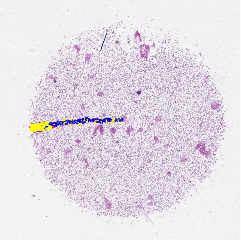
Learning to be EXACT 3
Table 1. Overview of the dataset including the file id, the number of cells per type
and the screened sample area. The top two rows represent the completely annotated
validation and test slides for the cross validation with a fixed train set.
id eosinophils mast cells neutrophils macrophages lymphocytes total screened image
1 21 511 3301 3934 14846 22613 100%
2 47 762 951 16748 10342 28850 100%
3 10 69 1321 3081 15666 20147 8%
4 20 37 2467 729 2144 5397 28%
5 8 116 4491 1639 3077 9331 43%
6 2 40 26 370 323 761 1%
108 1535 12557 26501 46398 87099 46%
from training to compare the accuracy of human experts with our algorithmic
approach.
2.1 Label generation and training pipeline
The dataset containing only six WSIs appears to be comparably small. However,
the cells in the WSIs are annotated by experts and subdivided into five classes
using SlideRunner [6], rendering it one of the largest manually annotated cytol-
ogy datasets to date. The dataset displays an extreme class imbalance, with the
rarest class of eosinophils representing only 0.12% of all annotations. A particular
challenge for training neuronal networks emerges from the sparse annotation of
four WSIs, as shown in the column ”screened” in table 1. To meet this challenge of
1 2 GT Prediction
0 0
100 100
200 200
4 3
300 300
400 400
500 500
5 6 600 600
700 700
800 800
0 100 200 300 0 100 200 300
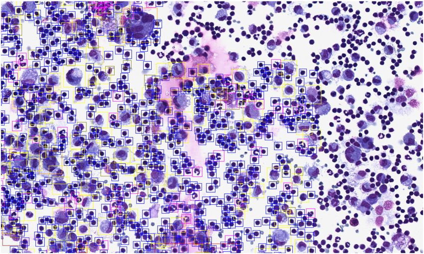
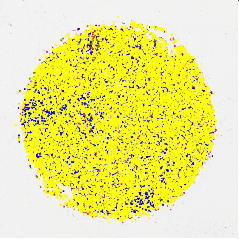
Fig. 1. Left: visualisation of the annotated regions/cells of the included slides. Center:
visualisation of the 16 traditional fully annotated sub-images for sampling in green and
624 cell-based sampling positions for our live sampling approach within the red area.
To prevent sampling from unannotated WSI areas, the red area is restricted to half of
the patch size to the annotation border. Right: The ground truth (GT) and the deep
learning based predictions; neutrophils (red), eosinophils (green), lymphocytes˜(blue),
macrophages (yellow), mast cells (purple).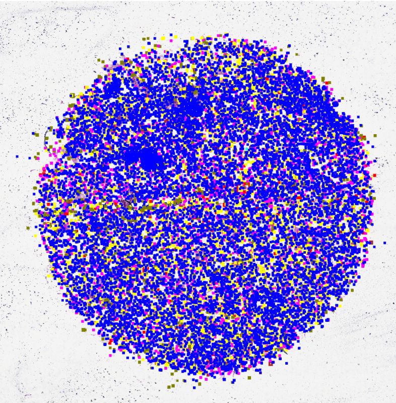
4                                 Marzahl et al.

partially annotated WSIs, we apply an online training approach using the open-
source online annotation platform EXACT [7]. EXACT supports a persistent
screening mode that allows experts to screen WSIs in a self-determined resolu-
tion. By reusing the information this screening mode provides, we are able to
track exactly which areas of the slide have already been annotated by the expert
and can download them into the training process via the provided REST-API.
    We propose the following online training pipeline for equine asthma. As a first
step, screened areas of the slides and associated annotations are downloaded from
the EXACT server via the REST-API. The training pipeline is then initialised
with the downloaded information, the network architecture and the loss function.
During training, new patches are sampled live from the WSI according to the
patch selection and sampling strategy described in the following sections and
are restricted by information about the screened area provided by EXACT. The
trained models can be applied to new data and the results can be synchronised
with the server for expert review.

2.2   Live patch selection and sampling strategy
To counteract the described class imbalance and partial annotations, we propose
the following sampling strategy which uses annotated cells as seeds for training
patches: Each training patch has a cell that was manually annotated by an
expert in its center. Consequently, only cells that have at least a distance of half
of the patch size to the border of the annotated region can be used as patch
centers (red area in Figure 1). Each training batch contains at least five patches
with each of the five cell types represented as patch center cell. The center cells
within each class are randomly chosen from the annotations. If the training batch
size is chosen larger than the number of cell types, a new cell type is randomly
selected with a probability proportional to 1 − pk where pk is the relative class
frequency of each cell type until the required batch size is reached. This results
in a sampling strategy which can freely choose the sampled patches and reduces
the possibility of sampling a given region repeatedly. This is highly desirable to
counter overfitting and works as an advanced augmentation technique.
    For comparison we extract all available fully annotated areas as sub-images of
the WSIs to simulate a traditional training pipeline. This results in a total of 1862
sub-images of which 851 belong to the two fully annotated test WSIs. Example
sub-images are visualised in Figure 1 on the right with green rectangles. For
training the same cell type-based sampling strategy to counteract the described
class imbalance is applied.

2.3   Object detection methods
Since the training strategy itself is the main contribution of this work, we use a
publicly available and for cytology optimised implementation [8] of the success-
ful RetinaNet [9] architecture. Different ResNet-variants [10] (ResNet-18, -34,
-50) pretrained on ImageNetare applied as backbone networks with appropri-
ate mini-batch sizes. The networks are trained using the sub-images-based and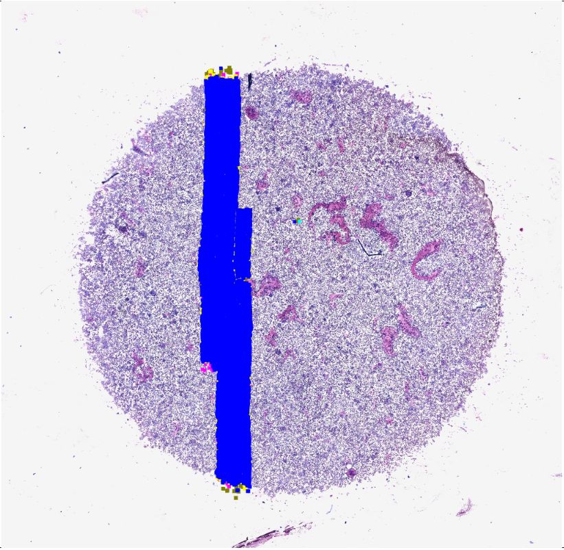
Learning to be EXACT             5

Table 2. The mean average precision for the five types of cells in respect to the number
of layers used for the ResNet backbone network (BB) and batch size (BS). The modes
represent our method working on partially annotated WSIs and a classical approach
with extracted sub-images.

    mode     BB BS eosinophils mast cell neutrophils macrophages lymphocytes ∅
    ours     18   16   0.93       0.85       0.88         0.89          0.81     0.87
 sub-image   18   16   0.69       0.72       0.68         0.80          0.71     0.72
    ours     34   16   0.91       0.86       0.90         0.89          0.78     0.87
 sub-image   34   16   0.70       0.71       0.68         0.80          0.72     0.72
    ours     50    6   0.92       0.80       0.90         0.89          0.81     0.86
 sub-image   50   6    0.72       0.69       0.68         0.81          0.75     0.73

the proposed live sampling-based approach with a patch size of 1024×1024 px.
Each of the two fully annotated WSIs (Table 1) are used once as the validation
set and once as the test set while keeping the training set static to allow for
a form of cross-validation given the limited amount of cases. During training,
data augmentation (rotation between zero and 90 degrees, horizontal and verti-
cal flips, random increase or decrease of intensity in the range of -20 to +20%)
is applied and the networks are trained until convergence on the validation set.
The initial learning rate is set to 1e-3 and reduced to 1e-4 and 1e-5 if the vali-
dation loss doesn’t decrease for three epochs. One epoch consists of 500 training
patches regardless of the WSI-based sampling mode or the extracted sub-images.
The object detection accuracy is measured as mean Average Precision (mAP)
according to the 2007 PASCAL VOC challenge.

3    Results
Independent of the backbone model or batch size, the accuracy of our WSI-based
sampling approach converge at an mAP of 0.87 (min=0.86, max=0.87, IoU=0.5,
epochs=73), outperforming the model trained on sub-images (mAP=0.72, min
= 0.72, max=0.73, IoU=0.5, epochs=23) as shown in Table 2. After 23 epochs
the sub-images-based training is terminated due to overfitting. The backbone
network of the model has no effect on the accuracy. The live sampling approaches
show the lowest performance for the lymphocytes, which are the smallest and
most clustered type of cells. The sub-images-based approach scores lowest on the
rare classes of eosinophils and mast cells due to overfitting.
   When applying the trained solution with a ResNet-18 as a backbone on the
image patches and ground-truth published in [blinded for peer review], we reach
a mean mAP across images of 0.76 with the proposed method and 0.63 with the
sub-images-based approach compared to the experts reaching a published mean
concordance of µ=0.73 mAP (min=0.56, max=0.82,σ=0.08).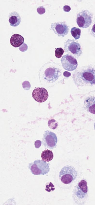
6                                 Marzahl et al.

        4   Discussion and outlook

        We demonstrated the creation of an object detection training pipeline which
        is able to use partially annotated WSIs efficiently and is superior to a simple
        sub-image-based approach. Our proposed approach allows for better sampling
        strategies and data augmentation for rare classes which massively reduces the
        chance to sample the same patch repeatedly and therefore mitigate overfitting,
        as apparent in the considerable difference in performance for eosinophils in the
        evaluation. This resulted in a object detection model with human like perfor-
        mance on a small set of example patches. However, this work has the limitation
        that only six images have been partially annotated by one expert, which needs
        to be addressed in further research. This work can be used as a baseline for
        further enhancements, like increasing the detection performance of small cells,
        optimising the non-maximum suppression algorithm for high quantities of cells
        but also to create new annotations in an expert-algorithm based manner on the
        remaining WSIs.

        Acknowledgement. CAB gratefully acknowledges financial support received
        from the Dres. Jutta & Georg Bruns-Stiftung für innovative Veterinärmedizin.

        References
         1. Bullone M, Lavoie JP. Asthma “of horses and men”—how can equine heaves help
            us better understand human asthma immunopathology and its functional conse-
            quences? Mol Immunol. 2015;66(1):97–105.
         2. Marzahl C, Bertram CA, Aubreville M, et al. Are Fast Labeling Methods Reliable?
            A Case Study of Computer-Aided Expert Annotations on Microscopy Slides. In:
            MICCAI. Cham: Springer International Publishing; 2020. p. 24–32.
         3. Huang J, Rathod V, Sun C, et al. Speed/accuracy trade-offs for modern convolu-
            tional object detectors. In: CVPR; 2017. p. 7310–7311.
         4. Kawazoe Y, Shimamoto K, Yamaguchi R, et al. Faster r-cnn-based glomerular
            detection in multistained human whole slide images. Imaging. 2018;4(7):91.
         5. Yang F, Yu H, Silamut K, et al. Parasite Detection in Thick Blood Smears Based
            on Customized Faster-RCNN on Smartphones. In: AIPR; 2019. p. 1–4.
         6. Aubreville M, Bertram C, Klopfleisch R, et al. SlideRunner. In: Bildverarbeitung
            für die Medizin 2018. Springer; 2018. p. 309–314.
         7. Marzahl C, Aubreville M, Bertram CA, et al. EXACT: A collaboration toolset for
            algorithm-aided annotation of almost everything. arXiv preprint arXiv:200414595.
            2020;.
         8. Marzahl C, Aubreville M, Bertram CA, et al. Deep Learning-Based Quantification
            of Pulmonary Hemosiderophages in Cytology Slides. Sci Rep. 2020;10(1):1–10.
         9. Lin TY, Goyal P, Girshick R, et al. Focal loss for dense object detection. In:
            Proceedings of the IEEE international conference on computer vision; 2017. p.
            2980–2988.
        10. He K, Zhang X, Ren S, et al. Deep residual learning for image recognition. In:
            CVPR; 2016. p. 770–778.

E3020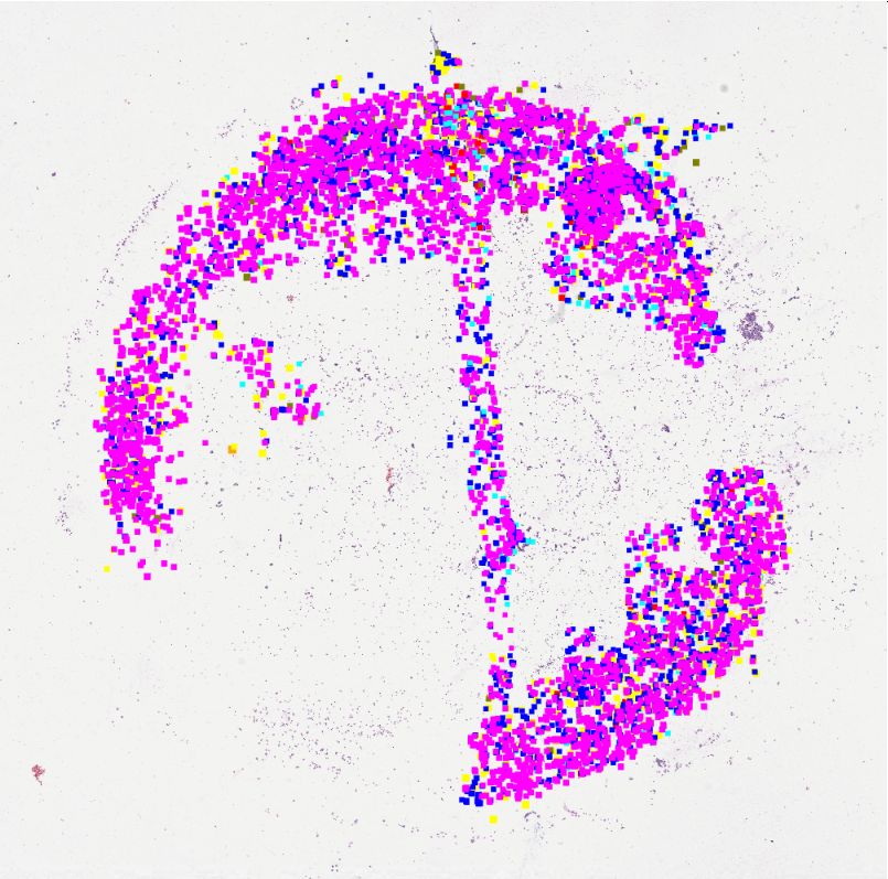
You can also read