Several different cytogenetic clones arising during treatment of Philadelphia positive chronic myeloid leukemia with tyrosine kinase inhibitors ...
←
→
Page content transcription
If your browser does not render page correctly, please read the page content below
Page 114                                         VOJNOSANITETSKI PREGLED                     Vojnosanit Pregl 2021; 78(1): 114–118.

CASE REPORT                                                                                               UDC: 616.155.392-08-036
                                                                                           https://doi.org/10.2298/VSP180723010D

Several different cytogenetic clones arising during treatment of
Philadelphia positive chronic myeloid leukemia with tyrosine kinase
inhibitors lead to the progression into Philadelphia negative acute
myeloid leukemia
             Različiti citogenetski klonovi nastali tokom lečenja Filadelfija pozitivne
           hronične mijelodne leukemije inhibitorima tirozin kinaze sa progresijom u
                                    Filadelfija negativnu akutnu mijeloidnu leukemiju

                            Marija Denčić-Fekete*, Danijela Leković*†, Vesna Djordjević*,
                          Jelica Jovanović*, Biljana Todorić-Živanović‡, Ljubomir Jaković*†,
                                                 Andrija Bogdanović*†

                       Clinical Center of Serbia, *Clinic for Hematology, Belgrade, Serbia; University of
                        Belgrade, †Faculty of Medicine, Belgrade, Serbia; ‡Military Medical Academy,
                                                        Belgrade, Serbia

Abstract                                                            was introduced, leading to a good molecular response and
                                                                    the disappearance/loss of the Ph+ clone with additional
Introduction. Additional karyotype abnormalities in the             abnormalities but with the appearance of the Ph- clone with
Philadelphia-positive (Ph+) clone can emerge during the             trisomy 8. Finally, after 5.5 years of nilotinib therapy, the
progression of chronic myeloid leukemia (CML) and are of-           Ph- clone with monosomy 7 occurred during the deep mo-
ten associated with the resistance to treatment with tyrosine       lecular response for BCR-ABL. At that time, the FISH anal-
kinase inhibitors (TKI). Sometimes, during the TKI treat-           ysis for trisomy 8 was negative, but the rise in blast count
ment, karyotype abnormalities can appear in the Philadelph-         was noticed in the bone marrow, and the diagnosis of the
ia-negative (Ph-) cells as well but do not seem to adversely        secondary AML was established soon after. Conclusion.
affect the outcome except for chromosome 7 abnormalities.           The achievement of the deep molecular response in CML
Case report. The patient presented was in the chronic               patients does not rule out regular cytogenetic testing of their
phase of Ph+ CML with highly diverse karyotype abnormal-            bone marrow. This is of crucial importance for detecting
ities. The abnormalities appeared in three unrelated clones         adverse karyotype abnormalities leading to the development
during the TKIs treatment, followed by the evolution of the         of the myelodysplastic syndrome and AML.
disease into acute myeloid leukemia (AML). The primary
Ph+ clone was revealed during the chronic phase of CML,             Key words:
and therapy with imatinib mesylate was commenced. After a           leukemia, myelogenous, chronic, bcr-abl positive;
three-year hematologic and cytogenetic remission period,            leukemia, myeloid, acute; karyotyping; enzyme
the evolution of the primary clone was noticed. Nilotinib           inhibitors; cytogenetics.

Apstrakt                                                            nosti prisutne u tri nezavisna klona sa evolucijom bolesti u
                                                                    akutnu mijeloidnu leukemiju (AML). Primarni Ph+ klon je
Uvod. Dodatne kariotipske abnormalnosti u Filadelfija-              otkriven tokom hronične faze CML i započeta je terapija
pozitivnom (Ph+) klonu mogu se javiti tokom progresije              imatinib mesilatom. Nakon tri godine hematološke i citoge-
hronične mijeloidne leukemije (CML) i često su povezane sa          netske remisije, uočena je evolucija primarnog klona. Zapo-
rezistencijom na terapiju tirozin kinaznim inhibitorima             četa je terapija nilotinibom koja je dovela do molekularnog
(TKI). Ponekad se tokom terapije TKI kariotipske abnor-             odgovora i povlačenja Ph+ klona sa dodatnim aberacijama,
malnosti javljaju i u Filadelfija-negativnim (Ph-) ćelijama, ali    ali i pojavljivanja novog Ph- klona sa trizomijom 8. Nakon
ne utiču na progresiju bolesti, izuzev abnormalnosti hromo-         5,5 godina lečenja nilotinibom i postizanja kompletnog
zoma 7. Prikaz bolesnika. Kod bolesnice u hroničnoj fazi            molekularnog odgovora, uočen je Ph- klon sa monozomi-
CML, tokom lečenja TKI uočene su kariotipske abnormal-              jom 7. Fluorescentna in situ hibridizacija (FISH) pokazala je

Correspondence to: Marija Denčić-Fekete, Clinical Center Serbia, Clinic for Hematology, Koste Todorovića 2, 11 000 Belgrade, Serbia.
E-mail: marijadfekete@yahoo.com
Vol. 78, No 1 VOJNOSANITETSKI PREGLED Page 115
odsustvo trizomije 8 i prisustvo monozomije 7. Istovreme- zbog mogućnosti razvoja sekundarnih maligniteta – mijelo-
no, registrovan je porast broja blasta u koštanoj srži i ubrzo displastičnog sindroma i AML.
je postavljena dijagnoza sekundarne AML. Zaključak. Pos-
tizanje kompletnog molekularnog odgovora primenom TKI Ključne reči:
terapije ne treba da isključi redovno citogenetsko testiranje leukemija, mijeloidna, hronična, bcr-abl pozitivna;
koštane srži bolesnika sa CML. Otkrivanje kariotipskih ab- leukemija, mijelocitna akutna; kariotip, određivanje;
normalnosti sa lošom prognozom je od velikog značaja enzimi, inhibitori; citogenetika.
Introduction RT-PCR. During the next three years, the patient remained in
a stable chronic phase with CCgR, on 400 mg of imatinib.
The BCR-ABL fusion gene, generating the Philadelphia However, in August 2005, the follow-up cytogenetics
chromosome (Ph), is a sole genetic abnormality in 90% of revealed complex translocation t(5;6;12) with t(9;22) (Figure
chronic myeloid leukemia (CML) patients in the chronic 1B) in all analyzed cells, without any clinical or laboratory
phase 1. With the progression of the disease, additional sign of disease progression. The tyrosine-kinase domain
karyotype changes in the Philadelphia positive (Ph+) clone mutations were negative by direct sequencing.
emerge and are often associated with resistance to imatinib
mesylate and/or nilotinib. The resistance can be a
consequence of one of the numerous mutations in the
tyrosine kinase domain or some other underlying mechanism
and is usually overwhelmed with some of the novel tyrosine
kinase inhibitor (TKI) drugs. Sometimes, during the TKI
treatment, karyotype changes in the Philadelphia negative
(Ph-) cells can appear 2. These aberrations, similar to those
frequently seen in the myelodysplastic syndrome (MDS) and
acute myeloid leukemia (AML), include trisomy 8 (+8), the
deletion or monosomy of chromosome 7 (del(7q)/-7), and
nullisomy of Y (-Y). However, chromosome abnormalities in
the Ph- clone do not seem to adversely affect the outcome
with the exception of the chromosome 7 abnormalities 1.
Monosomy 7 and del (7q) require frequent bone marrow
follow-up as several case reports indicate the development of
the MDS and subsequent AML 3–7.
A CML patient with highly diverse karyotype
abnormalities was presented in this paper. The abnormalities
appeared in three unrelated clones during the treatment with
imatinib mesylate and nilotinib. The patient mentioned
developed AML in the end.
Case report
A 44-year-old female was diagnosed with CML in the
chronic phase in April 2002. The cytogenetic analysis
revealed the translocation t(9;22) (q34;q11) (Ph
chromosome) in 20/20 mitoses (Figure 1A). The prognostic
Sokal and Hasford scores implied a low-risk patient at
presentation, without comorbidities or any other additional
treatment. The antileukemic therapy was commenced with
hydroxyurea and interferon with a subsequent increase in
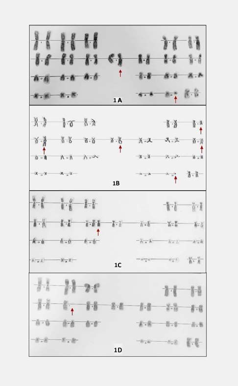
Fig. 1(A–D): A) (46,XX,t(9;22) (q34;q11) at presentation;
interferon dosage. Regardless of achieving hematological
B) [46,XX,t(5;6;12)(q14?;q21?;q23?),t(9;22)(q34;q11)
remission, no cytogenetic response was obtained after 6 imatinib (400 mg), TT 36 m];
months of the treatment. As the patient had no available stem C) 47,XX,+8 nilotinib (800 mg), TT 6 m; D) 45,XX,-7
cell donor, imatinib mesylate in a standard dose (400 mg a nilotinib (800 mg), TT 66 m.
day) was started in September 2002. Six months later, the
patient achieved partial cytogenetic response (PCgR) with The patient was treated with an escalated dosage of
53% Ph-negative metaphases. After 12 months, a complete imatinib (800 mg a day) since October 2006 without success.
cytogenetic response (CCgR) was achieved, although a b2a2 The patient was switched to nilotinib, 800 mg a day, from
transcript of the BCR-ABL fusion was detected by “nested” July 2008 through the “compassionate use program”.
Denčić-Fekete M, et al. Vojnosanit Pregl 2021; 78(1): 114–118.Page 116 VOJNOSANITETSKI PREGLED Vol. 78, No 1
However, due to hematologic and hepatic toxicity, after only was classified according to the standard of the UK Medical
a month, nilotinib was reduced to 400 mg. After 6 months of Research Council practice as complete (0% Ph+
400 mg nilotinib, the patient achieved CCgR again, but at 12 metaphases), major (1–34% Ph+), partial (35–65% Ph+
months, the reappearance of Ph+ clone with t(5;6;12) was metaphases), minor (66–95% Ph+) and no response (95–
noticed in 45% of metaphases, suggesting PCgR. Along with 100% Ph+). The cytogenetic clonal evolution was defined as
the Ph-positive cells, the Ph-negative clone with +8 was seen the presence of any abnormality other than a single Ph
in 10% of mitoses. After the recovery of blood counts and chromosome.
the hepatic function 6 months later, the patient was escalated
to the full dosage of nilotinib (800 mg) again. She achieved a Reverse transcription-polymerase chain reaction
major cytogenetic response [MajCgR, 10% of Ph+ with (RT-PCR) and ”nested“
t(5;6;12) clone] at 24 months of nilotinib treatment, and
finally, after 30 months on nilotinib, the patient achieved The total RNA was extracted from peripheral blood
CCgR. During the next 3 years on 800 mg of nilotinib, her cells according to the guanidine thiocyanate-phenol-
follow-up showed CCgR and a stable molecular response chloroform extraction method 8. Reverse transcription was
(MR3), though the Ph-negative clone with +8 was constantly performed on 1 μg of total RNA after heating at 65 ºC for 15
present in 10–30% of metaphases (Figure 1C). minutes. Reverse transcription was performed with the 1st
However, during the regular follow-up in February Strand cDNA Synthesis Kit for RT-PCR (AMV) (Roche
2014 (5.5 years of nilotinib therapy), profound neutropenia Diagnostics Corporation, Indianapolis, IN, USA) according
without anemia and thrombocytopenia was noticed to the manufacturer’s manual. The amplification was done
(hemoglobin 120 g/L, white blood cells 2.5 × 10⁹/L, 76% with slight modifications as described by Moravcová et al. 9.
lymphocytes, 4% blasts, 10% monocytes, platelets 258 × The FISH analysis for centromere regions of
109/L) together with elevated transaminases (alanine chromosomes 7 and 8 (CEP7 and CEP8) was performed on
aminotransferase ‒ ALT 86 U/L). Immediate bone marrow interphase nuclei and metaphase cells according to the
evaluation revealed dysplastic changes in erythroid and manufacturer’s instructions (Vysis/Abbott Laboratories, Des
megakaryocyte lineages together with 6% of blasts. The Plaines, IL).
karyotype revealed the poor quality of chromosomes, but the
clonal change with a loss of one chromosome from the C Discussion
group was evident in 60% of mitoses. The fluorescence in
situ hybridization (FISH) analysis with the BCR-ABL probe Our case demonstrates highly diverse karyotype
was negative both for the Ph chromosome and trisomy 8, but changes appearing one after another in three unrelated clones
the CEP7 probe revealed the monosomy of chromosome 7 in during the treatment with tyrosine kinase inhibitors.
80% of interphase nuclei. Complex aberrations in the Ph-positive clone emerged
The administration of nilotinib was stopped. Two during the management with imatinib, while +8 and -7
months later, the patient's bone marrow was hypocellular appeared separately in the Ph-negative clones during the
with less dysplasia than at the previous examination but with nilotinib treatment.
the rise in blast count (12%). The cytogenetic examination Karyotype changes in the Ph+ clone emerged 40
confirmed -7 in all analyzed cells (20/20) and the absence of months after the imatinib therapy was started as the only sign
trisomy 8 (Figure 1D). Real-time qualitative polymerase of disease relapse. This distinctive karyotype included
chain reaction (RQ-PCR) for BCR-ABL revealed a deep complex translocation and the rare event of centromere
molecular response, MR4. fission, which were previously published 10, 11. A negative
After one month, in April 2014, further evaluation search for mutations by Sanger sequencing in the kinase
revealed leukemic progression and development of AML domain further contributed to the complexity of the case.
(30% of blasts) confirmed by the flow cytometry Only after the introduction of a more potent TKI
immunophenotype (HLA-DRmed, CD34high, CD117med, treatment, nilotinib, the Ph+ clone slowly decreased, but
CD13high, CD33med, CD7+). The patient was treated with the trisomy 8 in the Ph-negative cells appeared. The CCgR was
antileukemic treatment (3 + 7 regimen) without success, achieved after 30 months on nilotinib, while +8 remained
followed by the ‟salvage” protocol FLAG-Ida without and existed in up to 30% of analyzed cells during the next
achieving any morphological or cytogenetic response. 2.5 years of follow-up.
Unfortunately, the patient died of aplasia during the Nota bene, the Ph-negative clones are less frequent in
treatment. patients treated with second-generation TKIs and after the
failure of imatinib due to higher pressure on leukemic and
Cytogenetic study and response criteria residual normal hematopoiesis 12. However, when present,
their type and frequency are very similar to those seen in
The cytogenetic study was performed on unstimulated patients on imatinib, as well as their incidence and effect in
bone marrow cells using a standard technique. The Giemsa- evolution to MDS/AML 12.
banded metaphases were analyzed, and the result was Our patient developed secondary AML after 66 months
reported by the International System for Human Cytogenetic of the nilotinib treatment. The cytogenetic and FISH analysis
Nomenclature standards, 2013. The cytogenetic response revealed -7 in 60% of metaphases and 80% of interphase
Denčić-Fekete M, et al. Vojnosanit Pregl 2021; 78(1): 114–118.Vol. 78, No 1 VOJNOSANITETSKI PREGLED Page 117
nuclei, respectively, along with the absence of BCR-ABL During the treatment with TKIs, it is highly important
and +8. The clone with -7 quickly progressed to 100% of the to reveal the biological diversity of the Ph-negative clones,
analyzed cells in two months, while RQ-PCR still showed a which in some patients can lead to disease transformation
stable MR4. Despite introducing a high-dose therapy for (clone with -7), while in others, it does not have the
AML, the patient died 6 months after the diagnosis of the propensity towards secondary hematological malignancy
secondary AML had been established. (clone with +8). Minimal investigations should include blood
The Ph-negative clones with -7 were described in the test results (cytopenia), bone marrow morphology (dysplastic
CML cases with a high propensity to evolve into changes and blast count), and cytogenetic (evidence of the
MDS/AML 13. However, there have been rare cases with -7 Ph-negative clones and -7). In cases with the additional Ph-
without disease evolution 4, 5, 14. negative clones, further evaluation of changes with the FISH
In several studies, factors contributing to the appearance and real-time PCR analyses are highly recommended.
of chromosomal aberrations in the Ph-negative clone have
been discussed. The previous cytotoxic treatment 13, the Conclusion
negative effect of TKIs on DNA repair mechanisms 15–17, or
the innate genetic instability in the CML marrow 18 are The evolution of karyotype and the occurrence of
described as potential causes of the Ph-negative clone diverse clones arising from the stem cell level in our patients,
appearance. However, among all the abnormalities, only those warrants the need for thorough follow-up and evaluation of
involving chromosome 7 [del(7q) and/or -7] bear a higher risk all related hematological and biological findings during the
of secondary malignancies 5, 19. We can conclude that while the treatment with tyrosine kinase inhibitors, including the
patient was in a stable chronic phase of CML, complex standard karyotype, although, some study groups tend to
chromosomal aberrations in the Ph-positive cells might reflect omit any bone marrow evaluation in the current monitoring
a highly unstable genome, which could contribute to a further schedule.
lower sensitivity to a subsequent alternative treatment and
thus, negatively affect overall survival. Acknowledgement
Other parameters that could lead to the development of
MDS/AML are pretreatment with interferon or hydroxyurea, a We would like to express our gratitude to Prof. Dr. J.
persistent aberration in the Ph-negative clone, and clone size Apperley, Imperial Colledge of Medicine, Hammersmith
> 50% 19. Unfortunately, our patient had all the negative features Hospital Campus, London, for generous help in performing
mentioned above in developing secondary malignant disease. mutation analysis.
R E F E R E N C E S
1. Baccarani M, Deininger MW, Rosti G, Hochhaus A, Soverini S, Ap- therapy with imatinib mesylate is frequently characterized by
perley JF, et al. European LeukemiaNet recommendations for trisomy 8. Leukemia 2002; 16(8): 1390–1393.
the management of chronic myeloid leukemia: 2013. Blood 8. Chomczynski P, Sacchi N. Single-step method of RNA isolation
2013; 122(6): 872‒84. by acid guanidinium thiocyanate-phenol-chloroform extrac-
2. Deininger MW, Cortes J, Paquette R, Park B, Hochhaus A, Baccarani tion. Anal Biochem 1987; 162(1): 156‒9.
M, et al. The prognosis for patients with chronic myeloid leu- 9. Moravcová J, Lukásová M, Starý J, Haskovec C. Simple competi-
kemia who have clonal cytogenetic abnormalities in Philadel- tive two-step RT-PCR assay to monitor minimal residual dis-
phia chromosome-negative cells. Cancer 2007; 110(7): 1509- ease in CML patients after bone marrow transplantation. Leu-
19. kemia 1998; 12(8): 1303‒12.
3. Perel JM, McCarthy C, Walker O, Irving I, Williams B, Kennedy GA. 10. Denčić-Fekete M, Đorđević V, Storlazzi C. T, Janković G, Bogdanović
Clinical significance of development of Philadelphia chromo- A, Jovanović J, et al. t(5;6;12) associated with resistance with
some negative clones in patients with chronic myeloid leuke- imatinib mesylate in chronic myeloid leukemia. Int J Hematol
mia treated with imatinib mesylate. Haematologica 2005; 90 2009; 89(4): 508‒12.
(Suppl): ECR25. 11. Storlazzi CT, Albano F, Dencić-Fekete M, Djordjević V, Rocchi M.
4. Bacher U, Hochhaus A, Berger U, Hiddemann W, Hehlmann R, Haf- Late-appearing pseudocentric fission event during chronic my-
erlach T, et al. Clonal aberrations in Philadelphia chromosome eloid leukemia progression. Cancer Genet Cytogenet 2007;
negative hematopoiesis in patients with chronic myeloid leu- 174(1): 61‒7.
kemia treated with imatinib or interferon alpha. Leukemia 12. Baldazzi C, Luatti S, Marzocchi G, Stacchini M, Gamberini C,
2005; 19(3): 460‒3. Castagnetti F, et al. Emergence of clonal chromosomal ab-
5. Kovitz C, Kantarjian H, Garcia-Manero G, Abruzzo LV, Cortes J. normalities in Philadelphia negative hematopoiesis in
Myelodysplastic syndromes and acute leukemia developing af- chronic myeloid leukemia patients treated with nilotinib af-
ter imatinib mesylate therapy for chronic myeloid leukemia. ter failure of imatinib therapy. Leukemia Res 2009; 33(12):
Blood 2006; 108(8): 2811‒3. e218‒20.
6. Terre C, Eclache V, Rousselot P, Imbert M, Charrin C, Gervais C, et 13. Ribeiro de Mello Conchon M, Bendit I, Ferreira P, Lima W, Kumeda
al. Report of 34 patients with clonal chromosomal abnormali- C, Dias L, et al. Emergence of abnormal clone with mono-
ties in Philadelphia negative cells during imatinib treatment of somy 7 in Philadelphia negative cells of CML patients treated
Philadelphia-positive chronic myeloid leukemia. Leukemia with tyrosine kinase inhibitors. Int J Hematol 2009; 89(1):
2004; 18(8): 1340-6. 123‒5.
7. Andersen MK, Pedersen-Bjergaard J, Kjeldsen L, Dufva IH, Brøndum- 14. McMullin MF, Humphreys M, Byrne J, Russell NH, Cuthbert RJ,
Nielsen K. Clonal Ph-negative hematopoiesis in CML after O’Dwyer ME. Chromosomal abnormalities in Ph- cells of pa-
Denčić-Fekete M, et al. Vojnosanit Pregl 2021; 78(1): 114–118.Page 118 VOJNOSANITETSKI PREGLED Vol. 78, No 1
tients on imatinib. Blood 2003; 102(7): 2700‒1; author reply 18. Tanaka H, Tanaka K, Oguma N, Ito K, Ito T, Kyo T, et al. Effect
2701. of interferon-α on chromosome abnormalities in treated
15. Bumm T, Müller C, Al-Ali HK, Krohn K, Shepherd P, Schmidt E, et chronic myelogenous leukemia patients. Cancer Genet Cyto-
al. Emergence of clonal cytogenetic abnormalities in Ph- cells genet 2004; 153(2): 133‒43.
in some CML patients in cytogenetic remission to imatinib but 19. Groves MJ, Sales M, Baker L, Griffiths M, Pratt N, Tauro S. Fac-
restoration of polyclonal hematopoiesis in the majority. Blood tors influencing a second myeloid malignancy in patients with
2003; 101(5): 1941‒9. Philadelphia-negative -7 or del(7q) clones during tyrosine ki-
16. Kharbanda S, Pandey P, Jin S, Inoue S, Bharti A, Yuan ZM, et al. nase inhibitor therapy for chronic myeloid leukemia. Cancer
Functional interaction between DNA-PK and c-Abl in re- Genet 2011; 204(1): 39‒44.
sponse to DNA damage. Nature 1997; 386(6626): 732‒5.
17. Fabarius A, Haferlach C, Müller M, Erben P, Lahaye T, Giehl M, et
al. Dynamics of cytogenetic aberrations in Philadelphia chro- Received on July 23, 2018.
mosome positive and negative hematopoiesis during dasatinib Revised on November 29, 2018.
therapy of chronic myeloid leukemia patients after imatinib Accepted on January 16, 2019.
failure. Haematologica 2007; 92(6): 834‒7. Online First January, 2019.
Mitić J, et al. Vojnosanit Pregl 2020; 77(3): 114–118.You can also read