Abstract A Systematic Review of the Value of a Bladder Scan in Cauda Equina Syndrome Diagnosis - Cureus
←
→
Page content transcription
If your browser does not render page correctly, please read the page content below
Open Access Review
                              Article                                                DOI: 10.7759/cureus.14441

                                             A Systematic Review of the Value of a Bladder
                                             Scan in Cauda Equina Syndrome Diagnosis
                                             Awf A. Alshahwani 1 , Joseph Boktor 2 , Amr Elbahi 3 , Purnajyoti Banerjee 3

                                             1. Trauma and Orthopaedics, Leicester University Hospital, Leicester, GBR 2. Trauma and Orthopaedics, Cardiff
                                             University Hospital, Cardiff, GBR 3. Trauma and Orthopaedics, Kettering General Hospital, Kettering, GBR

                                             Corresponding author: Awf A. Alshahwani, dr_awf1985@yahoo.com

                                             Abstract
                                             Cauda equina syndrome (CES) is one of the emergency conditions that can lead to devastating permanent
                                             functional disabilities, if misdiagnosed. Multiple studies have questioned the reliability of clinical
                                             assessment in diagnosing CES, whether some of the features should be considered to be potential red flags.
                                             Bladder dysfunction can reflect CE compromise. The post-void residual (PVR) volume bladder scan is useful
                                             in CES diagnosis, but to date there has been no single systematic review supporting its use. Furthermore,
                                             there is no clear cut-off point to consider PVR statistically significant. The aim of the study is to perform a
                                             systematic review of the current evidence behind the use of the PVR bladder scan as a diagnostic tool for
                                             CES diagnosis. This was a comprehensive search using Medline, PubMed and Embase. All articles included
                                             post-void bladder scans with the mentioned clear cut-off volume as a diagnostic parameter. A total of five
                                             study articles from 1955 fit with our inclusion and exclusion criteria. The total number of patients who had a
                                             bladder scan was 531. CES was confirmed in 85 cases. Bladder scan diagnosed 70 cases and excluded 327. The
                                             best results for both sensitivity and specificity in correlation with the sample of the study were for PVR more
                                             than 200 ml. Measuring the post-void urine volume using a bladder scan is an essential tool in the diagnosis
                                             of CES. There is a significant correlation between the PVR volume more than 200 ml and higher sensitivity
                                             and specificity.

                                             Categories: Orthopedics
                                             Keywords: cauda equina, bladder scan, pvr, screen, adjunct

                                             Introduction And Background
                                             Cauda equina syndrome (CES) is an emergency spinal pathology that can lead to devastating permanent
                                             functional disabilities. It is rare with an estimated incidence of 1 in 2000 [1]. Missing timely diagnosis of
                                             cauda equina compression can end up in irreversible bowel, bladder and sexual dysfunction. Missed CES
                                             represents the most common diagnoses associated with successful litigation and paying off huge sum of
                                             compensation to the victim thereby causing substantial financial strain in every health system in the world.
                                             In the United States, nearly half of the claims have been paid, and settlement costs millions of dollars per
                                             person [2,3].
Review began 03/31/2021
Review ended 04/05/2021                      There is a debate in the literature questioning the accuracy of clinical features in the diagnosis of CES
Published 04/12/2021
                                             including saddle area anesthesia, perineal loss of sensation, sphincter dysfunction, bilateral sciatica, and
© Copyright 2021                             motor and sensory function disturbances [4]. Magnetic resonance imaging (MRI) scan is essential to rule out
Alshahwani et al. This is an open access     cauda equina as no single or collective features will definitely confirm or exclude diagnosis [5]. The British
article distributed under the terms of the
                                             Association of Spine Surgeons (BASS) emphasized that an urgent MRI scan should be performed for
Creative Commons Attribution License
CC-BY 4.0., which permits unrestricted       suspected CES, and decompressive surgery should be undertaken at the earliest opportunity in confirmed
use, distribution, and reproduction in any   cases [6]. Early surgery is indicated to avoid the permanent functional disability and financial implications
medium, provided the original author and     associated with a missed diagnosis or inadequate management. Therefore, early diagnosis with an MRI scan
source are credited.
                                             is an essential tool to achieve this [7].

                                             As MRI scanning may not be available in peripheral hospitals with no specialist spinal surgical services, an
                                             alternative measure is to identify the patient who needs a transfer to the specialist spinal “hub” hospital
                                             where MRI scans can be undertaken especially after working hours. These hospitals usually have around-
                                             the-clock cover for spinal emergencies and might reduce the risk of further delay by undertaking an MRI
                                             scan and then surgery if necessary without the need for further consultations or transfer to another center
                                             [6]. Furthermore, exposing a patient to an unnecessary imaging study during a situation like coronavirus
                                             disease 2019 (COVID-19) pandemic and using the same machine for COVID-positive patient can result,
                                             theoretically at least, to an increased spread of the disease, in addition to the burden on the resources
                                             [8]. As bladder function can be assessed on a frequent basis through urination, post-void bladder volume
                                             represents a surrogate marker of the cauda equina function and the bladder musculature contractions as
                                             well [9]. Bladder emptying, which can be accurately assessed by a bladder scan, would be a good criterion
                                             [10].

                                             In this study, we have undertaken a systematic review, assessing the efficacy of a bladder scan used as a

                              How to cite this article
                              Alshahwani A A, Boktor J, Elbahi A, et al. (April 12, 2021) A Systematic Review of the Value of a Bladder Scan in Cauda Equina Syndrome
                              Diagnosis. Cureus 13(4): e14441. DOI 10.7759/cureus.14441
bedside clinical tool in possible diagnosis of cauda equina compression. Furthermore, we have delineated an
accurate cut-off point to highlight the possibility of CES and prioritize the patient for a subsequent MRI
scan.
Our research question is, “Is bladder scan a suitable tool for screening cauda equina syndrome
complementary to clinical examination?” and “What would be the expected significant post-void residual
(PVR) volume to proceed to the MRI scan?”
Review
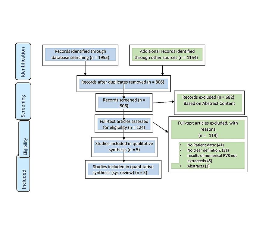
This systematic review was performed according to the Preferred Reporting Items for Systematic Reviews
and Meta-analyses (PRISMA) guidelines as shown in Figure 1.
FIGURE 1: PRISMA Flow Diagram
PRISMA, Preferred Reporting Items for Systematic Reviews and Meta-analyses; PVR, post-void residual
Starting on December 2019, till September 2020, a comprehensive search was done by two spinal surgeons
with the help of an experienced librarian. We looked at the databases including Embase, PubMed and
Medline. We included original peer-reviewed studies that included numeric values of PVR apparent in
bladder scans as a diagnostic tool and MRI scans done to confirm the diagnosis of CES. We excluded (1)
abstracts, case reports and studies mentioning high post-void residual volumes but no actual numeric values
reported and (2) studies that did not detail urine retention and volume but only mentioned bladder
dysfunction. Filtration of studies was done by reading the article title, abstract and for some studies, by
reading the full articles. The process was repeated by two authors and unrelated studies were removed after
revising.
The first electronic search detected a total of 1955 studies published from 2008 till July 2020. No studies were
found before 2008. Removal of duplicates was done and 806 studies remained and were further screened for
eligibility based on title and abstract; 120 articles were found eligible. After reading full text, only seven
studies met the inclusion criteria. Yet two of the seven were abstracts and were not fully published articles.
There was another randomized controlled trial that was stopped due to ethical approval withdrawal [11].
A total of five studies were included in this review. Table 1 shows the key features of these studies. Four of
the five studies were retrospective and one study was prospective. In the reviewed papers, the total number
of patients who had a bladder scan was 531. CES was confirmed in 85 cases. Bladder scan diagnosed 70 cases
2021 Alshahwani et al. Cureus 13(4): e14441. DOI 10.7759/cureus.14441 2 of 5and excluded 327. We looked at the sensitivity and specificity of each study and found the best results for
both sensitivity and specificity in correlation with the sample of the study would be at PVR >200 ml as
shown in Table 2.
Cases
Total With
Author &
Title Study Type No. of Positive Outcome
Year
Cases MRI for
CES
A PVR volume more than 500 ml with
Predictive value of clinical characteristics in
Domen et or without other clinical features of
patients with suspected cauda equina Retrospective 58 8
al. (2009) CES is an important predictor for
syndrome [12]
CES
No single clinical feature is adequate
Does rectal examination have any value in
Gooding et as standalone to discriminate with
the clinical diagnosis of cauda equina Retrospective 57 13
al. (2013) statistical significance to proceed to
syndrome? [5]
the MRI outcome
276
The clinical features and outcome of scan- but
negative and scan-positive cases in Hoeritzauer only No PVR records for two-thirds of the
Retrospective 15
suspected cauda equina syndrome: a et al. (2018) 65 patients
retrospective study of 276 patients [13] had a
scan
Bladder scans and postvoid residual volume The presence of red flags with a PVR
Venkatesan
measurement improve diagnostic accuracy Prospective 92 17 volume more than 200 ml obligates
et al. (2019)
of cauda equina syndrome [9] the necessity of an MRI scan
Use of the PVR volume ≥200 ml was
A prospective study of the role of bladder considerably more accurate in
scanning and post-void residual volume Katzouraki predicting CES. It is a useful adjunct
Prospective 260 32
measurement in improving diagnostic et al. (2020) to conventional clinical assessment
accuracy of cauda equina syndrome [14] and allows risk stratification in
managing suspected CES
TABLE 1: Key features of the five studies
PVR, post-void residual; CES, cauda equina syndrome
Author PVR Patients TP FN FP TN Sensitivity Specificity
Hoeritzauer et al. [13] >100 65 10 5 24 26 67% 52%
Venkatesan et al. [9] >200 92 16 1 19 56 94% 72%
Domen et al. [12] >500 58 6 2 0 50 100% 93%
Gooding et al. [5] >500 57 6 7 0 44 46.1% 100%
Katzouraki et al. [14] >200 260 32 2 75 151 94.1% 66.8%
TABLE 2: Data analysis from the studies, with sensitivity and specificity
PVR, post-void residual; TP, true positive; FN, false negative; FP, false positive; TN, true negative
CES was first described in the mid-50s by Jennett after correlating symptoms of back pain, sphincter
dysfunction with an intraoperatively confirmed disc prolapse [15]. The urgency of decompression surgery in
CES was emphasized by Ahn et al. who noticed in his meta-analysis better prognosis if decompression was
done within 48 hours, and even better prognosis was detected by Kohles et al. when surgery done earlier
2021 Alshahwani et al. Cureus 13(4): e14441. DOI 10.7759/cureus.14441 3 of 5within 24 hours [7,16].
Early decompression should be preceded by prompt workup with a confirmed diagnosis by an MRI scan as
soon as possible, and since this includes detecting potential patients even outside working hours and on
weekends, there must be a rationale to perform MRI scans in potential cases with all the economic and
logistic implications especially in peripheral hospitals where no MRI service is available after hours and over
weekends. In 2019, Dionne et al. concluded that known clinical red flag symptoms are not sensitive enough
to detect CES [6,17].
Mechanical compression of S2, S3 and S4 nerve roots can be picked up early as they can cause weakness of
the detrusor muscle in the bladder wall that controls voluntary bladder function and affects its emptying
capability. This can be picked up early with a bladder scan and measuring the PVR volume [10]. PVR volume
is utilized as a part of the screening pathway of management of back pain with bowel/bladder weakness to
identify the need to involve the spine service [18,19].
Domen et al., in 2009, showed that a PVR volume >500 ml had a sensitivity of 100% and a specificity of 93%.
The study involved 58 patients where all were inspected for red flags including PVR more than 500 ml; using
this high value as a cut-off point carries the risk of missing impending CES that might progress with time
and might lead to irreversible CES with retention with no expected recovery of the urinary function [12].
Gooding et al., four years later, used the same cut-off point when they included 57 patients with suspected
CES; 13 of them (23%) were found to have CES on MRI scan. Surprisingly, they showed much less impressive
conclusion with the sensitivity of PVR at 38% and the specificity 76% [5].
Hoeritzauer et al., in 2018, included 276 patients of whom only 65, a third of the patients, had recorded PVR,
as the study focused on differentiating MRI scan positive and negative CES. A cut-off point of PVR >100 ml
was used. The sensitivity and specificity were 67% and 52%, respectively [13].
In 2019, Venkatesan et al., in a prospective study hypothesized >200 ml PVR as the most reliable cut-off
point. The study included 92 patients, which is the largest number of patients involved amongst all studies.
The sensitivity was 94% and specificity 72%. The probability of CES was 43% (P200 ml. There is a need for a large-sample size prospective trial with a multi-center input to clearly
delineate the true role of bladder scan as a screening tool for CES. It is unclear how accurate a bladder scan is
to dictate the need for an urgent MRI scan in early cauda equina compression (incomplete CES); however, it
definitely represents a valuable adjunctive tool. Furthermore, a meta-analysis might help in determining the
optimal cut-off PVR to guide when we should proceed to an MRI scan.
Additional Information
Disclosures
Conflicts of interest: In compliance with the ICMJE uniform disclosure form, all authors declare the
following: Payment/services info: All authors have declared that no financial support was received from
any organization for the submitted work. Financial relationships: All authors have declared that they have
no financial relationships at present or within the previous three years with any organizations that might
have an interest in the submitted work. Other relationships: All authors have declared that there are no
other relationships or activities that could appear to have influenced the submitted work.
Acknowledgements
We would like to thank Margaret Theaker, Librarian, Kettering General Hospital, for her kind support.
References
2021 Alshahwani et al. Cureus 13(4): e14441. DOI 10.7759/cureus.14441 4 of 51. Terao T, Kato N, Ishii T, et al.: Spontaneous hemorrhage of a spinal ependymoma in the filum terminale
presenting with acute cauda equina syndrome: case report. NMC Case Rep J. 2016, 3:91-95.
10.2176/nmccrj.cr.2015-0295
2. Kavanagh M, Walker J: Assessing and managing patients with cauda equina syndrome . Br J Nurs. 2013,
22:134-137. 10.12968/bjon.2013.22.3.134
3. Gardner A, Gardner E, Morley T: Cauda equina syndrome: a review of the current clinical and medico-legal
position. Eur Spine J. 2011, 20:690-697. 10.1007/s00586-010-1668-3
4. Ahad A, Elsayed M, Tohid H: The accuracy of clinical symptoms in detecting cauda equina syndrome in
patients undergoing acute MRI of the spine. Neuroradiol J. 2015, 28:438-442. 10.1177/1971400915598074
5. Gooding BW, Higgins MA, Calthorpe DA: Does rectal examination have any value in the clinical diagnosis of
cauda equina syndrome?. Br J Neurosurg. 2013, 27:156-159. 10.3109/02688697.2012.732715
6. Germon T, Ahuja S, Casey ATH, Todd NV, Rai A: British Association of Spine Surgeons standards of care for
cauda equina syndrome. Spine J. 2015, 15:S2-S4. 10.1016/j.spinee.2015.01.006
7. Ahn UM, Ahn NU, Buchowski JM, Garrett ES, Sieber AN, Kostuik JP: Cauda equina syndrome secondary to
lumbar disc herniation: a meta-analysis of surgical outcomes. Spine (Phila Pa 1976). 2000, 25:1515-1522.
10.1097/00007632-200006150-00010
8. ACR guidance on COVID-19 and MR use . Accessed: January 6, 2021: https://www.acr.org/Clinical-
Resources/Radiology-Safety/MR-Safety/COVID-19-and-MR-Use.
9. Venkatesan M, Nasto L, Tsegaye M, Grevitt M: Bladder scans and postvoid residual volume measurement
improve diagnostic accuracy of cauda equina syndrome. Spine (Phila Pa 1976). 2019, 44:1303-1308.
10.1097/BRS.0000000000003152
10. Coombes GM, Millard RJ: The accuracy of portable ultrasound scanning in the measurement of residual
urine volume. J Urol. 1994, 152:2083-2085. 10.1016/s0022-5347(17)32314-5
11. Post-void bladder scanning in acute cauda equina syndrome . (2018). Accessed: January 6, 2021:
https://clinicaltrials.gov/ct2/show/NCT02806167.
12. Domen PM, Hofman PA, van Santbrink H, Weber WE: Predictive value of clinical characteristics in patients
with suspected cauda equina syndrome. Eur J Neurol. 2009, 16:416-419. 10.1111/j.1468-1331.2008.02510.x
13. Hoeritzauer I, Pronin S, Carson A, Statham P, Demetriades AK, Stone J: The clinical features and outcome of
scan-negative and scan-positive cases in suspected cauda equina syndrome: a retrospective study of 276
patients. J Neurol. 2018, 265:2916-2926. 10.1007/s00415-018-9078-2
14. Katzouraki G, Zubairi AJ, Hershkovich O, Grevitt MP: A prospective study of the role of bladder scanning
and post-void residual volume measurement in improving diagnostic accuracy of cauda equina syndrome.
Bone Joint J. 2020, 102-B:677-682. 10.1302/0301-620X.102B6.BJJ-2020-0195.R1
15. Jennett WB: A study of 25 cases of compression of the cauda equina by prolapsed intervertebral discs . J
Neurol Neurosurg Psychiatry. 1956, 19:109-116. 10.1136/jnnp.19.2.109
16. Kohles SS, Kohles DA, Karp AP, Erlich VM, Polissar NL: Time-dependent surgical outcomes following cauda
equina syndrome diagnosis: comments on a meta-analysis. Spine (Phila Pa 1976). 2004, 29:1281-1287.
10.1097/00007632-200406010-00019
17. Dionne N, Adefolarin A, Kunzelman D, et al.: What is the diagnostic accuracy of red flags related to cauda
equina syndrome (CES), when compared to magnetic resonance imaging (MRI)? A systematic review.
Musculoskelet Sci Pract. 2019, 42:125-133. 10.1016/j.msksp.2019.05.004
18. Todd NV: Quantifying the clinical aspects of the cauda equina syndrome - The Cauda Scale (TCS) . Br J
Neurosurg. 2018, 32:260-263. 10.1080/02688697.2018.1441975
19. Buell KG, Sivasubramaniyam S, Sykes M, Zafar K, Bingham L, Mitra A: Expediting the management of cauda
equina syndrome in the emergency department through clinical pathway design. BMJ Open Qual. 2019,
8:e000597. 10.1136/bmjoq-2018-000597
2021 Alshahwani et al. Cureus 13(4): e14441. DOI 10.7759/cureus.14441 5 of 5You can also read