The Effect of Abdominal and Spinal Muscles Fatigue in Spinal Postures - Opast
←
→
Page content transcription
If your browser does not render page correctly, please read the page content below
Research Article                                                   International Journal of Orthopaedics Research
The Effect of Abdominal and Spinal Muscles Fatigue in Spinal Postures
Mohammed Elmajee1, Mahmoud Al Hinai2, Ahmed Aljawadi3*, Lugman Elgayar4, Shoaib Khan5, Jamie A Court6,
Frances Arnall7, Anand Pillai8 and Irfan Siddique9
1
 ST4 Spine department, Royal Orthopaedic Hospital NHS Foundation
Trust, Birmingham, UK, B31 2AP

MSc student, Trauma and orthopaedic, University Of Salford, Salford,
2

Manchester

3
 Trauma and Orthopaedics, Wythenshawe Hospital, Manchester, UK, M23
9LT

Speciality registrar, Trauma and Orthopaedics, Wales Deanery, UK
4

ST3 Trauma and Orthopaedics, Arrowe Park Hospital, CH49 5PE
5
                                                                           *
                                                                               Corresponding author
                                                                           Ahmed Aljawadi, Department of Trauma and Orthopaedics, Wythenshawe
6
 ST4 Department of trauma and orthopaedics surgery, University, Hospital   Hospital, Manchester, UK, M23 9LT
of South Manchester, Wythenshawe, Manchester, M23 9LT
                                                                           Submitted: 23 Jan 2020; Accepted: 01 Feb 2020; Published: 17 Feb 2020
7
 Trauma & Orthopaedics Academic Module lead, School of Health Sciences
Allerton Building C711, University of Salford, Fredrick Road Campus M6
6PU

8
 Consultant Trauma and Orthopaedics, Department of trauma
and orthopaedics surgery, University Hospital of South Manchester,
Wythenshawe, Manchester, M23 9LT

9
 Department of Spinal Surgery, Salford Royal NHS Trust, Stott Lane,
Salford, M6 8HD

           Abstract
           Background: A Pre/Post-Test Cohort investigating the effect of spinal and abdominal muscles fatigue on spinal
           curvatures.

           Method and Results: The effect of spinal and abdominal muscle fatigue on pelvic tilt, trunk inclination and the lordotic
           angle, and on the rotation of the T6, L2 and L4 vertebras was investigated in 10 healthy individuals. Abdominal and
           spinal muscles fatigue had a significant effect (p
at a rate of 50 frames per second through simple movement (e.g. walking on a treadmill) for a period of 5 seconds. Consequently, about
250 static images are composed and rapidly transformed into a 3D demonstration of the patient’s spine. The images are combined and
formatted into a real-time 3D illustration of the shape and motion of the individual segments of the spine throughout the gait cycle.
Method
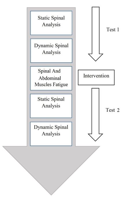
A (pre/post-test) cohort study was conducted in 10 healthy individuals to investigate the effect of spinal and abdominal muscles fatigue
on pelvic tilt (DL-DR ), trunk inclination (VP-DM) and the lordotic angle (ITL-ILS max) in a static spinal posture. Three different levels
of vertebrae were randomly chosen (T6, L2 and L4) to assess the effect of spinal and abdominal muscle fatigue on the rotations of these
vertebrae. Prior to the study, ethical approval was obtained. The inclusion criteria for participation in this study were male individuals,
aged between 18 to 45 years old, able to walk for 2 – 3 minutes on a treadmill, able to understand both written and spoken English
language, able to attend the University of Salford testing laboratory and had no history of any musculoskeletal diseases. Spinal muscles
fatigue was induced using the Sorenson test and abdominal muscles fatigue was made using the double leg lowering (DLL) test [4]. The
new technology, the DIERS formetric 4D dynamic system, was used as an outcome measure (Figure 1).
Figure 1: The Study Protocol
Results
All statistical analyses were performed using SPSS® (version 23.0, IBM, USA) software, by applying descriptive statistics [mean and
standard deviation (SD)] to represent the whole data set. The statistical significance of the results achieved was set at a value of p < 0.05.
In the static spinal analysis, abdominal and spinal muscles fatigue had a significant effect (p < 0.05) on pelvic tilt (DL-DR), trunk inclination
(VP-DM) and lordotic angle (ITL-ILS max) for the majority of the participants (Table I). The effect of fatigue differed between subjects,
with increases and decreases observed in all parameters.
In dynamic spinal analysis, abdominal and spinal muscle fatigue had less significant effect in peak to peak (p = 0.634) and on the mean
values of T6 (p = 0.055), L2 (p = 0.256, p = 0.676, respectively) and L4 (p = 0.75, p = 0.70, respectively) vertebra rotations of all
participants involved in the study. Table II demonstrates one of the three assessed vertebrae, T6.
Int J Ortho Res, 2020 www.opastonline.com Volume 3 | Issue 1 | 21Table 1: Description of the static spinal analysis of all participants
Participants Parameters Pre muscle fatigue Post muscle fatigue P value
Mean SD Mean SD
1 Pelvic Tilt DL-DR ° -5.050 0.219 -2.540 0.966 0.000
Trunk Inclination VP-DM ° -1.000 0.085 -0.075 0.176 0.002
Lordotic Angle ITL-ILS (max) 38.75 0.437 36.7667 0.459 0.000
2 Pelvic Tilt DL-DR ° -7.820 0.459 -7.560 1.025 0.754
Trunk Inclination VP-DM ° 1.250 0.206 0.733 0.201 0.002
Lordotic Angle ITL-ILS (max) 41.583 0.863 40.09 1.900 0.031
3 Pelvic Tilt DL-DR ° -1.50 0.00 1.950 1.690 0.000
Trunk Inclination VP-DM ° 8.470 0.080 6.290 0.144 0.002
Lordotic Angle ITL-ILS (max) 2.300 0.112 31.61 2.352 0.002
4 Pelvic Tilt DL-DR ° 2.300 0.112 31.61 2.352 0.002
Trunk Inclination VP-DM ° 1.200 0.239 0.325 0.333 0.002
Lordotic Angle ITL-ILS (max) 40.841 0.689 41.15 0.4699 0.058
5 Pelvic Tilt DL-DR ° -2.580 0.728 -2.240 0.124 0.129
Trunk Inclination VP-DM ° 1.910 0.406 -0.614 0.227 0.002
Lordotic Angle ITL-ILS (max) 33.725 0.422 37.40 0.447 0.000
6 Pelvic Tilt DL-DR ° -3.84 0.355 -2.24 0.260 0.000
Trunk Inclination VP-DM ° -0.266 0.137 3.208 0.09 0.002
Lordotic Angle ITL-ILS (max) 37.58 0.401 31.408 0.828 0.000
7 Pelvic Tilt DL-DR ° -3.320 1.854 -3.510 2.016 0.665
Trunk Inclination VP-DM ° 4.575 4.060 3.175 3.175 0.345
Lordotic Angle ITL-ILS (max) 29.50 2.462 31.00 1.289 0.051
8 Pelvic Tilt DL-DR ° -0.270 0.045 -1.590 0.962 0.007
Trunk Inclination VP-DM ° 3.009 0.165 7.025 0.252 0.002
Lordotic Angle ITL-ILS (max) 27.733 0.238 32.17 0.739 0.002
9 Pelvic Tilt DL-DR ° -0.500 0.987 -1.201 0.989 0.099
Trunk Inclination VP-DM ° 0.833 0.049 4.300 0.229 0.002
Lordotic Angle ITL-ILS (max) 29.833 0.192 26.333 0.439 0.000
10 Pelvic Tilt DL-DR ° 0.750 1.090 0.408 1.440 0.487
Trunk Inclination VP-DM ° 6.808 0.099 7.871 0.105 0.002
Lordotic Angle ITL-ILS (max) 37.316 0.476 34.991 1.856 0.009
Table 2: Peak to Peak (P-P) and Mean Values of T6 Vertebral Rotation (Pre- & Post- Fatigue) of all subjects
Pre-Fatigue Post-Fatigue
P-P Mean P-P Mean
Subject 1 7.2 4.2 -2.1 -3.77
Subject 2 4.9 7.5 -0.87 -1.26
Subject 3 5.8 6 -10.77 -10.17
Subject 4 9.8 9.5 -1.24 -2.67
Subject 5 3.8 2.9 1.82 1.42
Subject 6 4 5.9 3.6 -2.19
Subject 7 6.8 5.2 -0.11 -2.15
Subject 8 6 4.9 1.86 -1.14
Subject 9 4.5 8.3 -2.27 -3.87
Subject 10 4.6 6.3 -8.57 -7.02
Int J Ortho Res, 2020 www.opastonline.com Volume 3 | Issue 1 | 22Discussion contraction (MVC) to 30.6% MVC prior to fatigue as compared to
To the best knowledge of the authors, no previous studies have 14.6% MVC to 25.2% MVC post fatigue). It would be interesting
examined the effects of abdominal and spinal muscles fatigue to investigate the neuromuscular components that maintain the
(utilising Sorenson and DLL tests) on static parameters including stability of the spine. For example, how do the primary muscular
pelvic tilt (DL-DR), trunk inclination (VP-DM), lordotic angle stabilisers and the other secondary adaptive factors integrate? [12].
(ITL-ILS max) and dynamic parameters including T6, L2 and L4
vertebral rotations. In this study, fatigued-spinal and abdominal muscles failed to
maintain the neuromuscular control and the spinal stability; this
Suboptimal and excessive use of abdominal and spinal muscles forces was evident by the changes in the static spinal parameters. However,
can lead to certain changes in the biomechanics of the spine, which to ascertain which factor is responsible for these changes, that is,
may limit spinal stability and increase spinal load [5]. However, fatigued muscles or the failure of proprioceptive mechanisms or
previously published studies have applied empirical assessment to other adaptive mechanisms, such activation of another muscular
assess fatigue in spinal and abdominal muscles utilising tools such group, future larger scale studies incorporating tools to assess fatigue
as electromyography (EMG) [6-7]. In our study, the assessment of and proprioception could be considered.
fatigue was performed by measuring changes in six different spinal
parameters (three in static and three in dynamic positions). An Dynamic parameters
assessment of these parameters can inform health-care professionals An advantage of the DIERS system is the ability to analyse the spine
about real time structural changes in the spine during fatigue. This in dynamic postures. Assessment of vertebral rotation is of clinical
could assist our understanding of conditions such as Low Back Pain importance, particularly in diseases such as Adolescent Idiopathic
(LBP) and in different implications for ergonomic industry. Scoliosis (AIS) [13-14]. These assessments provide more insight
into the changes in vertebral column, aids in the assessment of the
Static Parameters significance of an intervention and assist surgeons in pre-surgical
In this study, 50% of the participants showed an increase in the planning [13].
mean degree of pelvic tilt (DL-DR). Conversely, they also showed
a decrease in the mean degree of pelvic tilt (DL-DR) which was Although the importance of assessing vertebral rotation in structural
observed in the other 50% participants. With regards to trunk deformities, such as scoliosis, has already been investigated and
inclination, six of the participants showed an increase in the mean emphasised with different radiological modalities, however, to the
degree of trunk inclination (VP-DM), with a decrease observed in authors’ best knowledge, the effect of abdominal and spinal muscles
the other four participants. Regarding lordotic angle, the results fatigue on vertebral rotation (T6, L2 and L4) in a dynamic spinal
of this study revealed that five of the participants experienced an posture has not been previously investigated. This assessment will
increase in mean lordotic angle (ITL-LLs max), whereas the other provide an insight into the effect of the integrity of these muscles
five participants had a decrease in the mean lordotic angle (ITL-LLs and the fatigue induced by the applied fatigue tests (Sorensen and
max). The results of the static parameters suggest that spinal and DLL tests) on musculoskeletal disorders, such as LBP. 3 vertebral
abdominal muscle fatigue have an effect on the above-mentioned levels T6, L2 and L4 were randomly selected for measurements in
parameters. From the data we can summarise the individuals this study to coincide with current research in this area. We assessed
experience less muscle control and the ability to adapt the spine to the effect of spinal and abdominal muscle fatigue on the rotations
the induced fatigue, as evidenced by the observed changes in pelvic of these vertebrae.
tilt. We hypothesis the effects could be worse in unhealthy or elderly
populations and patients with other de-conditioning lumbosacral The statistical data showed that abdominal and spinal muscles fatigue
disorders such as disco genic problems or LBP. had less significant difference in P-P values of T6, L2 & L4 vertebral
rotations (P value= 0.634, 0.75 & 0.256 respectively) or the mean
One of possible explanations for the results demonstrated above is values (p = 0.055, 0.70 & 0.676) of T6, L2 & L5 vertebral rotations
the neuromuscular control factors and co-activation of antagonistic pre- and post-abdominal and spinal muscles fatigue (Table 2).
muscular group, which has an important role to maintain mechanical
stiffness and spinal stability [5, 8]. Previous studies have identified Although the researchers had some difficulties in determination of max
that lumbar extensor muscle fatigue alters the onset and cessation P and min P values in the graphical representation of P-P values during
of myoelectric silence during the performance of flexion-extension 5 sec gait cycle of T6 vertebral rotation. However, in comparison with
tasks [9]. Moreover reported that lumbar extensor muscle fatigue T6 graphical representation, the data was easier to evaluate when L2
increased body sway during standing as a consequence of declined rotation was assessed. Eight of the participants had clear graphical
muscle proprioceptive acuity, impaired postural control and reduced representation of P-P values of L2 vertebral rotation. Furthermore,
trunk stability [10]. Furthermore, a subsequent study by revealed when the results of fatigue on T6 and L2 vertebral rotation are compared
an increase in trunk muscle co-contraction after lumbar muscle together, P-P value of L2 rotation was more statistically significant than
fatigue to compensate for the reduced stability and to increase trunk T6 rotation (P value of L2 = 0.256, P value of T6 = 0.634 respectively).
stiffness [11]. This may be explained by the difference in muscle recruitment in
the Sorensen test as this test has a task-dependency effect on lumbo-
Investigated the effect of fatigue tasks (3 min intense stair climbing) pelvic muscle fatigue. Whereby hip extensor muscles tend to fatigue
on spinal postures and trunk muscular activation patterns. After the simultaneously with the para-spinal muscles. Lifting the upper body
fatigue protocol, the researchers concluded that participants had mass during the Sorensen test is mostly dependant of lower lumbar
greater spinal flexion (16.3° maximum prior to fatigue as compared and pelvic muscles in addition to hip extensors, with less contribution
to 20.1° post fatigue) and reduced abdominal muscle co-activation of the muscles in the thoracic area and subsequently, less noticeable
post fatigue tasks (mean ranging from 16.6% maximum voluntary effects of fatigue in this area [15].
Int J Ortho Res, 2020 www.opastonline.com Volume 3 | Issue 1 | 23The outcomes of current study should be interpreted in light of few assessment of paraspinal muscle fatigue: an updated systematic
limitations. Firstly, the low number of participants. However, all review. Journal of manipulative and physiological therapeutics
of the included participants have shown changes in the parameters 37: 510-521.
measured, particularly static parameters. This could be considered 7. Arnall FA, Koumantakis GA, Oldham JA, Cooper RG (2002)
as a pilot study which will pave the way for future studies in this Between-days reliability of electromyographic measures of
field. Secondly, the possibility of a gender bias. Future studies which paraspinal muscle fatigue at 40, 50 and 60% levels of maximal
incorporate both genders are required to improve the generalization voluntary contractile force. Clinical Rehabilitation 16: 761-771.
of the results of such type of studies. Thirdly, this study included 8. Granata KP, Orishimo KF (2001) Response of trunk muscle
healthy participants only. Assessment of changes in the parameters coactivation to changes in spinal stability. Journal of
measured in un-healthy population is warranted which will assist biomechanics 34: 1117-1123.
researchers to understand and treat some lumbo-sacral disorders 9. Hu B, Ning X (2015) the changes of trunk motion rhythm
such a LBP. Finally, a technical issue was encountered during and spinal loading during trunk flexion and extension motions
dynamic spinal analysis as there were difficulties to control the time caused by lumbar muscle fatigue. Annals of biomedical
of 5 second digital motion image capture by DIERS system and engineering 43: 2112-2119.
the stride length of similar limb between pre and post abdominal 10. Davidson BS, Madigan ML, Nussbaum MA (2004) Effects
muscles fatigue. This could have affected dynamic data obtained of lumbar extensor fatigue and fatigue rate on postural sway.
and potentially the validity of the study. Future improvement of the European journal of applied physiology 93: 183-189.
DIERS system to overcome such difficulties with re-evaluation of 11. Granata KP, Slota GP, Wilson SE (2004) Influence of fatigue
the effect of fatigue on spinal postures is required. in neuromuscular control of spinal stability. Human factors
46: 81-91.
Conclusion 12. Gregory DE, Narula S, Howarth SJ, Russell C, Callaghan
The application of simple and quick fatigue tests resulted in changes JP (2008) the effect of fatigue on trunk muscle activation
in all the static parameters (pelvic tilt (DL-DR), trunk inclination patterns and spine postures during simulated firefighting tasks.
(VP-DM) and the lordotic angle (ITLILS max)) as measured by Ergonomics 51: 1032-1041.
the DIERS system, reaching statistical significance (p < 0.05) in 13. Lam GC, Hill DL, Le LH, Raso JV, Lou EH (2008) vertebral
nearly all participants. There was not a specific pattern for the rotation measurement: a summary and comparison of common
observed changes within the same parameter or between the three radiographic and CT methods. Scoliosis 3: 16.
different static parameters. In contrast to the changes noted in 14. Lafage V, Leborgne P, Mitulescu A, Dubousset J, Lavaste F, et
static parameters, fatigue did not induce the same effect noticed al. (2002) Comparison of mechanical behaviour of normal and
on dynamic parameters (T6, L2 and L4 vertebral rotation). To the scoliotic vertebral segment: a preliminary numerical approach.
authors’ best knowledge, this is the first study which applied the Studies in health technology and informatics 88: 340-344.
parameters mentioned above to investigate the effects of spinal and 15. Champagne A, Descarreaux M, Lafond D (2008) back and hip
abdominal muscles fatigue utilising Sorenson and DLL tests. Taking extensor muscles fatigue in healthy subjects: task-dependency
into consideration the statistically significant results obtained in effect of two variants of the Sorensen test. European Spine
statics parameters from this study it provides the basis for future Journal 17: 1721-1726.
studies
Acknowledgement
The authors declare that no part of this study has been taken
from existing published or unpublished materials without due
acknowledgement and that all secondary materials used herein has
been fully referenced.
References
1. Hawes MC, O Brien JP (2006) the transformation of spinal
curvature into spinal deformity: pathological processes and
implications for treatment. Scoliosis 1: 3.
2. Na Y, Kang S, Bae H, Kang M, Park J, (1996) The analysis
of spinal curvature in low back pain patients. J Korean Acad
Rehab Med 20: 669-674.
3. Hwang S, Park S, Kim Y (2009) Measurement Comparison about
Lumbar Lordosis: Radiography and 3D Motion Capture. World
Congress on Medical Physics and Biomedical Engineering,
Germany Springer 25: 1669-1671.
4. Demoulin C, Vanderthommen M, Duysens C, Crielaard J M
(2006) Spinal muscle evaluation using the Sorensen test: a
critical appraisal of the literature. Joint Bone Spine 73: 43-50. Copyright: ©2020 Ahmed Aljawadi, et al. This is an open-access article
5. Gardner Morse MG, Stokes IA (1998) the effects of abdominal distributed under the terms of the Creative Commons Attribution License,
muscle coactivation on lumbar spine stability. Spine 23: 86-91. which permits unrestricted use, distribution, and reproduction in any
6. Bandpei MAM, Rahmani N, Majdoleslam B, Abdollahi I, Ali medium, provided the original author and source are credited.
SS, et al. (2014) Reliability of surface electromyography in the
Int J Ortho Res, 2020 www.opastonline.com Volume 3 | Issue 1 | 24You can also read