Role of robotic natural orifice specimen extraction surgery in colorectal neoplasms - Nature
←
→
Page content transcription
If your browser does not render page correctly, please read the page content below
www.nature.com/scientificreports
OPEN Role of robotic natural orifice
specimen extraction surgery
in colorectal neoplasms
Hongliang Yao, Tiegang Li, Weidong Chen, Sanlin Lei, Kuijie Liu, Bo Liu & Jiangjiao Zhou*
Natural orifice specimen extraction surgery (NOSES) is especially suitable for colorectal surgery.
Until now, most of the reports published were about laparoscopic NOSES, the reports about robotic
NOSES are extremely rare. This study aims to explore the safety and feasibility of robotic NOSES
for colorectal neoplasms. All patients underwent robotic NOSES from March 2016 to October 2019
in our hospital were enrolled for retrospective analysis. Clinicopathological data including patient
characteristics, perioperative information and pathological information were collected and analyzed.
According to the distance between tumor and anus or whether neoadjuvant chemoradiotherapy
(nRCT) is performed, we grouped the cases and studied its influence on robotic NOSES. Also, we
compared the previous reports on laparoscopic NOSES with our study and revealed advantages
of robotic NOSES in terms of safety and feasibility. A total of 180 patients were enrolled. The
average distance from the lower edge of the tumor to the anus was (8.64 ± 3.64) cm and maximum
circumferential diameter (CDmax) of specimen was (3.5 ± 1.6) cm. In terms of safety, the average
operation time, intraoperative blood loss, and postoperative hospital stay were (187.5 ± 78.3) min,
(47.4 ± 34) mL, and (11.3 ± 7.5) days, respectively. In terms of feasibility, the average number of lymph
node harvested was (14.8 ± 5). Robotic NOSES shows advantages in terms of safety and feasibility
compared with laparoscopic NOSES. This procedure could not only be a safe procedure but also could
achieve good oncological outcomes.
Total mesorectal resection (TME) and laparoscopic surgery are the major breakthroughs for colorectal cancer
surgery1. However, an incision must still be made at the abdominal wall to remove the s pecimen2. Natural orifice
specimen extraction robotic surgery (NOSES) can solve this problem. NOSES is designed to use laparoscopic
instruments or soft endoscopy and other equipment to complete intra-abdominal surgery. No auxiliary incision is
made at the abdominal wall, and the specimen is removed through the natural lumen (anus or vagina). Compared
with traditional laparoscopic surgery, the most evident advantage of NOSES is the avoidance of auxiliary incision,
which could meet the esthetic needs of patients, such as actors and gymnasts, engaged in specific occupations.
To date, NOSES can be used for colorectal, stomach, small intestine, hepatobiliary, and gynecological t umors3.
Many reports had also verified the effectiveness and safety of NOSES for colorectal n eoplasms4,5.
Although the procedure of NOSES could be performed by laparoscopy, the da Vinci Surgical System still has
its unique advantages due to its high-definition field of vision and seven degrees of freedom of the operation
arm. Our department is one of the centers which previously carried out robotic NOSES for colorectal cancer
in China6. This study aimed to summarize single-center experience in robotic NOSES for sigmoid and rectal
neoplasms through a retrospective analysis of the collected cases.
Materials and methods
Inclusion and exclusion criteria of cases. From March 2016 to October 2019, all patients with sigmoid
and rectal neoplasm and underwent robotic NOSES in the General Surgery Department of The Second Xiangya
Hospital, Central South University were considered for retrospective analysis.
Inclusion criteria: (1) age ≥ 18 years old; (2) diagnosis of sigmoid and rectal neoplasm by biopsy via colo-
noscopy or benign neoplasm located in rectum that could not be resected locally through the anus; (3) written
informed consent of patients; (4) expected removal of the specimen through the anus indicated by preoperative
evaluation.
Department of General Surgery, The Second Xiangya Hospital of Central South University, Changsha 410011, China.
*
email: zhoujiangjiao@csu.edu.cn
Scientific Reports | (2021) 11:9818 | https://doi.org/10.1038/s41598-021-89323-z 1
Vol.:(0123456789)www.nature.com/scientificreports/
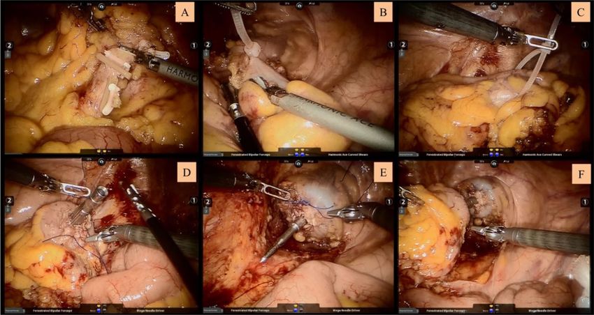
Figure 1. Surgical procedure. (A) The inferior mesenteric artery and vein were isolated and clipped by
absorbable vascular clamps; (B) Rectum was ligated with self-locking nylon bandage; (C) Sigmoid colon was
ligated with self-locking nylon bandage; (D) Suture the stump of sigmoid colon and put the orvil into the
sigmoid colon; (E) Suture the stump of rectum; (F) Complete the anastomosis.
Exclusion criteria: (1) emergency operation due to gastrointestinal obstruction, perforation, or bleeding; (2)
metastasis of the lung, bone, or liver that cannot be removed simultaneously; (3) contraindications for robotic
surgery.
This work is in accordance with the declaration of Helsinki and is approved by the Ethics Committee of
the Second Xiangya Hospital, Central South University.
Information collection. The statistical information included the following: (1) patient characteristics: gen-
der, age, chief complaint, comorbidity, history of laparotomy, body mass index (BMI), American Society of Anes-
thesiologists (ASA) classification, distance from the lower edge of the tumor to the anus (lower group < 5 cm,
5 ≤ middle group < 10 cm, upper group ≥ 10 cm)7, and presence or absence of nRCT (surgery was performed
4–6 weeks after nRCT); (2) perioperative information: operation time, intraoperative blood loss, protective
ileostomy, conversion to laparotomy, postoperative hospital stay, postoperative complications, reoperation, and
total costs; (3) pathological information: histological type, differentiation, maximum circumferential diameter
(CDmax) of specimen, depth of tumor invasion, the number of lymph nodes harvested, and the number of
metastatic lymph nodes.
Surgical procedure. After successful general anesthesia, the patient assumed the Trendelenburg position.
Five trocars were used (Fig. 1): one 12 mm trocar located at 3 cm above the umbilicus for robotic camera and
another 12 mm trocar located at the right midclavicular line 1 cm above the umbilicus for assistant; three 8 mm
trocar for robotic arms (R1 located at the right anterior superior iliac spine, R2 located at the left midclavicular
line 1 cm above the umbilicus, and R3 located at the left anterior superior iliac spine). First, an ultrasonic scalpel
via R1 was used to separate the internal and external peritoneum of sigmoid colon, and the left ureter was prop-
erly protected. The inferior mesenteric artery and vein were isolated and clipped by absorbable vascular clamps
(Fig. 1A). The left colic artery was preserved. The rectum was completely isolated until 2–5 cm to the lower edge
of the tumor. Then, the colorectum was ligated with self-locking nylon bandage (Fig. 1B,C). After the rectum was
cut off by an ultrasonic scalpel, the assistant inserted an endoscope-sterile sleeve for protection of the specimen
into the pelvic cavity through the anus, and the resected specimen was pulled out through the anus. The assistant
sent an orvil through the anus, and the operator sutured the stump of the sigmoid colon and placed the orvil into
the sigmoid colon (Fig. 1D). After the operator sutured the stump of the rectum (Fig. 1E), the assistant placed the
curved intraluminal stapler through the anus to complete the anastomosis (Fig. 1F). Then, the assistant injected
air into the rectum through the anus. If air leakage occurred from the anastomosis or serous membrane eversion,
suture can be performed. Perioperative management followed the international g uidelines8.
Statistical analysis. Data processing and statistical analysis were performed using SPSS (version 22.0, SPSS
Inc., Chicago, IL, USA). Measurement data were expressed as the mean ± standard deviation, and an independ-
Scientific Reports | (2021) 11:9818 | https://doi.org/10.1038/s41598-021-89323-z 2
Vol:.(1234567890)www.nature.com/scientificreports/
ent sample t-test or variance analysis was used to compare the mean values. Qualitative data were expressed
as frequency and percentage, and Pearson’s χ2 test or Fisher’s exact probability method was used. P < 0.05 was
considered statistically significant (two-tailed test).
Institutional review board statement. This study was reviewed and approved by the Ethics Committee
of the Second Xiangya Hospital, Central South University.
Informed consent statement. Informed consent was obtained from all patients for using their clinical
data in the study.
Results
Patient characteristics. This study included 180 patients. In terms of gender, men accounted for 60%
(108/180) and women for 40% (72/180). The mean age was (57.3 ± 13.1) years old, and the mean body mass
index (BMI) was (23.6 ± 3.3) kg/m2. The most common chief complaint was hematochezia in 81.1% of patients
(146/180). 165 patients were tested for carcinoembryonic antigen (CEA) and carbohydrate antigen (CA) 242,
and 133 patients were tested for CA19-9 before operation. The distance from the lower edge of the tumor to
the anus was based on the colonoscopy report, with an average of (8.64 ± 3.64) cm; those measuring less than
5, 5–10, and greater than or equal to 10 cm accounted for 7.2% (13/180), 54.4% (98/180), and 38.3% (69/180),
respectively. The proportion of patients with nRCT was 15% (27/180). Twenty-six patients had abdominal or
pelvic surgery history (14.4%). American Society of Anesthesiologists (ASA) classification levels 1, 2, 3, and 4
reached 3.3% (6/180), 50.5% (91/180), 44.4% (80/180), and 1.7% (3/180) of patients, respectively (Table 1).
Postoperative pathological information. In this study, the most common histological types were tubu-
lar adenocarcinoma(170/180, 94.4%), and the most common degree of differentiation of tubular adenocarci-
noma was moderately differentiated adenocarcinoma, accounting for 80.6% (137/170). The average CDmax of
specimen was (3.5 ± 1.6) cm, and the highest CDmax value was 12 cm. The number of lymph nodes harvested
in all cases ≥ 12 accounted for 73.9% (133/180), and the average number of lymph nodes harvested for each case
was (14.8 ± 5). Based on the depth of tumor invasion, Tis stage was 1.2% (2/173), T1 stage was 8.1% (14/173), T2
stage was 26.6% (46/173), T3 stage was 37.6% (65/173), and T4 stage was 26.6% (46/173). Lymph node metas-
tasis accounted for 33.5% (58/173), and simultaneous liver metastasis was observed in one case. No positive
resection margin was observed (Table 2).
Perioperative outcomes. All 180 cases completed robotic NOSES successfully, and no conversion to
laparotomy was observed. The average medical cost was 110,046 ± 35,003 CNY. The mean operative time was
(187.5 ± 78.3) min, and the mean intraoperative blood loss was (47.4 ± 34) mL. Most patients underwent robotic
TME for the rectum or complete mesocolic excision (CME) for the sigmoid (171/180). All specimens were
removed through the anus. Cases for protective ileostomy accounted for 6.7% (12/180). The average postop-
erative fasting time was (4.2 ± 3.9) days, and the average postoperative hospital stay was (11.3 ± 7.5) days. The
incidence of postoperative complications was 11.7% (21/180). The incidence of anastomotic leakage was 4.4%
(8/180), and the other complications included abdominal infection, lung infection, anastomotic bleeding, and
abdominal hemorrhage. The reoperation rate was 2.2% (4/180), which was achieved with ileostomy in all cases
after anastomotic leakage. The other 17 patients with complications were managed with anti-infective therapy,
ultrasound-guided percutaneous drainage, colonoscopy, and endoscopic treatment. No postoperative anal dys-
function nor 90-day death was observed (Table 3).
Influence on robotic NOSES by the distance between tumor and anus or nRCT. The safety and
feasibility were compared between three groups based on the distance from the lower edge of the tumor to the
anus. Table 4 shows the results. Close distance from the lower edge of the tumor to the anus means long opera-
tion time, high intraoperative blood loss, and high incidence of anastomotic leakage. The difference was statisti-
cally significant (P < 0.05). nRCT had no effect on the safety and feasibility for robotic NOSES. The results are
shown in Table 5.
Discussion
In 2007, French doctor Marescaux completed the first truly scar free operation in the world, and transvaginal
cholecystectomy, which minimally invasive surgery requirements, has entered a new era3. NOSES is a kind of
operation that can realize the concept of “no scar” surgery to a certain limit. It is especially suitable for colorectal
surgery. Incisions in the oral cavity, rectum, vagina, and other natural orifices for appendectomy, cholecystec-
tomy, and nephrotomy are often necessary to remove specimens from the natural lumen. The rectum has to be
disconnected during colorectal surgery. The rectum and anus could be natural orifices for specimen extraction
and could be used to avoid performing any artificial incision, rendering them with evident natural advantage.
Based on the orifice for specimen extraction, NOSES could be divided into transanal and transvaginal NOSES.
However, all cases presented in these reports were transanal NOSES. We excluded the specimen removal from the
vagina for the following reasons. First, transvaginal NOSES could only be applicable for female patients. Second,
incision on the vaginal wall may increase the risk of postoperative complications and sexual dysfunction. Third,
transvaginal NOSES is also limited by ethics. In June 2017, China NOSES Alliance was established and released
Expert consensus of natural orifice specimen extraction surgery in colorectal neoplasm (2017 edition)9 to promote
Scientific Reports | (2021) 11:9818 | https://doi.org/10.1038/s41598-021-89323-z 3
Vol.:(0123456789)www.nature.com/scientificreports/
Cases (n) Ratio (%)
Gender
Male 108 60
Female 72 40
Age
20–39 16 8.9
40–59 79 43.9
60–79 75 41.7
≥ 80 10 5.6
Chief complaint
Hematochezia 146 81.1
increased times in defecation 25 13.9
Abdominal discomfortable 2 1.1
Anal distention 2 1.1
Routine examination 5 2.8
Systemic disease
Hypertension 41 22.8
Diabetes 16 8.9
Cardiovascular diseases 10 5.6
Respiratory diseases 6 3.3
Cerebrovascular disease 7 3.9
Cirrhosis 5 2.8
Other 9 5
ASA score
1 6 3.3
2 91 50.5
3 80 44.4
4 3 1.7
History of abdominal surgery
Yes 26 14.4
No 154 85.6
nCRT
Yes 27 15
No 153 85
Distance from the lower edge of the tumor to the anus
< 5 cm 13 7.2
5-10 cm 98 54.4
≥ 10 cm 69 38.3
CEA(ng/mL)
Normal 136 82.4
Higher 29 17.6
CA19-9(ng/mL)
Normal 124 93.2
Higher 9 6.8
CA242(ng/mL)
Normal 157 95.2
Higher 8 4.8
Table 1. Patient characteristics.
the application of NOSES. The International Alliance of NOSES also issued International consensus on natural
orifice specimen extraction surgery (NOSES) for colorectal cancer in 2 0197.
Many reports are available on laparoscopic NOSES for colorectal cancer4,10; compared with conventional
laparoscopic surgery, laparoscopic NOSES is a safe procedure and can achieve similar oncological outcomes.
Wolthuis et al. conducted a clinical trial comparing the short-term effects of conventional laparoscopic surgery
and laparoscopic NOSES; their results showed that the postoperative pain of patients in the NOSES group was
alleviated significantly11. According to the report of an international multicenter study including 412 cases of
conventional laparoscopic radical resection of rectal cancer and 356 cases of laparoscopic NOSES, no difference
was observed in the operation time, the number of lymph nodes harvested, incidence of anastomotic leakage,
Scientific Reports | (2021) 11:9818 | https://doi.org/10.1038/s41598-021-89323-z 4
Vol:.(1234567890)www.nature.com/scientificreports/
Cases (n) Ratio (%)
Histology type
Tubular adenocarcinoma 170 94.4
Mucinous adenocarcinoma 3 1.7
Adenoma 5 2.8
Endometriosis 1 0.6
Neuroendocrine tumor 1 0.6
Differentiation
Highly 3 1.8
Moderately 137 80.6
Poorly 30 17.6
CDmax
< 3 cm 59 32.8
3-5 cm 84 46.7
≥ 5 cm 37 20.6
T staging
Tis 2 1.2
T1 14 8.1
T2 46 26.6
T3 65 37.6
T4 46 26.6
Lymph node harvested
< 12 47 26.1
≥ 12 133 73.9
Lymph node metastasis
Yes 58 33.5
No 115 66.5
Liver metastasis of adenocarcinoma
Yes 1 0.6
No 172 99.4
Resection margins
Positive 0 0
Negative 180 100
Table 2. Pathological information.
and length of postoperative hospital stay. Incision infection and incisional hernia were also observed in the
conventional laparoscopic radical resection group10. A retrospective study of 718 cases of colorectal cancer in
79 hospitals in China showed the following results: incidence of anastomotic leakage of 3.5%, reoperation rate
of 3.6%, and average intraoperative blood loss less than 100 mL; these findings fully demonstrated the safety of
laparoscopic NOSES for rectal c ancer12. Two meta-analysis involving 1435 and 837 patients also showed that
compared with conventional laparoscopic surgery, NOSES may be a safe procedure and can significantly reduce
the duration of hospital stay, accelerate the postoperative recovery with good cosmetic results, result in less
postoperative pain and limited complications, and achieve similar oncological o utcomes13,14.
Numerous studies have reported robot-assisted radical resection of colorectal c ancer10,15–19. However, in most
reports, the specimens were extracted through a small abdominal incision; meanwhile, the reports on robotic
NOSES are rare. Three retrospective analysis reports exist, and the rest are case r eports20–23. Compared with the
previous reports on laparoscopic NOSES, robotic NOSES in our hospital revealed advantages in terms of safety
and feasibility5,12. In terms of the safety of robotic NOSES, our results showed that the average operation time
was (187.5 ± 78.3) min, intraoperative blood loss was (47.4 ± 34) mL, no conversion to laparotomy occurred,
postoperative hospital stay was (11.3 ± 7.5) days, the incidence of anastomotic leakage was 4.4%, the reoperation
rate was 2.2%, and no 90-day death was observed. In terms of the feasibility, the average number of lymph nodes
harvested was (14.8 ± 5), and no case with positive margin was observed (Table 6).
Either laparoscopic or robotic NOSES could be performed in our d epartment24,25. As per our experience,
especially in ultra-low rectal cancer, robotic NOSES has advantage compared with laparoscopic NOSES. For
ultra-low rectal tumor, if the rectum is cut off directly by an ultrasonic scalpel during laparoscopic surgery,
suturing of the pouch is difficult. However, if the rectum is to be amputated with an endoscopic linear cutter,
the procedure may not be completed due to the narrow pelvic cavity. However, the problems mentioned above
could be resolved by robotic NOSES. The ultra-low rectum can be sutured directly after the rectum is amputated
by an ultrasonic scalpel. If the anastomosis is unsatisfactory, we can use the da Vinci Robot System to sew and
strengthen the anastomosis directly to reduce the incidence of anastomotic leakage. However, it is important to
Scientific Reports | (2021) 11:9818 | https://doi.org/10.1038/s41598-021-89323-z 5
Vol.:(0123456789)www.nature.com/scientificreports/
Cases (n) Ratio (%)
Surgical procedure
TME or CME 171 95
TME + Subtotal colectomy 1 0.6
TME + Resection of liver metastasis 1 0.6
TME + Resection of partial transverse colon 1 0.6
TME + Ovariectomy 2 1.1
TME + Hysterectomy 2 1.1
TME + Left partial nephrectomy 1 0.6
TME + Ureteral repair 1 0.6
Protective ileostomy
Yes 12 6.7
No 168 93.3
Clavien-Dindo classification
I 1 0.6
II 10 5.6
IIIa 6 3.3
IIIb 3 1.7
IVb 1 0.6
Anastomotic leakage
Yes 8 4.4
No 172 95.6
Reoperation
Yes 4 2.2
No 176 97.8
Postoperative anal function
Abnormal 0 0
Normal 180 100
90-day mortality
Yes 0 0
No 180 100
Table 3. Perioperative outcomes.
< 5 cm 5–10 cm ≥ 10 cm P
Safety
Operative time (min) 263.9 ± 178.6 184 ± 61.9 178 ± 61.7 0.001
Blood loss (mL) 54.6 ± 48.4 45.2 ± 29.8 49.3 ± 36.8 0.546
Conversion to laparotomy (%) 0 0 0 N/A
Postoperative hospital stay (days) 16.5 ± 12.4 12 ± 8.3 9.4 ± 3.3 0.002
Anastomotic leakage (n, %) 1/13,7.7% 7/98,7.1% 0/69,0% 0.048
Reoperation (n, %) 0,0% 4/98,4.1% 0,0% 0.266
90-day mortality (%) 0 0 0 N/A
Feasibility
Lymph node harvested 13 ± 3.1 14.5 ± 5.3 15.5 ± 4.7 0.192
Positive resection margins (%) 0 0 0 N/A
Table 4. Influence on robotic NOSES by the distance between the lower edge of the tumor to the dentate line.
realize that a close distance from the lower edge of the tumor to the anus with long operation time and postop-
erative hospital stay indicates high incidence of anastomotic leakage. In addition, either received nRCT has no
effect on perioperative safety.
This study showed that T4 stage cases accounted for 26.6%, and the proportion of patients whose maximum
diameter of tumor is greater than or equal to 5 cm was 20.6%, which is inconsistent with the recommendations
of the International consensus on natural orifice specimen extraction surgery (NOSES) for colorectal cancer7. Based
on the recommendations, cases of T2 and T3 stage tumors should be appropriate for NOSES, whereas T4 stage
Scientific Reports | (2021) 11:9818 | https://doi.org/10.1038/s41598-021-89323-z 6
Vol:.(1234567890)www.nature.com/scientificreports/
With nRCT No nRCT P
Safety
Operative time (min) 184.8 ± 76.1 187.9 ± 78.9 0.846
Blood loss (ml) 57.4 ± 39 45.7 ± 32.9 0.098
Conversion to laparotomy (%) 0 0 N/A
Postoperative hospital stay (days) 11.7 ± 5.8 11.3 ± 7.7 0.774
Anastomotic leakage (n, %) 6/158,3.8% 2/27,7.4% 0.076
Reoperation (n, %) 0,0% 4/153,2.6% > 0.999
90-day mortality (%) 0 0 N/A
Feasibility
Lymph node harvested 14 ± 5.6 14.9 ± 4.9 0.423
Positive resection margins (%) 0 0 N/A
Table 5. Influence on robotic NOSES by nRCT.
Robotic NOSES Wang5 Xu12
Safety
Operative time (min) 187.5 ± 78.3 198.9 ± 55.2 210.5 ± 39.4
Blood loss (mL) 47.4 ± 34 73.7 ± 54.2 61.8 ± 23.1
Conversion to laparotomy (%) 0 0 0
Postoperative hospital stay (d) 11.3 ± 7.5 12.3 ± 4.1 12.1 ± 4.0
Anastomotic leakage (n, %) 8,4.4% 9,4.43% 25,3.5%
Reoperation (n, %) 4,2.2% 2,1% 23,3.2%
90-day mortality (%) 0 0 0
Feasibility
Lymph node harvested 14.8 ± 5 12.9 ± 5.1 13.4 ± 3.5
Positive resection margins (%) 0 0 0
Table 6. Comparison between robotic NOSES and laparoscopic NOSES.
cases are not recommended given the difficulty of guaranteeing a negative CRM. However, we believe that for
experienced teams in colorectal minimally invasive surgery, robotic NOSES for T4 stage cases is safe while strictly
observing the principle of tumor-free technique. In terms of the CDmax of specimen, the consensus suggested
that if the specimen is to be removed through the anus, then the CDmax should be less than 3 cm. However,
our data show that the proportion of CDmax of specimen exceeding 3 cm accounted for 67.3%, that exceeding
5 cm accounted for 20.6%, and the largest reached 12 cm. We believe that if CDmax is limited to specimens not
exceeding 3 cm, more than a half of patients will lose the opportunity for NOSES. Based on our experience,
indications for NOSES could be appropriately relaxed. On the premise of full anal dilation, most specimens
with CDmax of less than 5 cm can be removed through the anus without specimen damage. This procedure will
not lead to laceration of the rectum and anus. In addition, the specific situation should be analyzed in detail.
When the specimens are extracted from the anus during NOSES, one end of the rectum is clamped by surgical
forceps, and the specimens are extracted from the anus along the longitudinal axis of the rectum. Therefore, if
the CDmax is along the longitudinal axis of the rectum, specimens can still be easily extracted from the anus
despite a CDmax exceeding 5 cm. On the contrary, a CDmax vertical to the longitudinal axis of the rectum will
increase the difficulty of pulling out the specimen from the anus. In this study, the largest CDmax of specimen
extracted from the anus was 12 cm. However, this length was measured along the longitudinal axis of the rectum,
and its short axis was less than 2 cm. Thus, the specimen was a long strip that can be naturally removed through
the anus. Whether the specimen can be extracted from the anus is also affected by other factors. For example, if
the distance between the lower edge of the tumor and the anus is relatively close, dilation of the rectum, which
needs to be protected during specimen extraction, is unnecessary. Thus, the CDmax of specimens could be large.
Otherwise, if the distance is relatively long, the free rectum in the pelvic cavity must be dilated during specimen
extraction, which will lead to laceration of the rectum and anus if the CDmax of specimen is notably large. As
a promising minimally invasive technique, we appeal to all surgical colleagues working on robotic NOSES to
promote the development of robotic NOSES in the world for the benefit of colorectal patients.
Our analysis has several limitations. First, progression-free survival and overall survival of all selected cases
need to be verified through a long follow-up. Second, this research is a retrospective study. Thus, the integrity
and homogeneity of research data cannot be guaranteed.
Scientific Reports | (2021) 11:9818 | https://doi.org/10.1038/s41598-021-89323-z 7
Vol.:(0123456789)www.nature.com/scientificreports/
Data availability
No additional data are available.
Received: 10 December 2020; Accepted: 23 April 2021
References
1. Marks, J. H. et al. Outcomes in 132 patients following laparoscopic total mesorectal excision (TME) for rectal cancer with greater
than 5-year follow-up. Surg. Endosc. 30, 307–314 (2016).
2. Cho, M. S. et al. Minimally invasive versus open total mesorectal excision for rectal cancer: Long-term results from a case-matched
study of 633 patients. Surgery 157, 1121–1129 (2015).
3. Zorron, R. et al. NOTES. Transvaginal cholecystectomy: Report of the first case. Surg. Innov. 14, 279–283 (2007).
4. Park, J. S. et al. Long-term outcomes after Natural Orifice Specimen Extraction versus conventional laparoscopy-assisted surgery
for rectal cancer: A matched case-control study. Ann. Surg. Treat. Res. 94, 26–35 (2018).
5. Yuliuming, W. et al. Retrospective study of 203 cases of colorectal neoplasms treated by natural orifice specimen extraction surgery.
Chin. J. Colorectal. Dis. 29, 32–37 (2019).
6. Yao, H. et al. Safety and feasibility of robotic natural orifice specimen extraction surgery in colorectal neoplasms during the initial
learning curve. Front. Oncol. 10, 1355 (2020).
7. Guan, X. et al. International consensus on natural orifice specimen extraction surgery (NOSES) for colorectal cancer. Gastroenterol.
Rep. (Oxf) 7, 24–31 (2019).
8. Gustafsson, U. O. et al. Guidelines for perioperative care in elective colorectal surgery: Enhanced recovery after surgery (ERAS((R)))
Society Recommendations: 2018. World J. Surg. 43, 659–695 (2019).
9. Alliance, C. N. Expert consensus of natural orifice specimen extraction surgery in colorectal neoplasm (2017 edition). Chin. J.
Colorectal Dis. 6, 266–273 (2017).
10. Liu, Z. et al. A multicenter study evaluating natural orifice specimen extraction surgery for rectal cancer. J. Surg. Res. 243, 236–241
(2019).
11. Wolthuis, A. M. et al. Randomized clinical trial of laparoscopic colectomy with or without natural-orifice specimen extraction.
Br. J. Surg. 102, 630–637 (2015).
12. Xu, G. et al. Retrospective study of 718 colorectal neoplasms treated by natural orifice specimen extraction surgery in 79 hospitals.
Chin. J. Colorectal Dis. 6, 469–477 (2017).
13. Liu, R. J. et al. Safety and oncological outcomes of laparoscopic NOSE surgery compared with conventional laparoscopic surgery
for colorectal diseases: A meta-analysis. Front. Oncol. 9, 597 (2019).
14. Ma, B. et al. Laparoscopic resection with natural orifice specimen extraction versus conventional laparoscopy for colorectal disease:
A meta-analysis. Int. J. Colorectal Dis. 30, 1479–1488 (2015).
15. Mian, W. et al. Comparison of short-term outcomes between robotic-assisted and laparoscopic proctectomy for rectal cancer: A
case-control study. Chin. J. Colorectal. Dis. 4, 40–44 (2015).
16. Cao, C.-L. et al. Comparison of short-term outcomes between roboticassisted and laparoscopic surgery for rectal cancer. World
Chin. J. Digestol. 24, 2264–2269 (2016).
17. Zhen, Z. O. U. et al. Short-term outcomes of robotic versus laparoscopic radical resection for middle and low rectal cancer: A
single-center randomized, controlled study. Chin. J. Gen. Surg. 27, 408–413 (2018).
18. Liu, W. H. et al. Short-term outcomes of robotic versus laparoscopic total mesorectal excision for rectal cancer: A cohort study.
Am. Surg. 85, 294–302 (2019).
19. Megevand, J. L. et al. TME for rectal cancer: Consecutive 70 patients treated with laparoscopic and robotic technique-cumulative
experience in a single centre. Updates Surg 71, 331–338 (2019).
20. Xuefeng, Z. et al. Totally robotic surgery for rectal cancer with transanal specimen extraction. Chin. J. Pract. Surg. 33, 871–873
(2013).
21. Hu, R. et al. Trans-natural orifice transuminal specimen extraction in robotic rectal cancer surgery: An analysis of 21 patients.
Chin. J. Pract. Surg. 34, 252–254 (2014).
22. Efetov, S. K. et al. Natural orifice specimen extraction (NOSE) surgery with rectal eversion and total extra-abdominal resection.
Tech. Coloproctol. 23, 899–902 (2019).
23. Minjares-Granillo, R. O. et al. Robotic left-sided colorectal resection with natural orifice IntraCorporeal anastomosis with extrac-
tion of specimen: The NICE procedure. A pilot study of consecutive cases. Am. J. Surg. 217, 670–676 (2019).
24. Zhou, J. et al. Outcome of robot-assisted pancreaticoduodenectomy during initial learning curve versus laparotomy. Sci. Rep. 10,
9621 (2020).
25. Zhou, J. J. et al. Analysis of robotic natural orifice specimen extraction surgery on 162 cases with rectal neoplasms. Zhonghua
weichang waike za zhi [Chin J Gastrointest Surg] 23, 384–389 (2020).
Acknowledgements
We are grateful for the help of colleagues in the operating room and anesthesiology department during robotic
NOSES.
Author contributions
J.Z. conceived and designed the article; H.Y., T.L., K.L., B.L., and S.L. performed the operation; W.C. and S.L.
analyzed the data; J.Z. wrote the study; T.L., W.C., and H.Y. proofread and revised the manuscript.
Funding
Changsha Municipal Natural Science Foundation NO.kq2014245.
Competing interests
The authors declare no competing interests.
Additional information
Correspondence and requests for materials should be addressed to J.Z.
Reprints and permissions information is available at www.nature.com/reprints.
Scientific Reports | (2021) 11:9818 | https://doi.org/10.1038/s41598-021-89323-z 8
Vol:.(1234567890)www.nature.com/scientificreports/
Publisher’s note Springer Nature remains neutral with regard to jurisdictional claims in published maps and
institutional affiliations.
Open Access This article is licensed under a Creative Commons Attribution 4.0 International
License, which permits use, sharing, adaptation, distribution and reproduction in any medium or
format, as long as you give appropriate credit to the original author(s) and the source, provide a link to the
Creative Commons licence, and indicate if changes were made. The images or other third party material in this
article are included in the article’s Creative Commons licence, unless indicated otherwise in a credit line to the
material. If material is not included in the article’s Creative Commons licence and your intended use is not
permitted by statutory regulation or exceeds the permitted use, you will need to obtain permission directly from
the copyright holder. To view a copy of this licence, visit http://creativecommons.org/licenses/by/4.0/.
© The Author(s) 2021
Scientific Reports | (2021) 11:9818 | https://doi.org/10.1038/s41598-021-89323-z 9
Vol.:(0123456789)You can also read