Strategy for surgical treatment of acute thoracic empyema in adults
←
→
Page content transcription
If your browser does not render page correctly, please read the page content below
Review Article
                                                                                                                                             Page 1 of 8

Strategy for surgical treatment of acute thoracic empyema in
adults
Makoto Endoh, Satoshi Shiono

Department of Thoracic Surgery, Yamagata Prefectural Central Hospital, Yamagata, Japan
Contributions: (I) Conception and design: All authors; (II) Administrative support: All authors; (III) Provision of study materials or patients: All
authors; (IV) Collection and assembly of data: All authors; (V) Data analysis and interpretation: All authors; (VI) Manuscript writing: All authors; (VII)
Final approval of manuscript: All authors.
Correspondence to: Makoto Endoh, MD. Department of Thoracic Surgery, Yamagata Prefectural Central Hospital, 1800, Ooazaaoyagi, Yamagata 990-
2292, Japan. Email: m-endoh@ypch.gr.jp.

                 Abstract: This review summarized the surgical management of empyema and treatment strategy by
                 comparing the results of surgical and non-surgical therapies for empyema. Despite improvement in
                 healthcare practices, mortality from pleural infection remains high. Treatment for thoracic empyema
                 depends on its stage at diagnosis, and suggested options involve the administration of antibiotics and
                 drainage of pleural effusion. Prevalence of patients requiring either fibrinolytic treatment into the thoracic
                 cavity or surgical decortication was also reported. Prompt response and initial surgical treatment to empyema
                 could reduce the severity and complications, shorten the hospital stay, and reduce medical costs.

                 Keywords: Acute thoracic empyema; treatment strategy; thoracoscopic surgery

                 Received: 13 March 2020. Accepted: 06 April 2020; Published: 25 February 2021.
                 doi: 10.21037/ccts.2020.04.02
                 View this article at: http://dx.doi.org/10.21037/ccts.2020.04.02

Introduction                                                                    the severity and complications, shorten the hospital stay,
                                                                                and reduce medical costs (7,8).
Acute thoracic empyema is defined as active inflammation
                                                                                   For drainage of pleural effusion, some trials proceeded
and effusion between the parietal and visceral pleural space.
                                                                                directly to surgical management if the initial pleural fluid
Despite improvement in healthcare practices, mortality
                                                                                aspirate was thick pus or there were extensive loculations on
from pleural infection remains high (1). Guidelines state
                                                                                imaging (7,9,10). However, to the best of our knowledge,
that treatment for thoracic empyema depends on its                              there is currently no clear consensus on the most effective
stage at diagnosis (2-4). Suggested treatment involves                          method: primary surgical intervention versus non-surgical
the administration of antibiotics and drainage of pleural                       management.
effusion (2-5).                                                                    Thus, this review aimed to introduce surgical
   A recent systematic review of 134 articles (totaling                         management for empyema and reconcile the treatment
227,898 patients) reported that patients with thoracic                          strategy by comparing the results between surgical and non-
empyema had long inpatient hospital stay [median 19 days,                       surgical therapies for empyema.
interquartile range (IQR) 13–27 days], and the median
(IQR) in hospital or 30-day mortality was 4% (1–11%,
                                                                                Treatment strategy
totaling 179,031 patients) (6). The prevalence of patients
requiring either fibrinolytic treatment (median 31%, IQR                        Treatment of empyema includes administration of
17–57%; 30,071 patients) or surgery (median 20%, IQR                            antibiotics, thoracic drainage with closed tubing
1–32%; 37,330 patients) was also reported. Empyema                              thoracostomy, intrathoracic administration of fibrinolytic
should be addressed promptly. Prompt response can reduce                        agents, and surgery.

© Current Challenges in Thoracic Surgery. All rights reserved.            Curr Chall Thorac Surg 2021;3:4 | http://dx.doi.org/10.21037/ccts.2020.04.02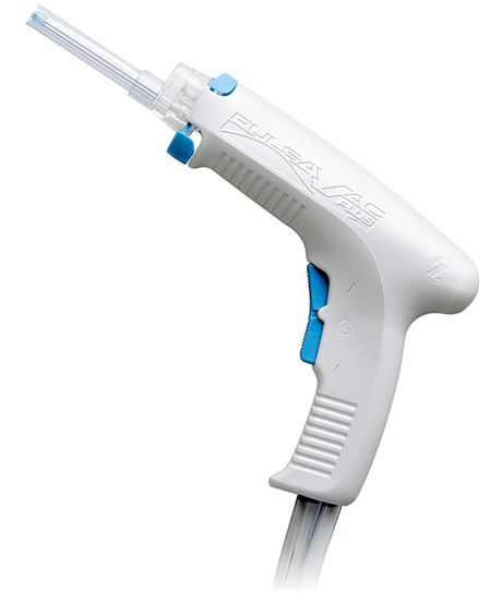
Page 2 of 8                                                                               Current Challenges in Thoracic Surgery, 2021

   The British Thoracic Society Pleural Disease Guideline              Operative technique
2010 indicated selecting patients for pleural fluid drainage
                                                                       Before surgery, chest computed tomography should be
in pleural infection (2). Patients with frankly purulent,
                                                                       performed to obtain anatomical information about the
turbid, or cloudy pleural fluid on sampling by thoracentesis
                                                                       location, size, extent of the empyema, and pleural surface
should receive prompt chest tube drainage of the pleural
                                                                       thickness (17).
space. The presence of organisms identified by positive
Gram stain and/or culture from a non-purulent pleural
                                                                       Video-assisted thoracic surgery (VATS)
fluid sampling indicates pleural infection and should lead to
                                                                       VATS, which is currently popular (18), consists of
prompt chest tube drainage (2).
                                                                       evacuation by suction, disruption of fibrous pleural
   The mortality benefit from intrathoracic administration
                                                                       septations and peeling off adhesions until the empyema
of fibrinolytic agent for empyema has not been reported;
                                                                       cavity becomes a single space. Chest tubes were inserted
thus further study was needed to explore this effect on
                                                                       at the end of the procedure through separate incisions for
mortality (2,6,11-14). This procedure is a known possible
cause of pneumothorax or hemothorax (15,16).                           thoraco-ports. The advantages of VATS are visualization of
   Patients who fail chest tube drainage are also additional           the entire thoracic cavity, removal of the purulent pleura,
candidates for surgical drainage. Delays in initiating                 and accurate placement of the thoracic drain with less
surgical drainage when indicated prolongs hospital stay and            surgical trauma, improved postoperative pain control, less
worsens clinical outcome. The goals in selecting a surgery             respiratory compromise and reduction in postoperative
are to rapidly establish an effective pleural drainage and             complications including 30-day mortality.
to promote lung re-expansion to obliterate the empyema                    However, despite effective placement of drainage tubes,
space.                                                                 improvement of empyema cannot be achieved if the drain is
                                                                       obstructed. In the past, we have used porous 24–32 Fr tubes;
                                                                       however, they drain pus only through their tips, which
Operative indication                                                   should be washed to clear the blockage when occluded.
In surgical treatment for empyema, “drainage and                       Recently, a drain has been developed with a drainage hole
dilatation of the lungs” is a basic concept. Pneumonia-                at the tip and a longitudinal groove at the side; this has
induced empyema is classified into three stages according              the advantages of both drains and is useful for drainage of
to its progression as stage I, parapneumonic effusion with             empyema. It is made of silicone and is soft; further, drainage
exudative effusion; stage II, fibrinopurulent stage marked by          of empyema may have a longer indwelling period, which
fibrinopurulent effusion; and stage III, chronic organizing            may reduce pain.
stage forming granulation tissue (pleural peel) (3).                      We placed the aforementioned drainage tube on the
   Surgery is performed mainly for stages II and III and               dorsal side and on the diaphragm (near the pulmonary
consists of aspiration of pleural effusion, destruction of the         ligament) at the time of thoracoscopic surgery and another
fibrin septum in the pleural cavity and single cavitation,             drain on the ventral side in cases with extensive empyema.
removal of purulent pleural effusion and inflammatory
substances, adequate pleural lavage, and placement                     Open thoracotomy (OT) approach
of drainage tubes. In stage III, additional removal of                 In the OT approach, decortication removes fibrous tissue
the thickened pleura and promotion of reinflation are                  and peels from the parietal and visceral pleura and pus
performed.                                                             from the pleural cavity (19). Theoretically, complete
   Patients in whom chest tube drainage was unsuccessful               decortication can improve intrathoracic infection and
are also candidates for surgical intervention. Conservative            expand the lungs. However, it should not be performed
treatment of empyema is challenging owing to host factors              solely to remove pleural thickening because pleural
and factors determining the disease stage, which possibly              thickening disappears spontaneously over several months
cause multiplication of the empyema cavities and decreased             (20,21). In a previous study, improving the numerical value
drainage. If the pleural effusion develops in the multiple             of the respiratory function at discharge and at 6 months had
loculae of the septum, medical treatment becomes difficult.            no significant effect on patients with or without residual
Performing drainage promptly during thoracoscopic                      pleural thickening, although pleural thickening markedly
surgery will help improve results.                                     reduced with regard to time at discharge and 6 months (21).

© Current Challenges in Thoracic Surgery. All rights reserved.   Curr Chall Thorac Surg 2021;3:4 | http://dx.doi.org/10.21037/ccts.2020.04.02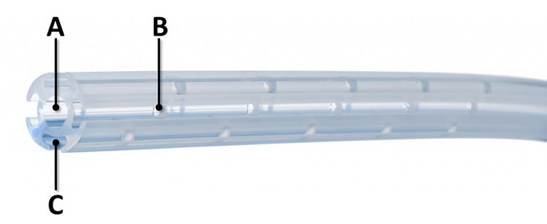
Current Challenges in Thoracic Surgery, 2021                                                                                    Page 3 of 8

Outcome of surgical treatment for empyema                              significant difference was found in the mortality rate
                                                                       between the two groups because patients who suffered
Surgical treatment versus non-surgical treatment for
                                                                       from treatment failure after the chest tube drainage were
empyema
                                                                       subsequently treated by surgical drainage and had successful
Recently, three studies using observational large-scale
                                                                       resolution of their empyema.
databases also revealed the actual clinical aspects of
empyema treatment (Table 1) (22-24).
                                                                       Outcomes of VATS and open thoracic surgery for
   Of the 17,533 patients treated for empyema in North
                                                                       empyema
America from 1987 to 2014 and in Canada from 1996 to
                                                                       Expert clinical opinion recommends that VATS should be
2015, 8,097 (VATS 4585 and OT 3512) in the surgical
                                                                       the first line approach (4). Experts mentioned that VATS
treatment group and 9436 in the non-surgical treatment
                                                                       and OT are logically parallel.
group were retrospectively examined for treatment results.
                                                                          Two prospective and seven retrospective studies of
The average age of patients was significantly higher in
                                                                       surgical treatment for stage II and III empyema involved
the non-surgical treatment group (61–62 years) than in                 a total of 1,954 patients. VATS and decortication by OT
the surgery group (53–56 years) (P
Page 4 of 8 Current Challenges in Thoracic Surgery, 2021
Table 1 Characteristics of adult patients in retrospective population-based cohort studies of thoracic empyema
Single-procedure Length of
Stage of Number of Procedure, number of Time to intervention, Initial treatment 30-day mortality Total mortality Readmission for empyema 90-day re-
Study Year Study period Region, country Age, year [IQR] treatment success hospital stay,
empyema patients patients [%] days [IQR] success [%] [%] [%] [%] intervention [%]
[%] days [IQR]
Farjah (22) 2007 1987–2004 Washington, USA Stage 2/3 4,424 OP 2,281 [52] 53±18 NA NA NA 16±13 123 [5] NA 68 [3] NA
Non-OP 2,143 [48] 62±19 13±11 356 [17] 126 [6]
PTable 3 Characteristics of patients in prospective studies analyzing surgical treatment
Tube Length of Primary Recurrence
Number Procedure, Age, Operative
Study Stage of drainage, postoperative treatment Conversion Mortality Morbidity of
Study Year of number of year time, min
design empyema days hospital stay, success [%] [%] [%] empyema
patients patients [range] [range]
[range] day [%] [%]
Chan (26) 2007 Prospective Stage 2/3 77 VATS 41 46±15 150±58 8±6 16±7 41 [100] 0 0 9 [22] NA
Open 36 49±16 228±84 9±4 21±14 36 [100] 0 8 [22]
P= PPage 6 of 8 Current Challenges in Thoracic Surgery, 2021
A B
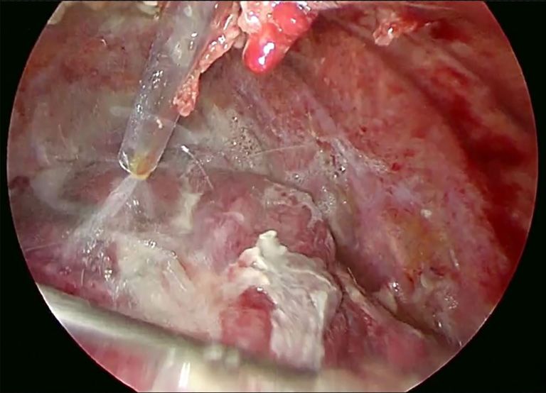
Figure 1 Pulsavac PlusTM (Zimmer Biomet G.K., Tokyo, Japan). (A) Washing device with pulsation to allow for debridement and suction in
the thoracic cavity. The tip of the device is 9 mm in size; (B) Pulsavac PlusTM was used for pulse lavage in the purulent thoracic cavity. The
lung coated with purulent content was washed with gentle water pressure, and the bony thorax can be washed with powerful water pressure;
therefore, the purulent tissue can be removed quickly and efficiently.
blood cell count and C-reactive protein levels). Patients
are observed for 24 h after drain removal is routinely
Figure 2 Naruke-type Thoraco-cottonTM (KENZMEDICO CO., performed.
LTD., Saitama, Japan). Cotton stick for the dissection of the tissue Dr. Satoshi Shiono: Could you show us the picture of the
and structures in the thorax. chest tube which you recently use?
Answer: Yes, I could. I have shown the Smart Coaxial
Drain (REDAX®, SENKO MEDICAL INSTRUMENT
A B Mfg. CO., LTD., Tokyo Japan) in Figure 3. The drain has
a drainage hole at the tip and a longitudinal groove on the
side.
C Conclusions
Figure 3 Smart Coaxial Drain (REDAX®, SENKO MEDICAL With the surgical treatment of complicated parapneumonic
INSTRUMENT Mfg. CO., LTD., Tokyo Japan). (A) Inner lumen; effusions and empyema, the treatment period will be
(B) small pores at the tube tip, 20 holes in total; (C) outer slit. shortened and medical cost will be reduced. However, there
are only small studies of immediate surgical treatment
versus chest tube drainage with fibrinolytics at the start of
organs, such as the lung, mediastinum or diaphragm can empyema treatment, but RCTs with increased number of
be washed with gentle water pressure, and the bony thorax cases are needed to draw definite conclusion.
can be washed with powerful water pressure; therefore, the
purulent tissue can be removed quickly and efficiently.
Acknowledgments
Dr. Satoshi Shiono: When do you remove the chest tubes
after surgery? Funding: None.
A n s w e r : Th e c hes t d ra i n c a n b e remo ved after
radiological confirmation of successful pleural drainage,
Footnote
that is, on observing a decrease in the size of the pleural
collection on the chest radiograph or computed tomography Provenance and Peer Review: This article was commissioned
and obtaining objective evidence of infectious resolution, by the Guest Editor (Satoshi Shiono) for the series
negativity of the pleural effusion culture for bacteria, and “Empyema” published in Current Challenges in Thoracic
decreasing levels of inflammatory markers (e.g., white Surgery. The article was sent for external peer review
© Current Challenges in Thoracic Surgery. All rights reserved. Curr Chall Thorac Surg 2021;3:4 | http://dx.doi.org/10.21037/ccts.2020.04.02Current Challenges in Thoracic Surgery, 2021 Page 7 of 8
organized by the Guest Editor and the editorial office. empyema therapy. Chest 1997;111:1548-51.
8. Thourani VH, Brady KM, Mansour KA, et al. Evaluation
Conflicts of Interest: Both authors have completed the of treatment modalities for thoracic empyema: a cost-
ICMJE uniform disclosure form (available at http:// effectiveness analysis. Ann Thorac Surg 1998;66:1121-7.
dx.doi.org/10.21037/ccts.2020.04.02). The authors have 9. Bilgin M, Akcali Y, Oguzkaya F. Benefits of early
no conflicts of interest to declare. SS serves as an unpaid aggressive management of empyema thoracis. ANZ J Surg
editorial board member of Current Challenges in Thoracic 2006;76:120-2.
Surgery from Sep 2019–Aug 2021. The series “Empyema” 10. Ahmed S, Azam H, Basheer I. Is open decortication
was commissioned by the editorial office without any superior to fibrinolytic therapy as a first line treatment
funding or sponsorship. in the management of pleural empyema? Pak J Med Sci
2016;32:329-32.
Ethical Statement: The authors are accountable for all 11. Maskell NA, Davies CW, Nunn AJ, et al. First Multicenter
aspects of the work in ensuring that questions related Intrapleural Sepsis Trial (MIST1) Group. U.K. Controlled
to the accuracy or integrity of any part of the work are trial of intrapleural streptokinase for pleural infection. N
appropriately investigated and resolved. Engl J Med 2005;352:865-74.
12. Tokuda Y, Matsushima D, Stein GH, et al. Intrapleural
Open Access Statement: This is an Open Access article fibrinolytic agents for empyema and complicated
distributed in accordance with the Creative Commons parapneumonic effusions: a meta-analysis. Chest
Attribution-NonCommercial-NoDerivs 4.0 International 2006;129:783-90.
License (CC BY-NC-ND 4.0), which permits the non- 13. Rahman NM, Maskell NA, Davies CW, et al. The
commercial replication and distribution of the article with relationship between chest tube size and clinical outcome
the strict proviso that no changes or edits are made and the in pleural infection. Chest 2010;137:536-43.
original work is properly cited (including links to both the 14. Rahman NM, Maskell NA, West A, et al. Intrapleural
formal publication through the relevant DOI and the license). use of tissue plasminogen activator and DNase in pleural
See: https://creativecommons.org/licenses/by-nc-nd/4.0/. infection. N Engl J Med 2011;365:518-26.
15. Ruiz A, Porcel JM, Madroñero AB, et al. Hemothorax
following administration of intrapleural alteplase.
References
Respiration 2006;73:715.
1. Bedawi EO, Hassan M, Rahman NM. Recent 16. Chai FY, Kuan YC. Massive hemothorax following
developments in the management of pleural infection: A administration of intrapleural streptokinase. Ann Thorac
comprehensive review. Clin Respir J 2018;12:2309-20. Med 2011;6:149-51.
2. Davies HE, Davies RJ, Davies CW, et al. Management of 17. Silen ML, Naunheim KS. Thoracoscopic approach to the
pleural infection in adults: British Thoracic Society Pleural management of empyema thoracis. Indications and results.
Disease Guideline 2010. Thorax 2010;65 Suppl 2:ii41-53. Chest Surg Clin N Am 1996;6:491-9.
3. Scarci M, Abah U, Solli P, et al. EACTS expert consensus 18. Chambers A, Routledge T, Dunning J, et al. Is video-
statement for surgical management of pleural empyema. assisted thoracoscopic surgical decortication superior
Eur J Cardiothorac Surg 2015;48:642-53. to open surgery in the management of adults with
4. Shen KR, Bribriesco A, Crabtree T, et al. The American primary empyema? Interact Cardiovasc Thorac Surg
Association for Thoracic Surgery consensus guidelines for 2010;11:171-7.
the management of empyema. J Thorac Cardiovasc Surg 19. Thurer RJ. Decortication in thoracic empyema.
2017;153:e129-46. Indications and surgical technique. Chest Surg Clin N Am
5. Redden MD, Chin TY, van Driel ML. Surgical versus 1996;6:461-90.
non-surgical management for pleural empyema. Cochrane 20. Neff CC, vanSonnenberg E, Lawson DW, et al.
Database Syst Rev 2017;3:CD010651. CT follow-up of empyemas: pleural peels resolve
6. Cargill TN, Hassan M, Corcoran JP, et al. A systematic after percutaneous catheter drainage. Radiology
review of comorbidities and outcomes of adult patients 1990;176:195-7.
with pleural infection. Eur Respir J 2019;54:1900541. 21. Jiménez Castro D, Díaz G, Pérez-Rodríguez E, et
7. Wait MA, Sharma S, Hohn J, et al. A randomized trial of al. Prognostic features of residual pleural thickening
© Current Challenges in Thoracic Surgery. All rights reserved. Curr Chall Thorac Surg 2021;3:4 | http://dx.doi.org/10.21037/ccts.2020.04.02Page 8 of 8 Current Challenges in Thoracic Surgery, 2021
in parapneumonic pleural effusions. Eur Respir J et al. VATS debridement versus thoracotomy in the
2003;21:952-5. treatment of loculated postpneumonia empyema. Ann
22. Farjah F, Symons RG, Krishnadasan B, et al. Management Thorac Surg 1996;61:1626-30.
of pleural space infections: a population-based analysis. J 29. Roberts JR. Minimally invasive surgery in the treatment
Thorac Cardiovasc Surg 2007;133:346-51. of empyema: intraoperative decision making. Ann Thorac
23. Semenkovich TR, Olsen MA, Puri V, et al. Current Surg 2003;76:225-30.
state of empyema management. Ann Thorac Surg 30. Luh SP, Chou MC, Wang LS, et al. Video-assisted
2018;105:1589-96. thoracoscopic surgery in the treatment of complicated
24. Nayak R, Brogly SB, Lajkosz K, et al. Outcomes of parapneumonic effusions or empyemas: outcome of 234
operative and nonoperative treatment of thoracic patients. Chest 2005;127:1427-32.
empyema: A population-based study. Ann Thorac Surg 31. Lardinois D, Gock M, Pezzetta E, et al. Delayed referral
2019;108:1456-63. and gram-negative organisms increase the conversion
25. Bagheri R, Tavassoli A, Haghi SZ, et al. The role thoracotomy rate in patients undergoing video-assisted
of thoracoscopic debridement in the treatment of thoracoscopic surgery for empyema. Ann Thorac Surg
parapneumonic empyema. Asian Cardiovasc Thorac Ann 2005;79:1851-6.
2013;21:443-6. 32. Tong BC, Hanna J, Toloza EM, et al. Outcomes of video-
26. Chan DT, Sihoe AD, Chan S, et al. Surgical treatment assisted thoracoscopic decortication. Ann Thorac Surg
for empyema thoracis: is video-assisted thoracic 2010;89:220-5.
surgery "better" than thoracotomy? Ann Thorac Surg 33. Marks DJ, Fisk MD, Koo CY, et al. Thoracic empyema: a
2007;84:225-31. 12-year study from a UK tertiary cardiothoracic referral
27. Muhammad MI. Management of complicated centre. PLoS One 2012;7:e30074.
parapneumonic effusion and empyema using different 34. Reichert M, Pösentrup B, Hecker A, et al. Thoracotomy
treatment modalities. Asian Cardiovasc Thorac Ann versus video-assisted thoracoscopic surgery (VATS) in
2012;20:177-81. stage III empyema-an analysis of 217 consecutive patients.
28. Angelillo Mackinlay TA, Lyons GA, Chimondeguy DJ, Surg Endosc 2018;32:2664-75.
doi: 10.21037/ccts.2020.04.02
Cite this article as: Endoh M, Shiono S. Strategy for surgical
treatment of acute thoracic empyema in adults. Curr Chall
Thorac Surg 2021;3:4.
© Current Challenges in Thoracic Surgery. All rights reserved. Curr Chall Thorac Surg 2021;3:4 | http://dx.doi.org/10.21037/ccts.2020.04.02You can also read