Enhanced Osseointegration of Porous Titanium Scaffold Implanted with Preload: An Experiment Study in Rabbits - SciELO
←
→
Page content transcription
If your browser does not render page correctly, please read the page content below

Int. J. Morphol.,
38(4):909-913, 2020.

      Enhanced Osseointegration of Porous Titanium Scaffold
    Implanted with Preload: An Experiment Study in Rabbits

                        Osteointegración Mejorada de Malla de Titanio Poroso Implantada
                                 con Precarga: Un Estudio Experimental en Conejos

                                       Linwei Lyu1,2; Ye Jing1,2; Jikun Wang3 & Chunqiu Zhang1,2

LYU, L.; JING, Y.; WANG, J. & ZHANG, C. Enhanced osseointegration of porous titanium scaffold implanted with preload: an
experiment study in rabbits. Int. J. Morphol., 38(4):909-913, 2020.

         SUMMARY: Porous titanium alloy scaffold was widely used in treating bone defect caused by traumatic injury and osteomyelitis,
which was incapable of self-healing. The implantation of scaffold produced stress shielding thereby forming osteolysis. The objective of
this study was to analysis trabecular morphological features of osseointegrated bone. 14 New Zealand rabbits were divided into two
groups, surgery group and healthy control group. 7 rabbits in surgery group were selected to perform 3D printed porous titanium alloy
scaffold implantation surgery with preload at the defect of femoral condyle for osseointegration. The other 7 rabbits in control group
were feed free. After 90 days healing, femoral condyles were extracted to perform micro-CT scanning with hydroxyapatite calibration
phantom. Mean bone mineral density (BMD), bone volume fraction (BV/TV), BS/TV (bone surface area ratio), Tb.Th (thickness of
trabeculae), Tb.N (number of trabeculae), Tb.Sp (trabecular separation) and DA (degree of anisotropy) were calculated from micro-CT
images. The results revealed that osseointegration inside and at the surface of scaffolds worked well from grey values of micro-CT
images. After 12 weeks healing, mean bone mineral densities (BMD) in surgery group and healthy control group were calculated as
800±20mg/cm3 and 980±90mg/cm3, respectively. This revealed that the strength of trabeculae in surgery group might lower than that in
the healthy group. Trabecular morphological parameters test showed that trabecular morphological parameters at the surface of scaffolds
in the surgery group deteriorated significantly. It was found from micro-CT images that ingrowth bone was filled with pores of scaffold.
Overall, the effect of osseointegration was promoted through the change of mechanical micro-environment in the scaffold region. Overall,
preload could improve osseointegration effect in the long-term after surgery. However, the trabecular morphology in the surgery group
was deteriorated, which might bring secondary fracture risk again.

          KEYWORDS: Osseointegration; 3D printed porous titanium alloy scaffold; preload; micro-CT; rabbit femoral condyle
defect.

INTRODUCTION

         Osseointegration was a research hotspot in treating                       Osseointegration, defined as bone ingrowth without
bone defect, trauma, infection and bone tumor in the clinical              interposed soft tissue, was found at bone to titanium screw
and research field of orthopedics (Wang et al., 2017a).                    implant contact surface (Carlsson et al., 1986).
According to WHO report in 2015, about 75 million people                   Osseointegration was found through microscopy after 3
suffered from trauma, in which 10 %-30 % victims were                      months surgery. In recent years, 3D printing technology of
unable to walk (Paka & Pokrowiecki, 2018). Bone autograft                  titanium alloys had been developed and matured. With the
was considered as a golden rule in bone defect repair (Dreifke             introduction of 3D printing technology in prosthesis, the
et al., 2015). However, in most cases, it was impossible to                manufacture of porous titanium alloy scaffold became
acquire patient bone autograft during the treatment (Winkler               possible. In vivo studies of rabbit scaffold implantation,
el al., 2018). Nowadays, porous titanium alloy scaffold was                significant osseointegration and bone ingrowth ability of
widely used in clinical with its advantages of good                        porous titanium alloy scaffold were found clearly through
mechanical properties, biocompatibility, and stable chemical               micro-CT images at four weeks after surgery (Chen et al.,
composition (Torres-Sanchez et al., 2017).                                 2017). Meanwhile, integration of ingrowth tissue with

1
  Tianjin Key laboratory for Advanced Mechatronic System Design and Intelligent Control, Tianjin University of Technology, China.
2
  National Demonstration Center for Experimental Mechanical and Electrical Engineering Education (Tianjin University of Technology), China.
3
  Institute of Automatic Control and Robotics, Faculty of Mechatronics, Warsaw University of Technology; Poland.

                                                                                                                                              909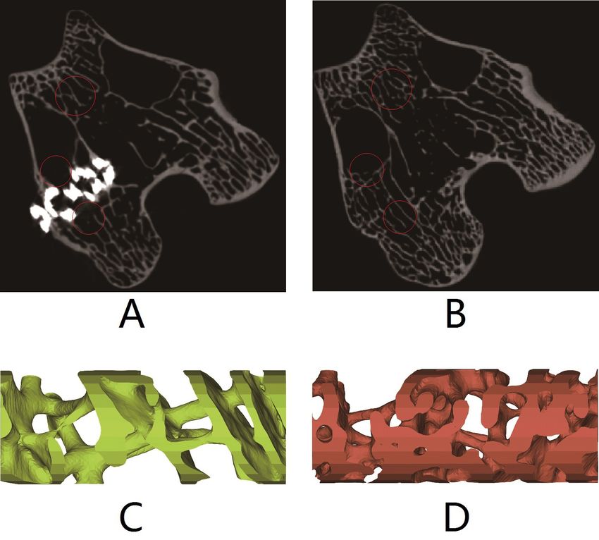
LYU, L.; JING, Y.; WANG, J. & ZHANG, C. Enhanced osseointegration of porous titanium scaffold implanted with preload: an experiment study in rabbits. Int. J. Morphol., 38(4):909-913, 2020.

scaffold could maintain good mechanical micro-environment                                                The objective of this study is to compare trabecular
and biocompatibility obviously (Wang et al., 2016).                                               morphological features after 90 days osseointegration in
                                                                                                  defect bone tissues with healthy trabecular tissues. All bone
        It was found from in vitro and in vivo experiments                                        tissues were test using micro-CT after 90 days
that 3D printed porous titanium alloy scaffold could promote                                      osseointegration. Trabecular morphological features in two
the effect of bone in-growth in the short-term after surgery.                                     groups were measured and compared at defect repair area.
Osseointegration period could be shrunk and through                                               The change trends of trabecular morphological features were
structural optimization and surface modification before                                           explored on ingrowth trabeculae long-term after surgery to
surgery (Salou et al., 2015; Srivasa et al., 2017; Ilea et al.,                                   provide applicable values for the improving mechanical
2019). In general, 3D printed scaffolds with high porosities                                      micro-environment in clinical and bone tissue engineering
and low stiffness were benefit for bone ingrowth compared                                         research.
with traditional implant (Wang et al., 2016). The porous
scaffold fabricated with approximate diameter of 500 µm and
porosity of 58 % by selective laser melting method showed                                         MATERIAL AND METHOD
significant bone in-growth as well as on-growth in rabbit
femoral condyles after four weeks to eight weeks (Srivasa et
al.). The effect of osseointegration was further improved after                                           Fourteen New Zealand rabbit with 2.5 months old and
surface modification. The pores of implant were completely                                        average weight of 2.5 kg were divided into two groups,
filled with bone within eight weeks in rabbit femur. The                                          including surgery group (7 rabbit) healthy control group (7
implant with anodized titanium dioxide nanotubes surface                                          rabbit). 7 porous titanium alloy scaffolds were fabricated by
presented higher but not significant bone-to-implant ratio and                                    3D printer with the diameter of 2mm, the height of 5 mm,
bone surface area at the distance of 0.5 mm than acid-etched                                      porosity of 70 %, pore diameter of 0.65 mm and wire diameter
surface or machined surface (Salou et al.). The porous scaffold                                   of 0.32 mm.
with mesh size of 800 um and coated by nano-hydroxyapatite
promoted the new bone formation and improved the                                                          Rabbits in surgery group were anesthetized with 10
osseointegration process best at the defect region when                                           %w/v chloral hydrate solution. Intraperitoneal dose was
compared with uncoated scaffolds (Ilea et al.).                                                   calculated with 3.5 ml per kilogram weight. Regions around
                                                                                                  right condyles of femur were shaved clearly and skins were
         The role of mechanical stimulation was increasingly                                      exposed. Limbs of rabbits were extended fixed on the
recognized in the pathogenesis of peri-prosthetic osteolysis                                      operation table. 2 ml lidocaine hydrochloride with
(Wang et al., 2017b). There were no significant differences                                       concentration of 0.02 g/ml was injected subcutaneously at
on osseointegration effect between loading group and                                              knee joint for local anesthesia after skin disinfection with
unloading group in the early time of postoperation (Reitmaier                                     iodine. An incision of 4-5 cm was made at lateral knee using
et al., 2018). In vivo mechanical stimulation could promote                                       scalpel to exposed femoral condyle. A Small amount of
bone ingrowth into metallic scaffold to enhance long-term                                         lidocaine hydrochloride would be sprayed while waking up
fixation after surgery (Willie et al., 2010). Implantation group                                  from anesthesia. Drilling location was set at the interosseous
coupled with the mechanical stimulation exhibited higher                                          eminence center of right femoral condyle of rabbit. Drilling
healing quality in contrast to the corresponding unloaded                                         dimension was with the diameter of 1.9 mm and depth of
group during 4 weeks and 8 weeks (Zhang et al., 2018).                                            5mm. A sterilized 3D printed porous titanium alloy scaffold
Bone-to-implant surface area in loading group was about                                           was implanted into drilling hole with preload produced by
10 % higher than unloading group at week 6, and more than                                         interference assembly. The implantation process was shown
30 % at week 26 (Luan et al., 2019).                                                              in Figure 1. The incision was sutured and disinfected with
                                                                                                  iodide. After waking up, rabbits were injected with
        Limited literatures were available on mechanical                                          enrofloxacin into the muscle of the operative leg with a dose
properties of ingrowth bone tissue. Pull-out test could reflect                                   of 0.2 ml /kg for three consecutive days. All rabbits were
trabecular mechanical properties of ingrowth bone tissue at                                       sacrificed at 90 days after surgery. The right femurs were
the surface of oral implant (Ilea et al.). The Young’s modulus                                    extracted and frozen in -40 ºC freezer.
of titanium alloy scaffold was significantly higher than bone
tissue around. The stress shielding produced by scaffold broke                                           All femurs in two groups were performed micro-CT
original mechanical micro-environment resulting in abnormal                                       scanning. Micro-CT scanning parameters were pixel size
bone remodeling In-growth bone tissue. Therefore, the                                             32 um, thickness 32 um, ROI 1500*1500 px, 1400 slices,
mechanical properties of ingrowth bone tissue must be                                             voltage 190 kV, current 130 uA. All micro-CT images were
influenced.                                                                                       imported into VG Studio MAX software to calculate
910
LYU, L.; JING, Y.; WANG, J. & ZHANG, C. Enhanced osseointegration of porous titanium scaffold implanted with preload: an experiment study in rabbits. Int. J. Morphol., 38(4):909-913, 2020.
trabecular morphological parameters at
the region of scaffolds, including BV/TV
(bone volume fraction), BS/TV (bone
surface area ratio ), Tb.Th (thickness of
trabeculae ), Tb.N (number of trabeculae),
Tb.Sp(trabecular separation), DA (degree
of anisotropy), BMD (bone mineral
density).
All trabecular morphological
parameters and bone mineral density data
were expressed as an average ± standard
deviation. Independent-sample T tests
were performed on each trabecular
morphological parameter and BMD with
significance level of 95 %. Significant
difference value P was considered of less
than 0.05. All data were statistically
analyzed and plotted in software SPSS
17.0.
RESULTS
Fig. 1. Surgery process of scaffold implantation of a rabbit. A. 3D printed porous titanium
scaffold. B. Surgery process of implantation. C. Stitching process after surgery. D. Right
femurs from rabbits after 90 days osseointegration.
Statistical analyses of trabecular
morphological parameters revealed that
there were significant differences between
surgery group and healthy control group,
which was shown in Table I as mean ±
standard deviation. BV/TV, Tb.Th and
Tb.N in surgery group were significantly
lower than that in healthy control group.
BS/BV and Tb.Sp in surgery group were
significantly higher than that in healthy
control group. There were no significant
differences in DA between surgery group
and healthy control group.
Typical micro-CT images and 3D
reconstruction models in surgery group
and healthy control group were shown in
Figure 2. Significant differences of
trabecular separation (Tb.Sp) were found
in Figures 2a and 2b with the red circle
markers.Three-dimensional
reconstructions were performed on the
micro-CT images. It was shown from
three-dimensional models in Figures 2c
Fig. 2. Typical trabecular morphology comparison of surgery group and healthy control and 2d that trabecular thickness (Tb.N) at
group. A. and B. Micro-CT images of femoral condyles from surgery group and healthy the surface of scaffold in the surgery group
control group, respectively. C and D. 3D reconstruction models in the area of femoral was significant lower than that in healthy
condyles from surgery group and healthy control group, respectively. control group.
911LYU, L.; JING, Y.; WANG, J. & ZHANG, C. Enhanced osseointegration of porous titanium scaffold implanted with preload: an experiment study in rabbits. Int. J. Morphol., 38(4):909-913, 2020.
Table I. Trabecular morphological parameters of rabbit femoral condyle.
Groups BV/TV BS/BV Tb.Th Tb.N Tb.Sp DA
Group A 0.34±0.06 11.61±2.27 0.18±0.05 0.77±0.21 1.19±0.35 0.44±0.10
Group B 0.47±0.05 7.15±1.82 0.29±0.06 1.65±0.27 0.33±0.05 0.36±0.10
Difference test 0.000 0.006 0.008 0.000 0.002 0.224
PLYU, L.; JING, Y.; WANG, J. & ZHANG, C. Enhanced osseointegration of porous titanium scaffold implanted with preload: an experiment study in rabbits. Int. J. Morphol., 38(4):909-913, 2020.
ludable. Se seleccionaron 7 conejos en el grupo de cirugía para Luan, H. Q.; Wang, L. T.; Ren, W. Y.; Chu, Z. W.; Huang, Y. F.; Lu, C. L. &
realizar una implantación de mallas de aleación de titanio poroso, Fan, Y. B. The effect of pore size and porosity of Ti6Al4V scaffolds on
impresas en 3D con precarga en el defecto del cóndilo femoral MC3T3-E1 cells and tissue in rabbits. Sci. China Technol. Sci., 62:1160-
8, 2019.
para la osteointegración. Los 7 conejos restantes del grupo control McArthur, B. A.; Scully, R.; Patrick Ross, F.; Bostrom, M. P. G. & Falghren,
se mantuvieron sin alimentación. Después de 90 días de curación, A. Mechanically induced periprosthetic osteolysis: a systematic review.
se extrajeron los cóndilos femorales para realizar una exploración HSS J., 15(3):286-96, 2019.
por micro-CT con un espectro de calibración de hidroxiapatita. Se Paka, K. & Pokrowiecki, R. Porous titanium implants: a review. Adv. Eng.
calcularon a partir de imágenes de micro-CTDensidad mineral ósea Mater., 20(5):1700648, 2018.
media (DMO), fracción de volumen óseo (BV / TV), BS / TV (re- Peng, W.; Xu, L.; You, J.; Fang, L. & Zhang, Q. Selective laser melting of
titanium alloy enables osseointegration of porous multi-rooted implants
lación de área de superficie ósea), Tb.Th (espesor de trabéculas),
in a rabbit model. Biomed. Eng. Online, 15(1):85, 2016.
Tb.N (número de trabéculas), Tb.Sp (trabecular separación) y DA Reitmaier, S.; Kovtun, A.; Schuelke, J.; Kanter, B.; Lemm, M.; Hoess, A.;
(grado de anisotropía). Los resultados revelaron que la Heinemann, S.; Nies, B. & Ignatius, A. Strontium(II) and mechanical
osteointegración dentro y en la superficie de los andamios funcio- loading additively augment bone formation in calcium phosphate
nó bien a partir de los valores grises de las imágenes de micro-CT. scaffolds. J. Orthop. Res., 36(1):106-17, 2018.
Después de 12 semanas de curación, las densidades medias de mi- Salou, L.; Hoornaert, A.; Louarn, G. & Layrolle, P. Enhanced osseointegration
nerales óseos (DMO) en el grupo cirugía y en el grupo control of titanium implants with nanostructured surfaces: an experimental study
in rabbits. Acta Biomater,, 11:494-502, 2015.
sano se calcularon como 800 ± 20 mg/cm3 y 980 ± 90 mg/cm3,
Srivas, P. K.; Kapat, K.; Dadhich, P.; Pal, P.; Dutta, J.; Datta, P. & Dharaa, S.
respectivamente. Esto reveló que la fuerza de las trabéculas en el Osseointegration assessment of extrusion printed Ti6Al4V scaffold
grupo de cirugía podría ser menor que la del grupo sano. La prue- towards accelerated skeletal defect healing via tissue in-growth.
ba de parámetros morfológicos trabeculares mostró que en el gru- Bioprinting, 6:8-17, 2017.
po de cirugía, la superficie de las mallas, se deterioraron Torres-Sanchez, C.; Al Mushref, F. R. A.; Norrito, M.; Yendall, K.; Liu, Y. &
significativamente. Se descubrió a partir de imágenes de micro- Conway, P. P. Torres-Sanchez, F.R.A. Al Mushref, M. Norrito, K. Yendall,
CT que el hueso en crecimiento estaba lleno de poros de andamio. Y. Liu, P.P. Conway. The effect of pore size and porosity on mechanical
properties and biological response of porous titanium scaffolds. Mater.
En general, el efecto de la osteointegración se promovió mediante
Sci. Eng. C Mater. Biol. Appl., 77:219-28, 2017.
el cambio del microambiente mecánico en la región de la malla. Wang, G.; Roohani-Esfahani, S. I.; Zhang, W.; Lv, K.; Yang, G.; Ding, X.;
En general, la precarga podría mejorar el efecto de osteointegración Zou, D.; Cui, D.; Zreiqat, H. & Jiang, X. Effects of Sr-HT-Gahnite on
a largo plazo después de la cirugía. Sin embargo, la morfología osteogenesis and angiogenesis by adipose derived stem cells for critical-
trabecular en el grupo de cirugía se deterioró, lo que podría traer un sized calvarial defect repair. Sci. Rep., 7:41135, 2017a.
nuevo riesgo de fractura secundaria. Wang, X.; Xu, S.; Zhou, S.; Xu, W.; Leary, M.; Choong, P.; Qian, M.; Brandt,
M. & Xie Y. M. Topological design and additive manufacturing of porous
metals for bone scaffolds and orthopaedic implants: A review.
PALABRAS CLAVE: Osteointegración; Mallas de alea- Biomaterials, 83:127-41, 2016.
ción de titanio poroso impreso en 3D; precarga micro-CT; De- Wang, Z.; Wang, C.; Li, C.; Qin, Y.; Zhong, L.; Chen, B.; Li, Z.; Liu, H.;
fecto del cóndilo femoral en conejo. Chang, F. & Wang, J. Analysis of factors influencing bone ingrowth into
three-dimensional printed porous metal scaffolds: A review. J. Alloys
Compd., 717:271-85, 2017b.
Willie, B. M.; Yang, X.; Kelly, N. H.; Han, J.; Nair, T.; Wright, T. M.; van der
REFERENCES Meulen, M. C. & Bostrom, M. P. Cancellous bone osseointegration is
enhanced by in vivo loading. Tissue Eng. Part C Methods, 16(6):1399-
406, 2010.
Carlsson, L.; Röstlund, T.; Albrektsson, B.; Albrektsson, T. & Brånemark, P. Winkler, T.; Sass, F. A.; Duda, G. N. & Schmidt-Bleek, K. A review of
biomaterials in bone defect healing, remaining shortcomings and future
I. Osseointegration of titanium implants. Acta Orthop. Scand., 57(4):285-
opportunities for bone tissue engineering: The unsolved challenge. Bone
9, 1986.
Chen, Y.; Frith, J. E.; Dehghan-Manshadi, A.; Attar, H.; Kent, D.; Soro, N. Joint Res., 7(3):232-43, 2018.
Zhang, Y.; Chen, Y.; Kou, H.; Yang, P.; Wang, Y. & Lu, T. Enhanced bone
D. M.; Bermingham, M. J. & Dargusch, M. S. Mechanical properties
healing in porous Ti implanted rabbit combining bioactive modification
and biocompatibility of porous titanium scaffolds for bone tissue
engineering. J. Mech. Behav. Biomed. Mater., 75:169-74, 2017. and mechanical stimulation. J. Mech. Behav. Biomed. Mater., 86:336-
Dreifke, M. B.; Jayasuriya, A. A. & Jayasuriya, A. C. Current wound healing 44, 2018.
procedures and potential care. Mater. Sci. Eng. C Mater. Biol. Appl.,
48:651-62, 2015. Corresponding author:
Ilea, A.; Vrabie, O. G.; Baban, A. M.; Miclus, V.; Ruxanda, F.; Sárközi, M.; Chunqiu Zhang
Barbu-Tudoran, L.; Mager, V.; Berce, C.; Boca, B. A.; et al. Tianjin Key laboratory for Advanced Mechatronic
Osseointegration of titanium scaffolds manufactured by selective laser
System Design and Intelligent Control
melting in rabbit femur defect model. J. Mater. Sci. Mater. Med., 30(2):26,
2019.
Tianjin University of Technology
Li, K.; Wang, C.; Yan, J.; Zhang, Q.; Dang, B.; Wang, Z.; Yao, Y.; Lin, K.; No. 391 Binshui Xidao
Guo, Z.; Bi, L.; et al. Evaluation of the osteogenesis and osseointegration Xiqing District
of titanium alloys coated with graphene: an in vivo study. Sci. Rep., Tianjin - CHINA Received: 07-02-2020
8(1):1843, 2018. Accepted: 10-03-2020
Liang, H.; Yang, Y.; Xie, D.; Li, L.; Mao, N.; Wang, C.; Tian, Z.; Jiang, Q. &
Shen, L. Trabecular-like Ti-6Al-4V scaffolds for orthopedic: fabrication
by selective laser melting and in vitro biocompatibility. J. Mater. Sci. E-mail: zhang_chunqiu@126.com
Technol., 35(7):1284-97, 2019. lvlw@tjut.edu.cn
913You can also read