Hearing Loss Case - Click to Begin - A Self-Directed Learning Module Department of Otolaryngology - Head & Neck Surgery - Western University
←
→
Page content transcription
If your browser does not render page correctly, please read the page content below
Hearing Loss Case
 A Self-Directed Learning Module

 Department of Otolaryngology – Head & Neck Surgery
Schulich School of Medicine & Dentistry, Western University

 Click to Begin
Case Presentation
A 32-year-old female teacher presents to
your family practice with a five year
history of slowly progressive bilateral
hearing loss.

You are the family physician, click through the module to diagnose and treat this
patient.

 Obtain a history
Back to Case Presentation
Patient History

 “My hearing has been getting worse for the past five years. I often have trouble hearing
 people’s voices and have to ask them to repeat themselves. The hearing loss is always
 there, along with some ringing in my ears. My hearing has been getting progressively
 worse, and I noticed more of a drop when I was pregnant. I have not had any dizziness or
 ear pressure and I haven’t tried anything for the hearing loss so far.

 Otherwise, I don’t have any other medical problems. I do not take any medications, or
 have any allergies.

 My mother had quite poor hearing, and so did her father. However, I don’t think they were
 given a diagnosis.”

 What is on your differential diagnosis so far?
Back to Case Presentation

What diagnoses are coming to mind?
Think VINDICATE! (Click on heading to test yourself)

 Vascular

 Endocrine/Metabolic Infectious

 ?
 Trauma/Toxins Neoplastic

 Autoimmune/Allergic Degenerative

 Congenital/Genetic Iatrogenic/Idiopathic

 With your DDx in mind, proceed to focused physical exam.
Back to Case Presentation

What diagnoses are coming to mind?
Think VINDICATE! (Click on heading to test yourself)
 Vascular - Unlikely
 X
 Why not?
 Bilateral and progressive
 Endocrine/Metabolic hearing loss. No pulsatile Infectious
 characteristic.

 ?
 Trauma/Toxins Neoplastic

 Autoimmune/Allergic Degenerative

 Congenital/Genetic Iatrogenic/Idiopathic

 With your DDx in mind, proceed to focused physical exam.
Back to Case Presentation

What diagnoses are coming to mind? X
Think VINDICATE! (Click on heading to test yourself) Infectious - Unlikely

 Vascular Why not?
 Five year history, general good health
 Endocrine/Metabolic
 DDx
 Luetic (otosyphilis) hearing loss

 ?
 Trauma/Toxins Neoplastic

 Autoimmune/Allergic Degenerative

 Congenital/Genetic Iatrogenic/Idiopathic

 With your DDx in mind, proceed to focused physical exam.
Back to Case Presentation

What diagnoses are coming to mind?
Think VINDICATE! (Click on heading to test yourself)

 Vascular

 Endocrine/Metabolic Infectious

 Neoplastic
 X

 ?
 Trauma/Toxins Why?
 Progressive history over years.

 DDx
 Autoimmune/Allergic Temporal bone neoplasm
 Degenerative
 Glomus jugulare tumor
 Cholesteatoma (*NOT cancerous;
 keratinizing squamous epithelium in
 Congenital/Genetic Iatrogenic/Idiopathic
 the temporal bone)

 With your DDx in mind, proceed to focused physical exam.
Back to Case Presentation

What diagnoses are coming to mind?
Think VINDICATE! (Click on heading to test yourself)

 Vascular

 Endocrine/Metabolic Infectious

 ?
 Trauma/Toxins Neoplastic

 Degenerative - Unlikely X
 Why not?
 Autoimmune/Allergic Young age and no fluctuating
 course

 Congenital/Genetic Iatrogenic/Idiopathic DDx
 Presbycusis
 Multiple Sclerosis
 With your DDx in mind, proceed to focused physical exam.
Back to Case Presentation

What diagnoses are coming to mind?
Think VINDICATE! (Click on heading to test yourself)

 Vascular

 Endocrine/Metabolic Infectious

 ?
 Trauma/Toxins X Neoplastic
 Iatrogenic/Idiopathic - Unlikely

 Why not?
 Autoimmune/Allergic No previous otologic surgery. NoDegenerative
 fluctuating course or associated
 vertigo.

 Congenital/Genetic DDx
 Meniere’s disease

 With your DDx in mind, proceed to focused physical exam.
Back to Case Presentation

What diagnoses are coming to mind?
Think VINDICATE! (Click on heading to test yourself)

 Vascular

 Endocrine/Metabolic Infectious

 ?
 Trauma/Toxins Neoplastic
 Congenital/Genetic X
 Why?
 Congenital less likely given prior
 Autoimmune/Allergic Degenerative
 normal hearing, but genetic
 diagnoses are possible.
 Iatrogenic/Idiopathic
 DDx
 Paget’s disease
 Osteogenesis imperfecta
 Otosclerosis With your DDx in mind, proceed to focused physical exam.
Back to Case Presentation

What diagnoses are coming to mind?
Think VINDICATE! (Click on heading to test yourself)

 Vascular

 Endocrine/Metabolic Infectious

 ?
 Trauma/Toxins Neoplastic

 Autoimmune/Allergic
 X
 Degenerative
 Why?
 Middle-aged female.

 DDx Congenital/Genetic Iatrogenic/Idiopathic
 Autoimmune inner ear disease

 With your DDx in mind, proceed to focused physical exam.
Back to Case Presentation

What diagnoses are coming to mind?
Think VINDICATE! (Click on heading to test yourself)

 Vascular

 Endocrine/Metabolic Infectious

 Trauma/Toxins - Unlikely
 X

 ?
 Why not? Neoplastic
 Unlikely based on history, but
 important to rule out on history.

 DDx
 Autoimmune/Allergic
 Ototoxic medication Degenerative
 Noise-induced hearing loss
 Head trauma
 Lead and mercury toxicity Congenital/Genetic Iatrogenic/Idiopathic
 Barotrauma
 Cerumen impaction
 With your DDx in mind, proceed to focused physical exam.
Back to Case Presentation

What diagnoses are coming to mind?
Think VINDICATE! (Click on heading to test yourself)

 Endocrine/Metabolic - Unlikely
 X Vascular

 Why not? Infectious
 Unlikely based on history and no
 suggestive associated symptoms.

 ?
 DDxTrauma/toxins Neoplastic
 Hypothyroidism

 Autoimmune/Allergic Degenerative

 Congenital/Genetic Iatrogenic/Idiopathic

 With your DDx in mind, proceed to focused physical exam.
Back to Case Presentation

Physical Exam (Click on the physical examinations.)

General inspection & Vitals

 Cranial Nerve Exams

 Head & Neck Exam

 Cerebellar Tests

 Proceed to investigations
Back to Case Presentation

Physical Exam (Click on the physical examinations.)

General inspection & Vitals
 Patient looks well and doesn't
 appear to be in any distress.

 Vitals:
 Cranial Nerve Exams HR: 80 bpm
 RR: 12 per minute
 BP: 122/80 mmHg
 Temperature: 37℃
 Head & Neck Exam

 Cerebellar Tests

 Proceed to investigations
Back to Case Presentation

Physical Exam (Click on the physical examinations.)

General inspection & Vitals
 Cranial Nerve Exams:

 CN I – VII, IX - XII : Within normal limits

 Cranial Nerve Exams CN VIII:
 Weber test: No lateralization
 Rinne test: Bone conduction > Air conduction

 Head & Neck Exam
 Review: Cranial Nerve Exams

 Review: Weber & Rinne tests

 Cerebellar Tests

 Proceed to investigations
Back to Case Presentation
 Review: Cranial Nerve Exams
Cranial Nerve Name Function Test

 I Olfactory Nerve • Smell • Test for sense of smell with coffee, alcohol swab, citrus, etc.

 II Optic Nerve • Vision • Visual acuity – Snellen’s eye chart
 • Visual fields – Confrontation testing
 • Pupillary reflexes – Direct and consensual response
 • Fundoscopy
 III Oculomotor Nerve • Motor innervation to most* extra-ocular muscles • H test & convergence
 • Pupillary reflex
 IV Trochlear Nerve • Motor innervation to superior oblique muscle* • H test : Look for ability to look ”down and out”

 V Trigeminal Nerve • Sensory innervation to the face • Corneal reflex
 • Motor innervation to muscles of mastication • Test sensory supply to the face – cotton swab
 • Test strength of muscles of mastication
 VI Abducens Nerve • Motor innervation to lateral rectus muscle* • H test: Look for ability to abduct eye

 VII Facial Nerve • Motor innervation to muscles of facial expression • Ask patient to do different facial expressions
 • Taste – anterior 2/3 tongue • Corneal reflex
 VIII Vestibulocochlear Nerve • Hearing & balance • Weber & Rinne tests

 IX Glossopharyngeal Nerve • Sensory innervation to the palate • Gag reflex
 • Taste – posterior 1/3 tongue • Say “Ahhh” – look for deviation of the uvula
 X Vagus Nerve • Motor supply to the pharynx • Gag reflex
 • PSNS supply to abdominal viscera
 XI Spinal Accessory Nerve • Motor innervation to trapezius and sternocleidomastoid • Shoulder shrug against resistance, head turn against resistance

 XII Hypoglossal Nerve • Motor supply to muscles of the tongue • Stick tongue out and move tongue from side-to-side

 Review: Weber & Rinne tests Back to Physical Exam
Back to Case Presentation

Review: Weber and Rinne tests
 1. Weber test
 • Strike a 512 Hz tuning fork and place on top of the patient’s head
 • A patient with normal hearing should hear the sound equally on both sides* (I.e. the sound shouldn’t lateralize to one ear)
 • *Note: A Rinne test is needed to confirm normal hearing, as a patient with bilateral conductive hearing loss would also
 have no lateralization of sound.

 2. Rinne test
 • Strike a 512 Hz tuning fork and place it on the mastoid bone behind the patient’s ear (Testing bone conduction (BC))
 • When the patient signals that they can no longer hear the sound, move the tuning fork next to the patients external auditory
 canal (Testing air conduction (AC))
 • A patient with normal hearing should hear the sound better through air conduction (AC > BC)
 • Rinne test might not be negative if the conductive hearing loss is very mild

Results from the Weber and Rinne test can be used to determine the type of hearing loss:

 Conductive Hearing Sensorineural Hearing
 Test Normal
 Loss Loss

 Weber Sound heard in Sound heard in Sound heard in good
 midline affected ear ear

 Rinne AC > BC BC > AC AC > BC

 Back
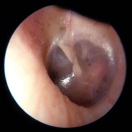
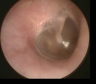
Back to Case Presentation

 Focused Head & Neck Exam
Inspection: Oral Cavity Exam: Nasal speculum exam: Otoscopy:
• No scars, asymmetry, enlarged • Turbinates, nasal mucosa, • External auditory canals have minimal
 thyroid or parotids, skin lesions and nasal septum normal cerumen
• No spontaneous nystagmus • No foreign bodies, discharge or mass

Palpation:
• No palpable lymph nodes,
 salivary glands palpable, thyroid
 not palpable • es

 Right Tympanic Membrane Left Tympanic Membrane

 Review: Otoscopy

 Back to Physical Exam
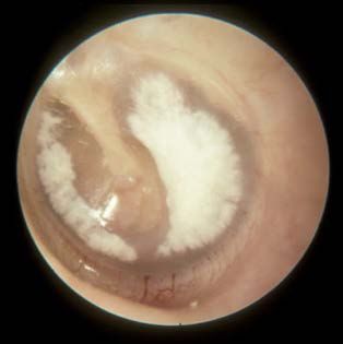
Back to Case Presentation

 Review: Otoscopy Examples of Abnormal TMs

 Normal Otoscopic Exam Findings:
 • Tympanic membrane (TM) should be intact and
 appear pearly grey or whitish/pinkish grey

 Bulging, red TM - Acute Otitis Media Cholesteatoma
 Normal TM – Right Side

Pars flaccida
 Lateral/short process of malleus

 Incus Manubrium of malleus

 Umbo Otitis media with effusion Myringosclerosis
 Cone of light
 Pars tensa
 Annulus

 Hemotympanum Retracted TM
 Back
Back to Case Presentation

Physical Exam (Click on the physical examinations.)

General inspection & Vitals

 Cerebellar Tests
 Cranial Nerve Exams
 • Finger-nose, heel-shin, and rapid
 alternating movements tests are normal

 • Gait (including tandem gait) is normal
 Head & Neck Exam
 • Rhomberg is normal

 Cerebellar Tests

 Proceed to investigations
Back to Case Presentation

 Investigations (Click on the buttons to see investigation results.)
 Cochlear/Acoustic Testing

 Vestibular Testing

 Temporal Bone CT Scan

Internal Auditory Canal MRI

 Continue to Diagnosis
Back to Case Presentation
 Cochlear/Acoustic Testing Back to investigations

 Audiogram Acoustic Reflex Tympanometry

 1
 0.8
 Left
 0.6

 Compliance (ml)
 0.4
 0.2
 0
 -400 -200 0 200 400
 Pressure at which peal compliance occurs
 (decaPascals)

 1
 0.8
 Right
 0.6

 Compliance (ml)
 0.4
 0.2
 0
 -400 -200 0 200 400
 Pressure at which peal compliance occurs
 (decaPascals)

 Test: Within normal limits? Test: Within normal limits? Test: Within normal limits?
Unsure? Review: Audiology Testing Unsure? Review: Acoustic Reflex Unsure? Review: Tympanometry
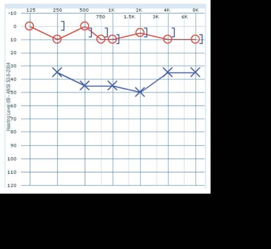
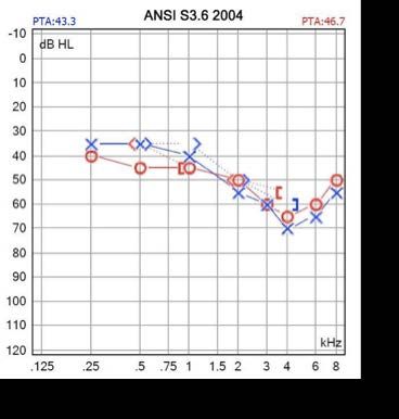
Back to Case Presentation
 Cochlear/Acoustic Testing Back to investigations

 Audiogram Acoustic Reflex Tympanometry

 1
 0.8
 Left
 0.6

 Compliance (ml)
 0.4
 0.2
 0
 -400 -200 0 200 400
 Pressure at which peal compliance occurs
 (decaPascals)

 1
 0.8
 Right
 0.6

 Compliance (ml)
 0.4
 0.2
 0
 X -400 -200 0 200
 Pressure at which peal compliance occurs
 400

No, the audiogram shows bilateral (decaPascals)

 conductive hearing loss.
 Test: Within normal limits? Test: Within normal limits?
Unsure? Review: Audiology Testing Unsure? Review: Acoustic Reflex Unsure? Review: Tympanometry
Back to Case Presentation
 Cochlear/Acoustic Testing Back to investigations

 Audiogram Acoustic Reflex Tympanometry

 1
 0.8
 Left
 0.6

 Compliance (ml)
 0.4
 0.2
 0
 -400 -200 0 200 400
 Pressure at which peal compliance occurs
 (decaPascals)

 1
 0.8
 Right
 0.6

 Compliance (ml)
 0.4
 0.2
 0

 X -400 -200 0 200
 Pressure at which peal compliance occurs
 400

 No, acoustic reflexes are absent (decaPascals)

 bilaterally.
 Test: Within normal limits? Test: Within normal limits?
Unsure? Review: Audiology Testing Unsure? Review: Acoustic Reflex Unsure? Review: Tympanometry
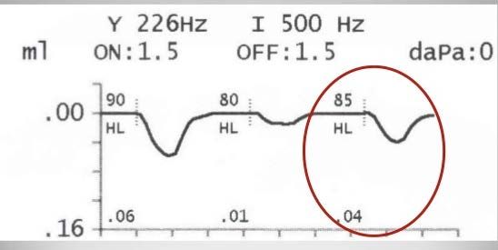
Back to Case Presentation
 Cochlear/Acoustic Testing Back to investigations

 Audiogram Acoustic Reflex Tympanometry

 1
 0.8
 Left
 0.6

 Compliance (ml)
 0.4
 0.2
 0
 -400 -200 0 200 400
 Pressure at which peal compliance occurs
 (decaPascals)

 1
 0.8
 Right
 0.6

 Compliance (ml)
 0.4
 0.2
 0
 -400 -200 0 200 400
 Pressure at which peal compliance occurs
 (decaPascals)
 X
 Yes, tympanometry is within
 Test: Within normal limits? Test: Within normal limits? normal limits.

Unsure? Review: Audiology Testing Unsure? Review: Acoustic Reflex Unsure? Review: Tympanometry
Back to Case Presentation
Review: Interpreting an Audiogram
 Low Pitch High Pitch
 Air conduction testing:
 Frequencies • Sound delivered through headphones or loudspeakers,
 being tested tests outer, middle, and inner ear.
 • Left ear = X
 • Right ear = O
 • Different symbols are used when “masking” is used.
 Soft

 Masking refers to noise presented to the non-test ear to
 Normal Hearing prevent it from hearing sound presented to the test ear.
 • Left ear =
 • Right ear = △
 Mild Hearing Loss
 Bone conduction testing:
 • Bone vibrator placed behind the ear to deliver sound
 Moderate Hearing Loss vibrations to the cochlea, bypassing the outer and middle
 ear.
 Moderately Severe Hearing Loss • Left ear = >
 • Right ear = <
 Severe Hearing Loss • Masking symbols [< >]
 • Left ear = ]
 • Right ear = [

 Profound Hearing Loss
 Loud

 Click to practice reading audiograms
How loud the sound needs
to be, in order to be heard
at that frequency
 Back
Back to Case Presentation
Review: Interpreting an Audiogram Back
 Air conduction testing compared to
 masked bone conduction testing.

 • Bone conduction: within normal
 Unmasked air conduction range
 audiogram. • Air conduction: mild – moderate
 hearing loss
 Interpretation:
 Normal hearing Interpretation:
 Conductive hearing loss.
 (I.e. Middle ear pathology)

 Right ear: Unmasked air and bone
 conduction testing
 Left ear: Masked air and bone
 conduction testing

 Interpretation:
 Masked air conduction Asymmetrical sensorineural hearing
 audiogram. loss.
 (I.e. Acoustic neuroma)
 Interpretation: Moderately
 severe hearing loss at high • Right ear: mild sensorineural
 frequencies. hearing loss at higher
 (I.e. Presbycusis) frequencies
 • Left ear: Mild to moderately
 severe hearing loss as move up
 frequencies
Back to Case Presentation
 Review: Acoustic Reflex Testing
The acoustic reflex is the reflexive contraction of the stapedius muscle, and subsequent stiffening of the tympanic
membrane (TM), in response to high-intensity sound or vocalization. *Anatomy reminder: stapedius is innervated
by CN VII.

In acoustic (stapedial) reflex testing, acoustic signals at varying frequencies (usually 500, 1000, or 2000 Hz) are
introduced into one ear and the acoustic impedance is measured in the both ears.

Acoustic Reflex Threshold (ART): Sound pressure level (SPL), in dB, from which a sound stimulus with a given
frequency will elicit the acoustic reflex.
 Frequency tested
 Reflexes may be absent or harder to
 illicit in patients with:
 • Conductive hearing loss
 • I.e. fixation of the ossicles
 Normal hearing: • Severe sensory hearing loss
 ART ~70-100 dB SPL • CN8 injury on side receiving sound
 • CN7 injury on side being measured

 Back
Back to Case Presentation

 Review: Tympanometry
 Tympanometry is an indirect test of middle ear function by the transmission/reflection of sound
 energy. A tympanogram plots compliance changes of the tympanic membrane (TM) versus air
 pressure in the external auditory canal.

 High peak = hypercompliant TM
 1
 • I.e. Ossicular discontinuity, monomeric TM (thin TM from
 healed TM perforation)
 0.8
 Normal tympanogram

 0.6 Shallow peak = stiff TM
Compliance (ml)

 • I.e. Otosclerosis, tympanosclerosis
 0.4
 No peak = non-mobile TM
 0.2 • I.e. Effusion, perforation

 0
 Peak shifted to a more negative pressure = retracted TM
 -400 -200 0 200 400 • I.e. Eustachian tube dysfunction, TM atelectasis
 Pressure at which peal compliance occurs (decaPascals)
 Back
Back to Case Presentation

 Investigations (Click on the buttons to see investigation results.)
 Cochlear/Acoustic Testing

 Unnecessary given no associated vestibular symptoms.
 Vestibular Testing

 Temporal Bone CT Scan

Internal Auditory Canal MRI

 Continue to Diagnosis
Back to Case Presentation

 Investigations (Click on the buttons to see investigation results.)
 Cochlear/Acoustic Testing

 Vestibular Testing
 Unnecessary given conductive loss with no associated
 vestibular or neurologic symptoms.

 Temporal Bone CT Scan

Internal Auditory Canal MRI

 Continue to Diagnosis
Back to Case Presentation

 Investigations (Click on the buttons to see investigation results.)
 Cochlear/Acoustic Testing Right temporal bone – Axial plane Left temporal bone – Axial plane

 Vestibular Testing

 Temporal Bone CT Scan

 Interpretation: Hypodense demineralized plaques (arrows)

Internal Auditory Canal MRI
 Review:
 Reading a temporal bone CT scan
 Continue to Diagnosis
Back to Case Presentation

 Review: Reading a Temporal Bone CT Scan
 Normal temporal bone CT scan – Coronal plane

 Magnified right temporal bone – Coronal plane
 Cochlea
Tip: Look for the
“ice cream cone”
 Malleus
 Malleus

 Incus
 Internal Incus
 IAC
 Auditory
 Canal
 (IAC) Footplate
Semicircular Canal of stapes
 at the
 Mastoid oval
 Air Cells window

 Stapes

 Back
Summary of findings Back to Case Presentation

 Chief Complaints:
 • 5 year history of progressive bilateral hearing loss
 • Exacerbated during pregnancy
 • Episodic tinnitus

 Physical Examination: Based on your findings,
 • Vitals and general inspection: normal
 • Cranial nerve exams:
 choose the most likely diagnosis:
 • CN I – VII, IX – XII were normal
 • CN VIII
 • Weber test: No lateralization
 a. Presbycusis
 • Rinne test: BC > AC
 • Head & neck exam: Normal
 • Otoscopy: Right ear Left ear
 b. Labyrinthitis

 c. Otosclerosis

 d. Paget’s Disease
 • Positive Schwartze sign on left
 • Cerebellar tests: Normal
 • Systems review: Normal e. Ménière’s Disease
 Investigations:
 • Acoustic/cochlear testing: f. Autoimmune inner ear disease
 • Bilateral conductive hearing loss, absent acoustic reflexes
 • Temporal bone CT scan:
 • Bilateral hypodense demineralised plaques noted at fissula ante
 fenestramx
Back to Case Presentation

Diagnosis
Correct! The most likely diagnosis is otosclerosis. Otosclerosis is characterized by
abnormal resorption and deposition of bone in the bony labyrinth and ossicles. Often
patients become symptomatic over time due to stapes fixation and associated conductive
hearing loss. Sensorineural hearing loss can also occur late in the disease progression. This
is called cochlear otosclerosis and can be seen with demineralization of cochlea on CT
(Double ring sign).

Otosclerosis is an autosomal dominant condition with incomplete penetrance. Symptoms
begin to manifest by 20-40 years old. Pregnancy can be associated with acceleration of
otosclerosis, as seen in our patient.

To note, definitive diagnosis of otosclerosis can only be made at the time of surgery or
during a histological study of the temporal bone.

Now that you have made the correct diagnosis, choose the best treatment for this patient.
 Treatments
Back to Case Presentation

Diagnosis
Incorrect. Presbycusis, also known as age-related hearing loss, is a
progressive and irreversible sensorineural hearing loss, usually occurring
after age 50.

While presbycusis can present in younger patients, our patient’s occupation
and lack of noise trauma in her history, makes age-related hearing loss highly
unlikely. Further, our patient’s audiogram showed a conductive hearing loss.

Please choose a different diagnosis.

 Back to diagnosis
Back to Case Presentation

Diagnosis

Incorrect. Our patient’s five year history of slowly progressive bilateral
conductive hearing loss and absence of vestibular symptoms is not
consistent with a diagnosis of labyrinthitis.

Labyrinthitis is an infection within the inner ear that usually presents with
sudden vertigo and sensorineural hearing loss.

Please choose a different diagnosis.

 Back to diagnosis
Back to Case Presentation

Diagnosis
Incorrect. Paget’s Disease is a rare autosomal dominant condition that
causes excessive breakdown and abnormal remodeling of bone.

Paget’s Disease involving the temporal bone could present similar to this
case. However, Paget’s typically causes sensorineural hearing loss due to
compression of CN VIII within the internal auditory canal. Further, there
were no signs of Paget’s Disease on the CT scan.

Please choose a different diagnosis.

 Back to diagnosis
Back to Case Presentation

Diagnosis
Incorrect. While our patient did report experiencing tinnitus, her audiogram showed a conductive hearing loss.
Further, there were no associated vestibular symptoms.

Diagnosis of Ménière’s Disease can be subdivided as definite versus probable. Below are the diagnostic criteria:
 Definite Ménière’s:
 • Two or more spontaneous episodes of vertigo 20 minutes and 12 hours
 • Low- to medium- frequency sensorineural hearing loss.
 • Fluctuating aural symptoms (hearing, tinnitus and/or fullness) in the affected ears.

 Probable Ménière’s
 • Episodic vestibular symptoms (vertigo or dizziness) 20 minutes to 24 hours
 • Fluctuating aural symptoms (hearing, tinnitus or fullness)

Please choose a different diagnosis.

 Back to diagnosis
Back to Case Presentation

Diagnosis
Incorrect. Autoimmune inner ear disease can manifest with progressive
bilateral hearing loss and tinnitus. However, since it is caused by
autoantibodies which attack the inner ear, it produces a sensorineural
hearing loss, which is typically accompanied by vestibular symptoms.

Please choose a different diagnosis.

 Back to diagnosis
Back to Case Presentation

Treatment for otosclerosis fits into 4 main categories.
Click on the links to learn about the treatments.

 Observation

 Medical management: Fluorides and Bisphosphonates

 Amplification: Hearing aid

 Surgery: Stapedotomy

 Continue to Quiz
Back to Case Presentation

 Observation

This option has the least risks and expense. It is preferred for
patients with mild conductive hearing loss (i.e. this case) with
minimal impact on quality of life.

If this option is chosen, the patient should be aware that the
hearing loss will continue to progress slowly, and that yearly
audiograms should be obtained.

 Continue to Quiz
 Back to treatments
Back to Case Presentation

 Medical management: Fluorides and Bisphosphonates

Sodium fluorides and bisphosphonates have been proposed as options
for medical therapy. These therapies theoretically prevent/slow the
progression of otosclerosis through suppression of bone remodeling.

To date, medical treatment for otosclerosis remain controversial and
has not been widely adopted.

 Continue to Quiz
 Back to treatments
Back to Case Presentation

 Amplification: Hearing aid

Since the majority of patients with otosclerosis have normal cochlear
function, they are good candidates for hearing aids.

Before a patient decides to have surgery, practitioners can encourage a
trial hearing aids. This may help defer surgery or alternatively allow
patients to appreciate the possible improvement following surgical
intervention. Hearing amplification does not halt the disease process.

It would be useful in this case to for the patient to try a hearing aid to
see if that would be a good solution for her.

 Continue to Quiz
 Back to treatments
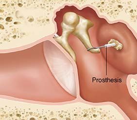
Back to Case Presentation

 Surgery: Stapedotomy

Most patients with conductive hearing loss Stapes Prosthesis
from otosclerosis are good surgical candidates.

Preferably, the patient would have air-bone
gap of >20dB and a good speech
discrimination.

Surgery for otosclerosis has evolved from total
extraction of the footplate (stapedectomy) to
creating a small hole in the stapes footplate
(stapedotomy). Prosthesis connecting the the
incus to the inner ear allows sound vibration to
be transmitted and corrects the conductive
hearing loss.
 Continue to Quiz
 Back to treatments
Back to Case Presentation

Quiz – Q1
Interpret the audiogram below:
 a. Normal hearing

 b. Unilateral sensorineural hearing loss

 c. Asymmetrical conductive hearing loss

 d. Bilateral sensorineural hearing loss

 e. Bilateral conductive hearing loss
Back to Case Presentation

 Quiz – Q1 Try again

 Interpret the audiogram below:
 a. Normal hearing

 Incorrect. Normal audiogram:

Review: Interpreting Audiograms
Back to Case Presentation

 Quiz – Q1 Try again

 Interpret the audiogram below:
 b. Asymmetrical sensorineural hearing loss

 Incorrect. Asymmetrical sensorineural hearing
 loss audiogram:
 (*Note air conduction = bone conduction)

Review: Interpreting Audiograms
Back to Case Presentation

 Quiz – Q1
 Interpret the audiogram below:
 a. Normal hearing

 b. Unilateral sensorineural hearing loss

 c. Unilateral conductive hearing loss

 d. Bilateral sensorineural hearing loss

 e. Bilateral conductive hearing loss

 Correct! This patient has normal
 hearing in their right ear and conductive
 hearing loss in their left ear.
Review: Interpreting Audiograms
 Proceed to Q2
Back to Case Presentation

 Quiz – Q1 Try again

 Interpret the audiogram below: d. Bilateral sensorineural hearing loss
 Incorrect. Bilateral sensorineural hearing loss
 audiogram:
 (*Note air conduction = bone conduction)

Review: Interpreting Audiograms
Back to Case Presentation

 Quiz – Q1 Try again

 Interpret the audiogram below:
 e. Bilateral conductive hearing loss
 Incorrect. Bilateral conductive hearing loss
 audiogram:
 (*Note air conduction does NOT equal bone
 conduction in both ears)

Review: Interpreting Audiograms
Back to Case Presentation
Review: Interpreting an Audiogram
 Low Pitch High Pitch
 Air conduction testing:
 Frequencies • Sound delivered through headphones or loudspeakers,
 being tested tests outer, middle, and inner ear.
 • Left ear = X
 • Right ear = O
 • Different symbols are used when the “masking” is used.
 Soft

 Masking refers to noise presented to the non-test ear to
 Normal Hearing prevent it from hearing sound presented to the test ear.
 • Left ear =
 • Right ear = △
 Mild Hearing Loss
 Bone conduction testing:
 • Bone vibrator placed behind the ear to deliver sound
 Moderate Hearing Loss vibrations to the cochlea, bypassing the outer and middle
 ear.
 Moderately Severe Hearing Loss • Left ear = >
 • Right ear = <
 Severe Hearing Loss • Masking symbols [< >]
 • Left ear = ]
 • Right ear = [

 Profound Hearing Loss
 Loud

 Click to practice reading audiograms
How loud the sound needs
to be, in order to be heard
 Back to Q1
at that frequency
Back to Case Presentation
Review: Interpreting an Audiogram Back
 Air conduction testing compared to
 masked bone conduction testing.

 • Bone conduction: within normal
 Unmasked air conduction range
 audiogram. • Air conduction: mild – moderate
 hearing loss
 Interpretation:
 Normal hearing Interpretation:
 Conductive hearing loss.
 (I.e. Middle ear pathology)

 Right ear: Unmasked air and bone
 conduction testing
 Left ear: Masked air and bone
 conduction testing

 Interpretation:
 Masked air conduction Asymmetrical sensorineural hearing
 audiogram. loss.
 (I.e. Acoustic neuroma)
 Interpretation: Moderately
 severe hearing loss at high • Right ear: mild sensorineural
 frequencies. hearing loss at higher
 (I.e. Presbycusis) frequencies
 • Left ear: Mild to moderately
 severe hearing loss as move up
 frequencies
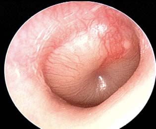
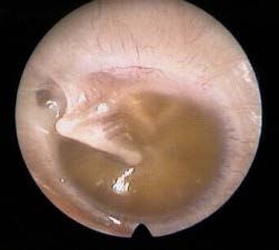
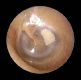
Back to Case Presentation

Quiz – Q2

Which of the following is a sign of severe otosclerosis on otoscopy?
Back to Case Presentation

 Quiz – Q2

 Which of the following is a sign of severe otosclerosis on otoscopy?
 Correct!

 Schwartze Sign Myringosclerosis Middle ear effusion Acute Otitis Media
Schwartze sign: Increased vascularity on the
promontory that is seen through the tympanic
membrane. This indicates active otosclerosis.
Only seen in ~10% of cases. Proceed to Q3
Back to Case Presentation

Quiz – Q2

Which of the following is a sign of severe otosclerosis on otoscopy?
 Incorrect – Please choose again

 Myringosclerosis

 Try again
Back to Case Presentation

Quiz – Q2

Which of the following is a sign of severe otosclerosis on otoscopy?
 Incorrect – Please choose again

 Middle ear effusion

 Try again
Back to Case Presentation

Quiz – Q2

Which of the following is a sign of severe otosclerosis on otoscopy?
 Incorrect – Please choose again

 Acute Otitis Media

 Try again
Back to Case Presentation

Quiz – Q3

Otosclerosis can only be confirmed at the time of surgery or through
histological analysis. What else is on the differential diagnosis for
progressive conductive hearing loss?
a. Osteogenesis Imperfecta
b. Presbycusis
c. Tympanosclerosis
d. Middle ear effusion
e. Meniere’s Disease
f. a, c, & d
g. b&e
Back to Case Presentation

Quiz – Q3

Otosclerosis can only be confirmed at the time of surgery or through
histological analysis. What else is on the differential diagnosis for
progressive conductive hearing loss?
a. Osteogenesis Imperfecta
b. Presbycusis Incorrect. Osteogenesis Imperfecta
c. Tympanosclerosis is on the DDx for otosclerosis,
d. Middle ear effusion however it is not the only correct
e. Meniere’s Disease answer. Please choose again.
f. a, c, & d
g. b&e

 Try again
Back to Case Presentation

Quiz – Q3

Otosclerosis can only be confirmed at the time of surgery or through
histological analysis. What else is on the differential diagnosis for
progressive conductive hearing loss?
a. Osteogenesis Imperfecta
b. Presbycusis
c. Tympanosclerosis Incorrect. Presbycusis is characterized by a
d. Middle ear effusion sensorineural hearing loss. Please choose again.
e. Meniere’s Disease
f. a, c, & d
g. b&e

 Try again
Back to Case Presentation

Quiz – Q3

Otosclerosis can only be confirmed at the time of surgery or through
histological analysis. What else is on the differential diagnosis for
progressive conductive hearing loss?
a. Osteogenesis Imperfecta Incorrect. Tympanosclerosis is on the
b. Presbycusis DDx for otosclerosis, but it is not the
c. Tympanosclerosis only correct answer.
d. Middle ear effusion
 When diagnosing progressive
e. Meniere’s Disease
 conductive hearing loss,
f. a, c, & d tympanosclerosis can typically be
g. b&e ruled in or out through findings on
 otoscopy (see right).

 Please choose again.
 Try again
Back to Case Presentation

Quiz – Q3

Otosclerosis can only be confirmed at the time of surgery or through
histological analysis. What else is on the differential diagnosis for
progressive conductive hearing loss?
 Incorrect. Middle ear effusion is on the DDx
a. Osteogenesis Imperfecta for otosclerosis, however it is not the only
b. Presbycusis correct answer.
c. Tympanosclerosis
d. Middle ear effusion When diagnosing progressive conductive
e. Meniere’s Disease hearing loss, middle ear effusion can
f. a, c, & d typically be diagnosed through otoscopy.
g. b&e On inspection of the tympanic membrane
 (TM), fluid or air-fluid levels behind the TM
 (see right).

 Please choose again. Try again
Back to Case Presentation

Quiz – Q3

Otosclerosis can only be confirmed at the time of surgery or through
histological analysis. What else is on the differential diagnosis for
progressive conductive hearing loss?
a. Osteogenesis Imperfecta
b. Presbycusis
c. Tympanosclerosis Incorrect. Meniere’s Disease is characterized by episodic
d. Middle ear effusion vertigo, sensorineural hearing loss, and tinnitus or aural
e. Meniere’s Disease fullness in the affected ear. Please choose again.
f. a, c, & d
g. b&e

 Try again
Back to Case Presentation

Quiz – Q3

Otosclerosis can only be confirmed at the time of surgery or through
histological analysis. What else is on the differential diagnosis for
progressive conductive hearing loss?
a. Osteogenesis Imperfecta Correct! Osteogenesis imperfecta, tympanosclerosis, and
b. Presbycusis middle ear effusion are on the DDx for otosclerosis.
c. Tympanosclerosis
d. Middle ear effusion Pathologies that impede sound transmission through the
e. Meniere’s Disease middle ear are on the DDx for otosclerosis. Other conditions
 on the DDx include:
f. a, c, & d
 • Chronic otitis media, with or without cholesteatoma
g. b&e • Trauma
 • Neoplasms of the middle ear or external auditory canal

 Proceed to Q4
Back to Case Presentation

Quiz – Q3

Otosclerosis can only be confirmed at the time of surgery or through
histological analysis. What else is on the differential diagnosis for
progressive conductive hearing loss?
a. Osteogenesis Imperfecta
b. Presbycusis
c. Tympanosclerosis Incorrect. Presbycusis and Meniere’s Disease are
d. Middle ear effusion associated with sensorineural hearing loss. Please choose
e. Meniere’s Disease again.
f. a, c, & d
g. b&e

 Try again
Back to Case Presentation

Quiz – Q4

What do you expect to find on the Weber and Rinne tests in someone
with left-sided otosclerosis (air bone gap >25dB)?
a. Weber: Lateralizes to left ear; Rinne both ears: AC > BC
b. Weber: No lateralization; Rinne both ears: AC > BC
c. Weber: No lateralization; Rinne both ears: BC > AC
d. Weber: Lateralizes to left ear; Rinne left ear: BC > AC

 Incorrect. This pattern is characteristic of Review: Interpreting
 sensorineural hearing loss in the right ear. Rinne and Weber tests.
 With the presenting air-bone gap, one
 would expect the Rinne test to be negative

 Try again
Back to Case Presentation

Quiz – Q4

What do you expect to find on the Weber and Rinne tests in someone
with left-sided otosclerosis (air bone gap >25dB)?
a. Weber: Lateralizes to left ear; Rinne both ears: AC > BC
b. Weber: No lateralization; Rinne both ears: AC > BC
c. Weber: No lateralization; Rinne both ears: BC > AC
d. Weber: Lateralizes to left ear; Rinne left ear: BC > AC

 Incorrect. This pattern is
 Review: Interpreting
 characteristic of normal hearing.
 Rinne and Weber tests.

 Try again
Back to Case Presentation

Quiz – Q4

What do you expect to find on the Weber and Rinne tests in someone
with left-sided otosclerosis (air bone gap >25dB)?
a. Weber: Lateralizes to left ear; Rinne both ears: AC > BC
b. Weber: No lateralization; Rinne both ears: AC > BC
c. Weber: No lateralization; Rinne both ears: BC > AC
d. Weber: Lateralizes to left ear; Rinne left ear: BC > AC

 Incorrect. This pattern is characteristic of Review: Interpreting
 conductive hearing loss in the both ears. Rinne and Weber tests.

 Try again
Back to Case Presentation

Quiz – Q4

What do you expect to find on the Weber and Rinne tests in someone
with left-sided otosclerosis (air bone gap >25dB)?
a. Weber: Lateralizes to left ear; Rinne both ears: AC > BC
b. Weber: No lateralization; Rinne both ears: AC > BC
c. Weber: No lateralization; Rinne both ears: BC > AC
d. Weber: Lateralizes to left ear; Rinne left ear: BC > AC

 Correct! This pattern is characteristic Review: Interpreting
 of left-sided conductive hearing loss. Rinne and Weber tests.

 Continue
Back to Case Presentation

Review: Weber and Rinne tests
 1. Weber test
 • Strike a 512 Hz tuning fork and place on top of the patient’s head
 • A patient with normal hearing should hear the sound equally on both sides* (I.e. the sound shouldn’t lateralize to one ear)
 • *Note: A Rinne test is needed to confirm normal hearing, as a patient with bilateral conductive hearing loss would also
 have no lateralization of sound.

 2. Rinne test
 • Strike a 512 Hz tuning fork and place it on the mastoid bone behind the patient’s ear (Testing bone conduction (BC))
 • When the patient signals that they can no longer hear the sound, move the tuning fork next to the patients external auditory
 canal (Testing air conduction (AC))
 • A patient with normal hearing should hear the sound better through air conduction (AC > BC)
 • Rinne test might not be negative if the conductive hearing loss is very mild

Results from the Weber and Rinne test can be used to determine the type of hearing loss:

 Conductive Hearing Sensorineural Hearing
 Test Normal
 Loss Loss

 Weber Sound heard in Sound heard in Sound heard in good
 midline affected ear ear

 Rinne AC > BC BC > AC AC > BC

 Back to Q4
Back to Case Presentation

Review: Weber and Rinne tests
 1. Weber test
 • Strike a 512 Hz tuning fork and place on top of the patient’s head
 • A patient with normal hearing should hear the sound equally on both sides* (I.e. the sound shouldn’t lateralize to one ear)
 • *Note: A Rinne test is needed to confirm normal hearing, as a patient with bilateral conductive hearing loss would also
 have no lateralization of sound.

 2. Rinne test
 • Strike a 512 Hz tuning fork and place it on the mastoid bone behind the patient’s ear (Testing bone conduction (BC))
 • When the patient signals that they can no longer hear the sound, move the tuning fork next to the patients external auditory
 canal (Testing air conduction (AC))
 • A patient with normal hearing should hear the sound better through air conduction (AC > BC)
 • Rinne test might not be negative if the conductive hearing loss is very mild

Results from the Weber and Rinne test can be used to determine the type of hearing loss:

 Conductive Hearing Sensorineural Hearing
 Test Normal
 Loss Loss

 Weber Sound heard in Sound heard in Sound heard in good
 midline affected ear ear

 Rinne AC > BC BC > AC AC > BC

 Back
Back to Case Presentation

 Congratulations! You have finished the hearing loss module.
Key points to remember:

• Otosclerosis is an autosomal dominant condition with incomplete penetrance.

• Typically presents in the 2nd to 4th decades of life
 • Unilateral or bilateral progressive conductive hearing loss Schwartze Sign
 • May experience tinnitus
 • Rare to experience vertigo
 • Hearing loss may be exacerbated by pregnancy

• Diagnosis
 • History
 • Audiometry: conductive hearing loss
 • Otoscopy: typically normal
 • Active cases: Hyperaemia of cochlear promontory (Schwartze sign) – 10% of patients
 • Temporal bone CT scan: Hypodense demineralized plaques at fissula ante fenestram
 • *Note: CT scan is not necessary for diagnosis of otosclerosis
 • Note: definitive diagnosis can only be made at the time of surgery or through histological analysis

• Three treatment options:
 • Observation
 • Hearing aid
 • Surgery

 Continue or return to the Review sections.
Back to Case Presentation
Module Review Sections
 Physical Exam: Investigations:

 Cranial Nerve Exams Audiograms

 Weber & Rinne tests Acoustic Reflex

 Otoscopy Tympanometry

 Imaging:

 Temporal bone CT scan

 Continue
Back to Case Presentation
 Review: Cranial Nerve Exams Back to Review
 Cranial Nerve Function Test

CNI – Olfactory Nerve • Smell • Test for sense of smell with coffee, alcohol swab, citrus, etc.
CN II – Optic Nerve • Vision • Visual acuity – Snellen’s eye chart
 • Visual fields – Confrontation testing
 • Pupillary reflexes – Direct and consensual response
 • Fundoscopy
CN III – Oculomotor Nerve • Motor innervation to most* extra-ocular muscles • H test & convergence
 • Pupillary reflex
CN IV – Trochlear Nerve • Motor innervation to superior oblique muscle* • H test : Look for ability to look ”down and out”

CN V – Trigeminal Nerve • Sensory innervation to the face • Corneal reflex
 • Motor innervation to muscles of mastication • Test sensory supply to the face – cotton swab
 • Test strength of muscles of mastication
CN VI – Abducens Nerve • Motor innervation to lateral rectus muscle* • H test: Look for ability to abduct eye

CN VII - Facial Nerve • Motor innervation to muscles of facial expression • Ask patient to do different facial expressions
 • Taste – anterior 2/3 tongue • Corneal reflex
CN VIII – Vestibulocochlear Nerve • Hearing & balance • Weber & Rinne tests

CN IX – Glossopharyngeal Nerve • Sensory innervation to the palate • Gag reflex
 • Taste – posterior 1/3 tongue • Say “Ahhh” – look for deviation of the uvula
CN X – Vagus Nerve • Motor supply to the pharynx • Gag reflex
 • PSNS supply to abdominal viscera
CN XI – Spinal Accessory Nerve • Motor innervation to trapezius and sternocleidomastoid • Shoulder shrug against resistance, head turn against resistance
CN XII – Hypoglossal Nerve • Motor supply to muscles of the tongue • Stick tongue out and move tongue from side-to-side
Back to Case Presentation

Review: Weber and Rinne tests Back to Review

 1. Weber test
 • Strike a 512 Hz tuning fork and place on top of the patient’s head
 • A patient with normal hearing should hear the sound equally on both sides* (I.e. the sound shouldn’t lateralize to one ear)
 • *Note: A Rinne test is needed to confirm normal hearing, as a patient with bilateral conductive hearing loss would also
 have no lateralization of sound.

 2. Rinne test
 • Strike a 512 Hz tuning fork and place it on the mastoid bone behind the patient’s ear (Testing bone conduction (BC))
 • When the patient signals that they can no longer hear the sound, move the tuning fork next to the patients external auditory
 canal (Testing air conduction (AC))
 • A patient with normal hearing should hear the sound better through air conduction (AC > BC)
 • Rinne test might not be negative if the conductive hearing loss is very mild

Results from the Weber and Rinne test can be used to determine the type of hearing loss:

 Conductive Hearing Sensorineural Hearing
 Test Normal
 Loss Loss

 Weber Sound heard in Sound heard in Sound heard in good
 midline affected ear ear

 Rinne AC > BC BC > AC AC > BC
Back to Review Back to Case Presentation

 Review: Otoscopy Examples of Abnormal TMs

 Normal Otoscopic Exam Findings:
 • Tympanic membrane (TM) should be intact and
 appear pearly grey or whitish/pinkish grey

 Bulging, red TM - Acute Otitis Media Cholesteatoma
 Normal TM – Right Side

Pars flaccida
 Lateral/short process of malleus

 Incus Manubrium of malleus

 Umbo Otitis media with effusion Myringosclerosis
 Cone of light
 Pars tensa
 Annulus

 Hemotympanum Retracted TM
Back to Case Presentation
Review: Interpreting an Audiogram Back to Review
 Low Pitch High Pitch
 Air conduction testing:
 Frequencies • Sound delivered through headphones or loudspeakers,
 being tested tests outer, middle, and inner ear.
 • Left ear = X
 • Right ear = O
 • Different symbols are used when the “masking” is used.
 Soft

 Masking refers to noise presented to the non-test ear to
 Normal Hearing prevent it from hearing sound presented to the test ear.
 • Left ear =
 • Right ear = △
 Mild Hearing Loss
 Bone conduction testing:
 • Bone vibrator placed behind the ear to deliver sound
 Moderate Hearing Loss vibrations to the cochlea, bypassing the outer and middle
 ear.
 Moderately Severe Hearing Loss • Left ear = >
 • Right ear = <
 Severe Hearing Loss • Masking symbols [< >]
 • Left ear = ]
 • Right ear = [

 Profound Hearing Loss
 Loud

 Click to practice reading audiograms
How loud the sound needs
to be, in order to be heard
at that frequency
Back to Case Presentation
Review: Interpreting an Audiogram Back to Review
 Air conduction testing compared to
 masked bone conduction testing.

 • Bone conduction: within normal
 Unmasked air conduction range
 audiogram. • Air conduction: mild – moderate
 hearing loss
 Interpretation:
 Normal hearing Interpretation:
 Conductive hearing loss.
 (I.e. Middle ear pathology)

 Right ear: Unmasked air and bone
 conduction testing
 Left ear: Masked air and bone
 conduction testing

 Interpretation:
 Masked air conduction Asymmetrical sensorineural hearing
 audiogram. loss.
 (I.e. Acoustic neuroma)
 Interpretation: Moderately
 severe hearing loss at high • Right ear: mild sensorineural
 frequencies. hearing loss at higher
 (I.e. Presbycusis) frequencies
 • Left ear: Mild to moderately
 severe hearing loss as move up
 frequencies
Back to Case Presentation
 Review: Acoustic Reflex Testing Back to Review

The acoustic reflex is the reflexive contraction of the stapedius muscle, and subsequent stiffening of the tympanic
membrane (TM), in response to high-intensity sound or vocalization. Anatomy reminder: Stapedius is innervated
by CN VII.

In acoustic (stapedial) reflex testing, acoustic signals at varying frequencies (usually 500, 1000, or 2000 Hz) are
introduced into one ear and the acoustic impedance is measured in the both ears.

Acoustic Reflex Threshold (ART): Sound pressure level (SPL), in dB, from which a sound stimulus with a given
frequency will elicit the acoustic reflex.
 Frequency tested
 Reflexes may be absent or harder to
 illicit in patients with:
 • Conductive hearing loss
 • Severe sensory hearing loss
 Normal hearing: • CN8 injury on side receiving sound
 ART ~70-100 dB SPL • CN7 injury on side being measured

 Reflexes may also be absent if there is
 fixation of the ossicles.
Back to Case Presentation

 Review: Tympanometry Back to Review

 Tympanometry is an indirect test of middle ear function by the transmission/reflection of sound
 energy. A tympanogram plots compliance changes of the tympanic membrane (TM) versus air
 pressure in the external auditory canal.

 High peak = hypercompliant TM
 1
 • I.e. Ossicular discontinuity, monomeric TM (thin TM from
 healed TM perforation)
 0.8
 Normal tympanogram

 0.6 Shallow peak = stiff TM
Compliance (ml)

 • I.e. Otosclerosis, tympanosclerosis
 0.4
 No peak = non-mobile TM
 0.2 • I.e. Effusion, perforation

 0
 Peak shifted to a more negative pressure = retracted TM
 -400 -200 0 200 400 • I.e. Eustachian tube dysfunction, TM atelectasis
 Pressure at which peal compliance occurs (decaPascals)
Back to Case Presentation

 Review: Reading a Temporal Bone CT Scan Back to Review

 Normal temporal bone CT scan – Coronal plane

 Magnified right temporal bone – Coronal plane
 Cochlea
Tip: Look for the
“ice cream cone”
 Malleus
 Malleus

 Incus
 Internal Incus
 IAC
 Auditory
 Canal
 (IAC) Footplate
Semicircular Canal of stapes
 at the
 Mastoid oval
 Air Cells window

 Stapes
Back to Case Presentation

Module Authors
• Kylen Van Osch, Meds 2020 & Peng You MD
• Module adapted from: Jason Beyea MD PhD FRCSC

 You may now exit, return to Review sections or retake the Quiz
You can also read