Aesculap Barré-Instruments - Instruments for Radical prostatectomy
←
→
Page content transcription
If your browser does not render page correctly, please read the page content below
Aesculap® Barré-Instruments
Instruments for Radical prostatectomy

Surgical Instruments

Barré-Instruments

2
Aesculap Surgical Instruments

     Content

1. Introduction                                                                     4

2. General recommendations                                                          5

3. Surgical technique                                                               6
           3.1 Incision and exposure                                                6
           3.2 Exposure of the prostate apex                                        6
           3.3 Preservation of the striated sphincter                               7
           3.4 Nerve sparing                                                        10
           3.5 Dissection of the seminal vesicles and section of the bladder neck   12
           3.6 Excision of the seminal vesicles                                     13
           3.7 Visual inspection of the specimen                                    13
           3.8 Vesicourethral anastomosis                                           14

4. References                                                                       15

5. Instruments                                                                      16

6. Index                                                                            26

                                                                                                                    3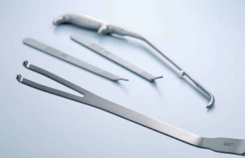
Barré-Instruments

    Introduction

                    Surgery in Motion
                    "Open Radical Retropubic Prostatectomy"
                    Dr. Christian Barré, Clinique Jules Verne, Nantes, France

                    Eur Urol. 2007 Jul;52(1):71-80. Epub 2006 Dec 11.

                    We describe a surgical procedure for radical retrobubic
                    prostatectomies that we have used in a prospective series
                    of 231 patients with localised prostate cancer (mean age
                    63 yr; range 46-75 yr). Nerve-sparing was performed in
                    148 of 231 patients. We insist on three points: (1) high-
                    quality preservation of the sphincter, with the sphincter
                    divided to keep its anatomic environment intact; (2) high-
                    precision retrograde dissection of the neurovascular bund-
                    les in the mid-gland prostate zone for early recovery of
                    erectile function (the dissection of the bundles is described
                    for each prostate zone [apex, mid-gland, base]); and (3) the
                    need to standardise each step of the procedure for a repro-
                    ducable protocol independent of individual patient anato-
                    my. These three measures will ensure the best cancer
                    control with the best functional results. The rate of positive
                    margins for patients with organ-confined (pT2) cancer was
                    3.7% (2.4% related to an invaded striated sphincter). The
                    quality of the excised specimens was further checked by
                    noting the frequency of capsular incision (2.6%) and of
                    inked benign prostatic glands (2.6%). By 12 mo, 94% of
                    patients were fully continent and 70.5% had recovered the
                    ability to maintain an erection for satisfactory sexual inter-
                    course without the need for medication.

4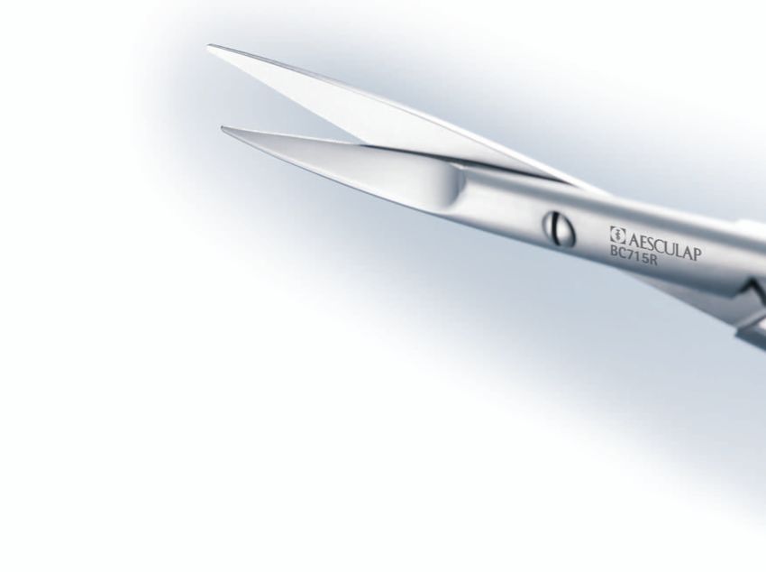
Aesculap Surgical Instruments

 General recommendations

Parameter             Recommendation

Patient positioning   Place patient in dorsal decubitus position.
                      Tilt the operating table at umbilicus level.
                      Above the umbilicus, the patient's body
                      should be in the Trendelenburg position
                      forming an angle of about 30° with the lower
                      part of the body which remains horizontal [7].

Instruments           Use specialised instrument: prostatic
                      retractor, urethral retractor, dorsal vein
                      complex clamp, nerve-sparing dissector,
                      nerve-sparing scissors, long, angled scalpels
                      (0°, 12°, 25°).

Bleeding control      Maintain mean arterial BP between 55 and
                      65 mm Hg during nerve sparing for a blood-
                      less operating field if no cardiovascular or
                      hepatic.

                                                                                                  5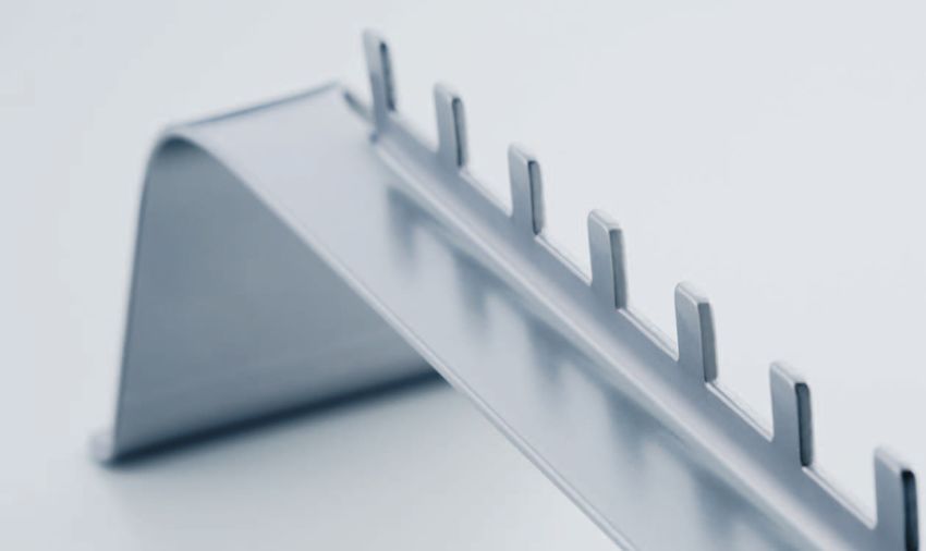
Barré-Instruments

     Surgical technique                                            Exposure of the prostate apex

We describe the steps of an RP protocol developed on          ❙ Make an incision in the endopelvic fascia extending
the basis of a personal series of >1500 RPs and charact-        from the prostate base to the puboprostatic ligament
erised by: (1) high-quality preservation of the sphincter       (Fig. 1a). Transect the puboprostatic ligaments.
within its anatomic environment, using a surgical knife       ❙ Free the levator ani muscle fibres at the prostate apex.
and not scissors for high-precision dissection; (2) retro-      This is a more delicate manoeuvre because of the
grade dissection of the neurovascular bundles; and (3)          depth of dissection, the thickness of thefibres, and the
a standard procedure foreach step of the RP, thus yiel-         presence of venous pedicles originating from the pel-
ding a reproducible protocol independent of individual          vic sidewall (Fig. 1b and c).
patient anatomy.                                              ❙ Dissect similarly on both sides.

     Incision and exposure

❙ Perform a conventional incision and exposure.
  Perform a bilateral pelvic lymphadenectomy except in
  patients with low Gleason grade biopsies (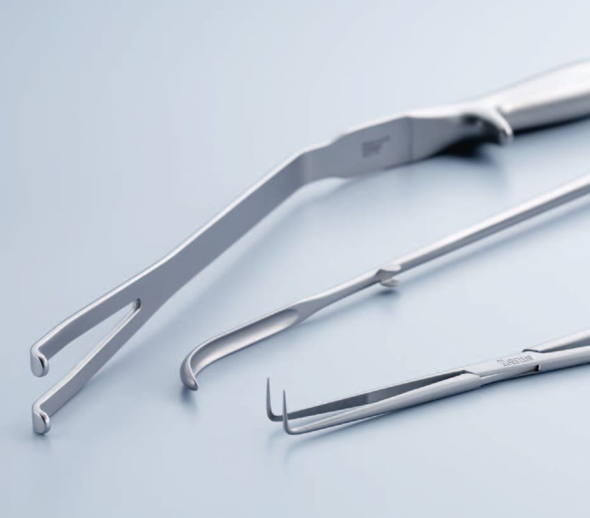
Aesculap Surgical Instruments

     Preservation of the striated sphincter

The striated urethral sphincter is located at the centre of
an anatomic unit [9] ("sphincteral complex") encased by
the dorsal vein complex, lateral pelvic fascia and Denon-
villiers’ fascia (Fig. 2a). The urethra should not be disso-
ciated from this environment as any dissection isolating
an urethral tube from adjoining structures will weaken
the sphincter. The sphincteral complex is divided in five
stages: the superficial then deep part of the dorsal vein
                                                               Fig. 2a Beginning of dorsal vein complex section
complex which covers the sphincter fibres, the ventral
then dorsal half of the urethra, and finally Denonvilliers’
fascia.

❙ Stop backbleeding with an x-shaped stitch
  (absorbable suture 0) over the anterior surface
  of the prostate.
❙ Slide the blades of a prostatic retractor (BT680R)
  along each side of the dorsal vein complex.
❙ Clamp the dorsal vein (15°-angled clamp EF167R)
  over only 1.5 cm to be above the striated sphincter.
  The remaining dorsal vein complex covering the
  sphincter fibres should be below the clamp. [Fig. 2b].

                                                               Fig. 2b

                                                                                                                             7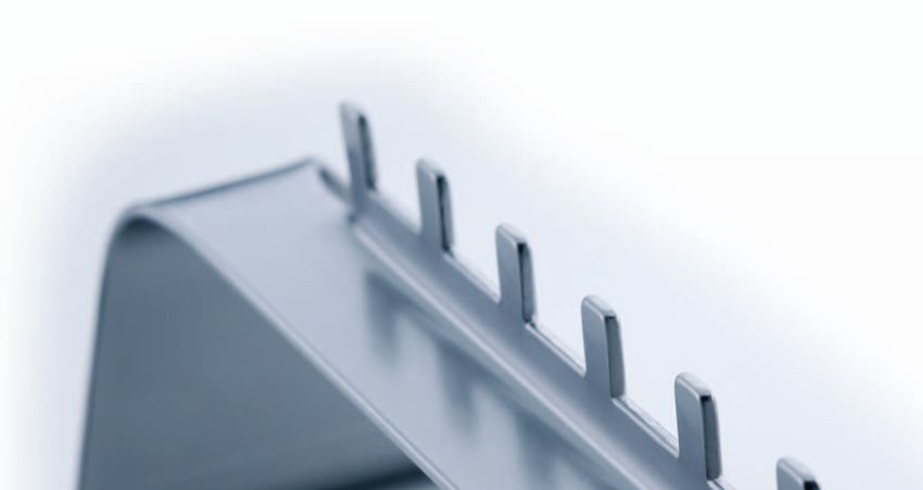
Barré-Instruments

     Preservation of the striated sphincter

❙ Divide the dorsal vein complex with a sharp surgical                     The incision should be made with great care and stop
  knife (BB176R - BB178R) [Fig. 2c]. When two thirds of                    as soon as the muscle fibres are visible. The roof of
  the section is complete, displace the prostatic retractor                the sphincter, with its fibres moving up towards the
  a few centimetres back against the anterior surface of                   prostate apex, should now be perfectly exposed.
  the prostate. Exert gentle pressure on the retractor to                  Make a crown-shaped 4/0 running suture on the
  obtain a horizontal urethral plane and thus good                         dorsal vein complex and on each side of the lateral
  exposure [Fig. 2d, 2e].                                                  pelvic fascia [Fig. 3b].
❙ Control bleeding by a continuous U-shaped suture                       ❙ Divide the urethra at 1 mm from the apex, down to
  behind the clamp [Fig. 2f].                                              the catheter, leaving a little muscle over the apex
❙ Divide the remaining part of the dorsal vein complex                     [Fig. 3c, 3d].
  and the lateral pelvic fascia covering the front and                   ❙ Grab the urethral catheter with forceps and bring its
  sides of the sphincter with an angled scalpel with a                     distal end into the surgical field. Clamp, then cut the
  rounded blade. Start the incision medially and proceed                   catheter to provide traction.
  towards the lateral pelvic fascia until 1 mm from the                  ❙ Remove the prostatic retractor and place a urethral
  prostate apex [Fig. 3a].                                                 retractor to expose the urethra. Gently push the pro-
                                                                           state backwards and tighten the urethral mucosa.

    Fig. 2c                                          Fig. 2d                                            Fig. 2e

    Fig. 2f                                          Fig. 3a Completion of dorsal vein                  Fig. 3b
                                                             complex section and anterior
                                                             sphincter section

8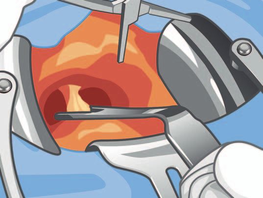
Aesculap Surgical Instruments

  Place 3-0 absorbable sutures as landmarks on the               In cases of posterior overhang [10], follow the con-
  anterior edge of the urethra in anticipation of anato-         tours of the prostate apex with care.
  mosis [Fig. 3e]. Gentle traction on the retractor            ❙ Incise the remaining layers of Denonvilliers’ fascia
  straightens the urethra and exposes the urethral               transversally with a pointed bistoury, a few milli-
  mucosa buldges over the posterior sphincter fibres.            meters from the apex, in a narrow midline position
❙ Divide the urethral mucosa, submucous chorion, and             in order not to injure the nerves located laterally
  smooth muscle (of variable thickness) with a pointed           [Fig. 4c].
  blade, by tracking the posterior striated fibres of the      ❙ Expose the median rectal prostate plane with
  sphincter (Fig. 4a). Once divided, they slide over the         Metzenbaum scissors, leaving the Denonvilliers' fascia
  plane of the striated fibres and retract. The fibres are       on the prostate [11]. If this plane, which is crucial for
  shaped like a ‘‘U’’ with the bottom of the ‘‘U’’ inserted      initiating nerve sparing, is difficult to find, as when
  in Denonvilliers fascia.                                       Denonvilliers' fascia is stuck to the pre-rectal fascia,
❙ Divide the posterior sphincter fibres of the prostate          free the apex over a few millimetres and then expose
  apex with a rounded blade. This must remove the                the plane.
  superficial layers of Denonvilliers’ fascia to which the
  sphincter fibres are attached [Fig. 4b].

   Fig. 3c                                           Fig. 3d                                     Fig. 3e

   Fig. 4a Posterior sphincter section               Fig. 4b                                     Fig. 4c

                                                                                                                                        9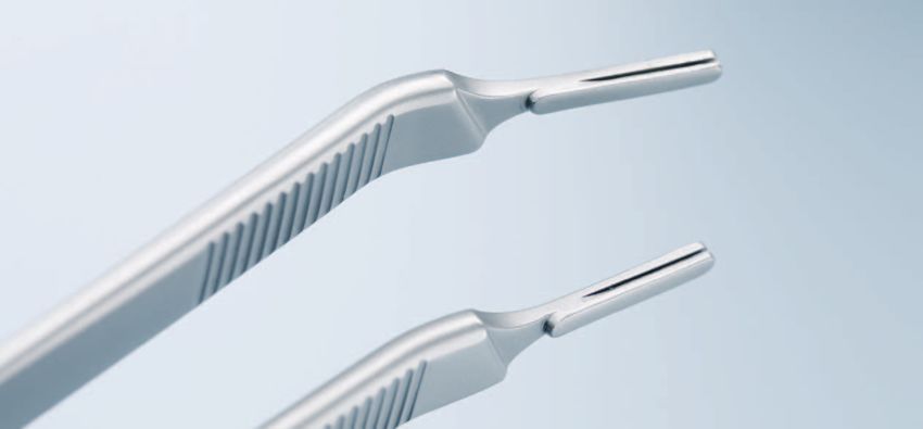
Barré-Instruments

      Nerve sparing

The neurovascular bundles run along the vascular pedi-               There are two nerve-sparing techniques:
cles coming from the terminal branch of the inferior                 ■ Antegrade dissection [Fig. 5b] starts at the lateral
vesical artery. As noted by Walsh, the nerves follow the               surface of the prostate [14,16], proceeds along the
vessels which act as a guide to dissection [1]. However,               posterolateral contour, and ends at the posterior
the vessels do not follow the posterolateral prostate                  edge. There is a degree of uncertainty associated
contour in a straight line but curve up its lateral surface.           with this technique as dissection can start either
The bundles lie between the parietal and the visceral                  above the neurovascular bundle (risk of creating
fasciae (lateral extension of Denonvilliers' fascia) [1]               intrafascial dissection) or below the bundle (risk of
[Fig. 5a]. Dissection should take place in this interfascial           injury to the nerves).
space. A thin layer of connective tissue should be left on           ■ Retrograde dissection [Fig. 5c] starts at the posterior
the prostate to prevent the risk of positive margins,                  surface of the prostate. The medial border of the
especially in cases of unsuspected focal extracapsular                 bundle is exposed after the plane between the rectum
extension (ECE) [12,13]. The visceral fascia should be                 and the prostate in the midline has been developed.
present on the excised specimen.                                       ❙ Dissect along the posterolateral surface of the
Although it is easier to preserve the bundles intact by                  prostate following Denonvilliers' fascia, then the
intrafascial dissection, this is not recommended as there                lateral prostate visceral fascia. This exposes and
is a high risk of positive margins from dissection in con-               isolates all the prostatic pedicles coming from
tact with gland tissue [Fig. 5a].                                        the neurovascular bundles. The bundles are thus
                                                                         progressively freed from the prostate up to their
                                                                         extremity on the lateral surface.
                                                                       ❙ Apply very gentle traction on the urethral catheter
                                                                         and gently roll the prostate over on its side to
                                                                         obtain good exposure of the neurovascular bund-
                                                                         les. Use tailor-made nerve-sparing dissector
                                                                         (BJ02R) and scissors (BC715R) for dissection.

     Fig. 5a Surgical anatomy of prostate fasciae [23].
             Surgical approach to sparing the neurovascular bundle

            p.f.       parietal fascia or levator ani fascia
            l.p.v.f.   lateral prostate visceral fascia
            p.p.v.f.   posterior prostate visceral fascia or
                       Denonvilliers' fascia

            Arrow 1 intrafascial dissection
            Arrow 2 interfascial dissection
            Arrow 3 extrafascial resection                              Fig. 5b Antegrade dissection        Fig. 5c Retrograde Dissection

10
Aesculap Surgical Instruments
The ease of dissection varies according to prostate ■ Base [Fig. 6 a3d]
zone [Fig. 6a]: In this zone, the neurovascular bundle pedicles are
■ Apex [Fig. 6a1b] further away from the prostate and travel towards
The risk of nerve injury is low in this zone as the the pelvic floor. It is easier to leave some connective
nerves lie in an external plane. The divided pedicles tissue when dividing each pedicle.
correspond mainly to the lateral attachment points
of Denonvilliers fascia and the levator fibres that Dissection ends once the posterior surface of the
were not released when freeing the apex. seminal vesicle, covered with the Denonvilliers’ fascia,
■ Mid-gland [Fig. 6 a2c] emerges.
This zone holds the greatest risk of injury to the
nerves and of positive margins. Free, then incise the This dissection technique has simplified our indications
parietal fascia overlying the nerve bundle. for nerve-sparing surgery. The decision to resect the
Expose the small prostate vessels by exerting very neurovascular bundle on the side with a 4 + 3 Gleason
gentle traction on the bundle. Isolate each prostatic score [17] or with a lobe induration on digital rectal
vessel, millimetre by millimetre, always leaving a thin examination is always made preoperatively. The decision
layer of connective tissue on the visceral fascia. to resect the neurovascular bundle [18] is made intra-
Should the visceral fascia have been inadvertently operatively in cases of
incised, dissect immediately 1 or 2 mm further away fibrosis of the prostate
with the tip of fine pointed scissors. Ensure haemo- fasciae that bar safe
stasis with small titanium ligature clips. Because at dissection and in patients
the top of the curve formed by the vessels, the nerves in whom nomograms
are in close contact with the visceral fascia, there predict a high risk of cap-
is hardly any space for dissection. The nerves may sule breach and in whom
sometimes have to be left over 1 or 2 mm to avoid insufficient connective
penetrating the visceral fascia. The quality of dissec- tissue can be left for the
tion in the mid-gland zone is critical for early sake of safety.
recovery of erectile function and to prevent the risk
of possible positive surgical margins. Binocular loops
may prove useful but have not been tested. Fig. 6a Three dissection zones:
a1b apex, a2c mid-gland, a3d base
Fig. 6b Fig. 6c Fig. 6d
11Barré-Instruments
Dissection of the seminal vesicles and division of the bladder neck
❙ Divide the thick prostate base pedicles covering the ❙ Divide both lateral vesicoprostatic junctions using the
lateral surface of the seminal vesicle. tightened surgical loop as a guide (Fig. 7c). Each divi-
❙ Use forceps to grab the seminal vesicle. Develop the sion starts with the retrovesical fat and comes gradu-
plane between the seminal vesicle and the posterior ally into contact with the posterior surface of the
bladder neck with Metzenbaum scissors, whilst remai- bladder neck. Join up both divisions on the anterior
ning in contact with the seminal plane (Fig. 7a), pro- surface of the bladder neck.
ceeding as far as possible. ❙ Perform the anterior vesicoprostatic division,
❙ Interrupt the procedure on the right-hand side. leaving a thin layer of bladder tissue on the prostate
Perform nerve sparing and seminal vesicle dissection base to avoid the risk of positive surgical margins.
on the left-hand side in the same fashion. Proceed with the incision until the Foley catheter is
❙ Push back the prostate to expose the posterior surface exposed. Incise the posterior bladder neck wall,
of the seminal vesicles. Insert a dissector inside the pushing back a median lobe if necessary [Fig. 7d].
seminal vesicles/bladder neck plane, leaving a surgical ❙ In cases of high-grade lesions of the prostate base,
loop as a landmark [19] [Fig. 7b]. it is preferable to resect the bladder neck [20,21].
❙ Push the prostate towards the lower part of the
surgical field.
Fig. 7a Dissection of seminal vesicles. Fig. 7b
Bladder neck section.
Excision of seminal vesicles
Fig. 7c Fig. 7d
12Aesculap Surgical Instruments
Excision of the seminal vesicles Visual inspection of the specimen
Traction of the prostate by the Foley catheter exposes ❙ Check the quality of the excision macroscopically,
the seminal vesicles and the vas deferens covered with paying special attention to the apex. There must be a
the thin anterior sheet of Denonvilliers’ fascia [Fig. 7e]. very small ring of sphincter muscle fibres around the
❙ Transect and ligate each vas deferens as far distally as urethra and the Denonvilliers’ fascia must be identifi-
possible from the prostate. able posteriorly.
❙ Remove the seminal vesicles in their entirety. ❙ Check that there is a small layer of connective tissue
In most cases, the tip of the seminal vesicles is above at the posterolateral edge indicating absence of cap-
the plane of the neurovascular bundles and there is sular incision.
no risk of injury to the nerves. In some cases, it is
necessary to tighten the specimen to move the lower
extremity of the seminal vesicles away from risk of
injury. Dissect in contact with the seminal vesicles,
neither too deep nor too wide. This may be a bit
awkward but can be done in patients with long
vesicles with tips extending beneath the plane of
the neurovascular bundles.
❙ Further haemostasis may be necessary notably on
the bladder neck. However, haemostasis near the
neurovascular bundles can cause irreversible neuro-
logical injury and no coagulation should be performed
at this level.
Fig. 7e
13Barré-Instruments
Vesicourethral anastomosis
❙ Check bladder neck opening. Use a linear posterior ❙ Join the edges together without excessive tension
"tennis racket" closure for wide bladder neck to prevent tearing or ischemia of the sphincter.
openings. Insert the urethral catheter and inflate the balloon.
❙ Insert a CH 16 Foley catheter to locate the urethral Tie all five posterior stitches on the inside in a
lumen before and after each suture passage. tensionless knot (Fig. 8b).
❙ Tighten both the anterolateral landmark sutures. ❙ Join the edges together without excessive tension to
Place 2 further anterior stitches (3-0 absorbable prevent tearing or ischaemia of the sphincter.
suture) from the outside in and 5 posterior stitches Insert the urethral catheter and inflate the balloon.
from the inside out, within a plane anterior to the Tie all four anterior sutures.
rectal plane of the neurovascular bundles (22) [Fig. Check that the anastomosis is watertight by filling
8a]. Whenever the urethra adjoins the rectal plane, the bladder with 120 cc saline.
avoid including the posterolateral angle of the urethra ❙ Position a suction drain on the anterior surface of the
by placing 2 stitches on each side, away from the bladder, avoiding direct contact with the anastomosis.
angle. Perform a conventional closure.
❙ Pass vesical sutures and bring the bladder smoothly in
contact with the urethra by sliding it along the poste-
rior sutures. Perform urethrovesical stitching. Tie all 5
posterior stitches on the inside in a tensionless knot
[Fig. 8b].
Fig. 8a Urethrovesical anastomosis Fig. 8b Urethrovesical anastomosis
14Aesculap Surgical Instruments
References
[1] Walsh P. Anatomic radical retropubic prostatectomy. [17] Sokoloff MH, Brendler CB. Indications and contraindications for
In: Walsh P, Retik A, Vaughan E, Wein A, eds. Campbell’s Urology. nerve sparing radical prostatectomy.
Philadelphia: Saunders; 1998. p. 2565-88. Atlas Urol Clin Nth Am 2001;28:535-43.
[2] Villers A. Extracapsular tumor extension in prostatic cancer: [18] Villers A, Stamey TA, Yemoto C, Rischmann P, McNeal JE.
pathways of spread and implications for radical prostatectomy. Modified extrafascial radical retropubic prostatectomy tech-
Monographs in Urology 1994;15:61-77. nique decreases frequency of positive surgical margins in T2
[3] Rosen MA, Goldstone L, Lapin S, Wheeler T, Scardino PT. cancersBarré-Instruments
The performance and progress of open retropubic approach in radical prostatectomy
Radical prostatectomy is one of the standard treatments even more significant. Success in cancer control and
for localised prostate cancer. However, this surgery is function preservation requires a detailed knowledge
still one of the most difficult in the field of urology be- of surgical anatomy and a rigorous surgical technique.
cause it has to achieve two objectives: reducing positive To improve the dorsal venous plexus control and ureth-
margins rates and retaining postoperative continence ral division and to allow a precise interfacial dissection
and erectile function. The increasing incidence of pro- in nerve sparing, Dr. Barré has designed and created
state cancer at young adults makes these objectives specific instruments which considerably simplify the
surgical technique.
16Aesculap Surgical Instruments
BT680R
❙ Prostate retractor
❙ 360 mm, 14 3/8”
BT680R
17Barré-Instruments
EF167R
❙ Dorsal vein complex clamp
❙ 210 mm, 8 1/4”
EF167R
18Aesculap Surgical Instruments
BB176R-BB178R
❙ No. 3 XL
❙ 250 mm, 10”
12 ° 25 °
BB176R BB178R BB177R
19Barré-Instruments
BT681R
❙ Urethra retractor
❙ 390 mm, 15 1/2”
BT681R
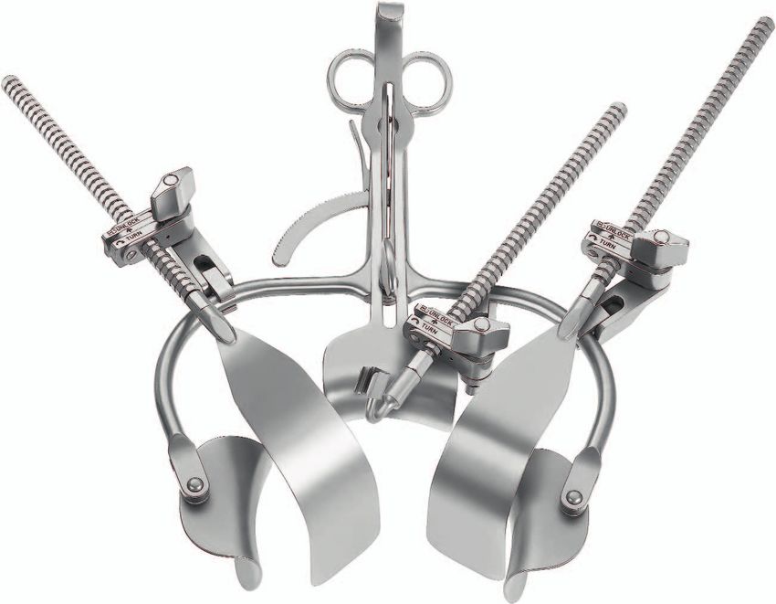
BJ029R
❙ Nerve sparing dissector
❙ 280 mm, 11”
1/
1
BJ029R
20Aesculap Surgical Instruments
BC715R sharp/sharp
❙ Nerve-sparing scissors
❙ 280 mm, 11”
1/
1
BC715R
21Barré-Instruments
BV927R
BV927R
22Aesculap Surgical Instruments
Barré Prostatectomy Retractor
BV921R
consisting of:
Quantity Product code Description
1 BV922R BARRÉ Retactor only
1 BV924R Pair of Blades with ball snap
closure, 50 x 70 mm
3 BV849R Connecting clamps
3 BV840R Notch rods
1 BV925R Fiber Light holder, for fiber
light carrier OP801R
2 BV926R Lateral Blade malleable,
190 x 50 mm
1 BV929R Center Blade, 54 x 70 mm
BV840R
BV922R
BV929R
BV849R
BV925R
BV926R BV921R
BV924R
23Barré-Instruments
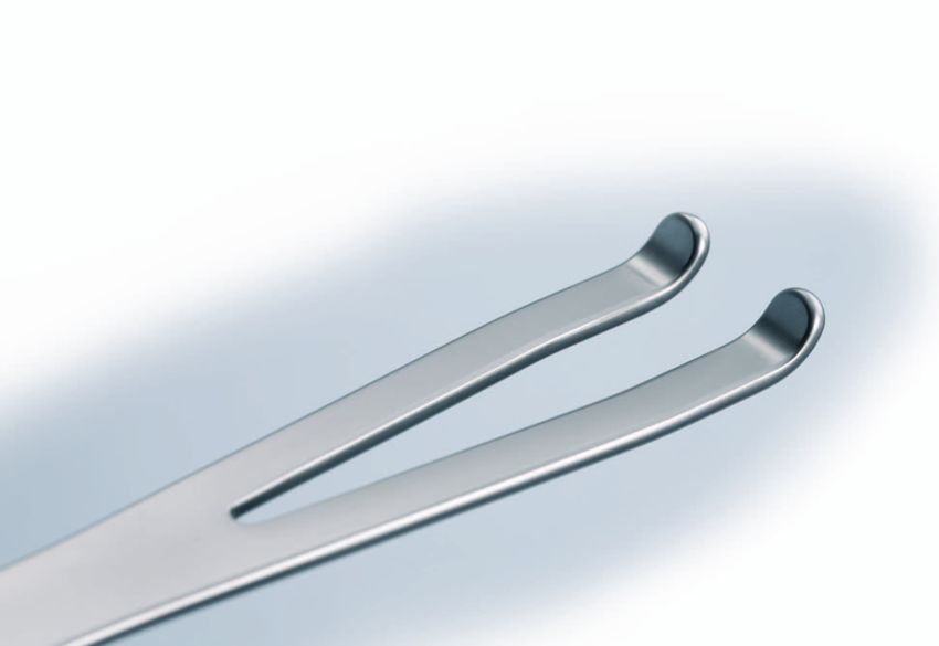
BT682R BT683R
❙ Dissecting Hook ❙ Dissecting Hook
❙ 260 mm, 10 1/4” ❙ 260 mm, 10 1/4”
1/ 1/
1 1
BT682R BT683R
24Aesculap Surgical Instruments
Additional Instruments
BJ110R GK685R
❙ Nerve sparing dissector ❙ Bipolar forceps
❙ 280 mm, 11” ❙ 260 mm, 10 1/4”
1/ 1/
1 1
BJ110R GK685R
25Index
Art. no. Page Art. no. Page
BB176R 19 EF167R 18
BB177R 19
BB178R 19 GK682R 24
GK683R 24
BC715R 21 GK685R 25
BJ029R 20
BJ110R 25
BT680R 17
BT681R 20
BV840R 23
BV849R 23
BV921R 23
BV922R 23
BV924R 23
BV925R 23
BV926R 23
BV927R 22
BV929R 23
26Aesculap Surgical Instruments
Notice
27The main product mark ’Aesculap’ is a
registered mark of Aesculap AG.
Subject to technical changes. All rights reserved.
This brochure may only be used for the exclusive
Aesculap AG | Am Aesculap-Platz | 78532 Tuttlingen | Germany purpose of obtaining information about our
products. Reproduction in any form partial or
Phone +49 0 74 61 95-0 | Fax +49 0 74 61 95-26 00 | www.aesculap.com otherwise is not permitted.
Aesculap – a B. Braun company Brochure No. C82802 1110/1/3You can also read