Embryonic Development of Siberian Sturgeon Acipenser baerii under Hatchery Conditions: An Image Guide with Embryological Descriptions
←
→
Page content transcription
If your browser does not render page correctly, please read the page content below
Original Article
                                                                                                              Fish Aquat Sci 16(1), 15-23, 2013

Embryonic Development of Siberian Sturgeon Acipenser
baerii under Hatchery Conditions: An Image Guide with
Embryological Descriptions
Chulhong Park, Sang Yoon Lee, Dong Soo Kim and Yoon Kwon Nam*
Department of Marine Bio-Materials & Aquaculture, Pukyong National University, Busan 608-737, Korea

Abstract
Normal embryonic development at a constant temperature (18°C) has been described for the Siberian sturgeon Acipenser baerii
(Acipenseriformes). Hormone-induced spawning and artificial insemination were performed to prepare embryonic batches for
embryologic examination. After insemination, early cleavages of the Siberian sturgeon embryos continued for 7 h post-fertilization
(HPF), showing the typical pattern of uneven holoblastic cleavage. Blastulation and gastrulation began at 9 HPF and 19 HPF,
respectively. Epiboly formation (2/3 covered) was observed at 25 HPF during gastrulation. Neurulation was initiated with the for-
mation of a slit-like neural groove from the blastopore at 33 HPF. During neurulation, the primary embryonic kidney (pronephros)
and s-shaped heart developed. The embryos underwent progressive differentiation, which is typical of Acipenseriform species. A
mass hatching was observed at 130 HPF, and the average total length of the hatched prolarvae was 10.5 mm. The hatched prolarvae
possessed a typical pigment plug (yolk plug). The results of this study are valuable not only as a reference guide for the artificial
propagation of Siberian sturgeon in hatcheries but also as the basis for the derivation of developmental gene expression assays for
this species.

Key words: Acipenser baerii, Siberian sturgeon, Embryonic development, Uneven holoblastic cleavage

Introduction

   The Acipenseriformes (Chondrostei) represent a group of                        genetics and genomic studies (Cho et al., 2007; Kim et al.,
primitive, ray-finned fish, comprising Acipenseridae (stur-                       2009; Akbarzadeh et al., 2011).
geons) and Polyodontidae (paddlefishes) (Bemis et al., 1997;                         In addition to the interest in their evolutionary aspects,
Birstein et al., 1997). They are often referred to as ‘living                     sturgeons have long been recognized as very valuable natural
fossils’ of the actinopterygian lineage and have distinctive                      resources for fisheries in Northern Hemisphere countries, par-
morphologic and/or developmental attributes (Billard and                          ticularly with regard to caviar production. However, over the
Lecointre, 2001). Furthermore, some sturgeon species exhibit                      last few decades, wild sturgeon stocks have faced considerable
repeated rounds of genome duplication, resulting in diversi-                      threats to their natural habitats as a result of various anthropo-
fication into different ploidy levels among sturgeon species                      genic and industrial activities (Pikitch et al., 2005). Currently,
(Blacklidge and Bidwell, 1993; Kim et al., 2005). Based on                        all extant sturgeon species are considered to be ‘critically en-
these unique and interesting characteristics, this extant primi-                  dangered’ and are included in the Convention on International
tive fish group is regarded as a useful model for evolutionary                    Trade in Endangered Species (CITES) list, which applies to

 Open Access      http://dx.doi.org/10.5657/FAS.2013.0015                         Received 5 November 2012;       Revised 23 November 2012
This is an Open Access article distributed under the terms of the Creative        Accepted 12 January 2013
Commons Attribution Non-Commercial License (http://creativecommons.
org/licenses/by-nc/3.0/) which permits unrestricted non-commercial use,           *Corresponding Author
distribution, and reproduction in any medium, provided the original work
is properly cited.                    pISSN: 2234-1749 eISSN: 2234-1757           E-mail: yoonknam@pknu.ac.kr

Copyright © The Korean Society of Fisheries and Aquatic Science              15                                                    http://e-fas.org
Fish Aquat Sci 16(1), 15-23, 2013

global trading. Thus, sustainable, aquaculture-based produc-          At 12 h after the primary injection, each female was injected
tion has become mandatory for the commercial exploitation             again with LHRHa at 90-135 μg/kg BW as a resolving dose.
of sturgeons and their products (Karpinsky, 2010; Webb and            The male broodfish were injected with LHRHa at 100 μg/kg
Doroshov, 2011).                                                      BW. The temperature of the water during the induced spawn-
   In Korea, the introduction of sturgeon species into the            ing was adjusted to 15.0 ± 0.5°C in both years. After injection,
aquaculture domain was first noted in 1999 for the Siberian           the fish were incubated in individual spawning tanks until a
sturgeon Acipenser baerii (Seong and Baik, 1999). Early suc-          low number of ovulated eggs was released, which usually oc-
cessful production of the Siberian sturgeon fingerlings using         curred around 36 h after the second injection of hormone.
adult brooders on Korean fish farms was achieved by both pri-
vate and nonprofit organizations in the early 2000s (personal         Artificial insemination and egg incubation
communication). These pioneering works have encouraged
the progressive expansion of Siberian sturgeon culturing in              When ovulation followed by spawning was identified, milt
Korea over the past decade, although the efficiency and ca-           was collected from the males using a silicon tube-connected
pacity of farming practices for Siberian sturgeon remain to be        aspirator and stored at 2-4°C until used. After milt collection,
improved. The establishment of effective guidelines for em-           the eggs were removed from the spawned females by hand
bryonic and larval development is one of the most important           stripping and Caesarean section. The eggs were mixed with
prerequisites for developing an optimal protocol for reproduc-        milt (diluted 1:200 with water) for 150 s, and then rinsed with
tive control in hatchery management. Despite its importance,          fresh water. To remove the adhesiveness of the fertilized eggs,
comprehensive information on the embryonic development of             the eggs were mixed continuously with Fuller’s earth (Sigma-
the Siberian sturgeon, guided by a complete set of relevant           Aldrich, St. Louis, MO, USA) and washed with fresh water
image data, has not become available to date, with the excep-         several times for 30 min. The fertilized eggs were placed in
tions of brief and general descriptions in the literature. The        McDonald incubation jars until hatched. Dead embryos were
objective of the present study was to provide a comprehensive         removed at 6-h intervals. The water temperature was main-
image-based guide to the embryologic development of Sibe-             tained at 18.0 ± 0.5°C until hatching.
rian sturgeons that are artificially propagated under hatchery
conditions.                                                           Sampling of biological specimens for develop-
                                                                      mental staging

Materials and Methods                                                    In 2011, the development of embryos from two mating
                                                                      pairs was examined. Based on a modified version of the devel-
Broodfish and hormonal artificial spawning                            opmental stages defined for Russian sturgeon, Acipenser gul-
                                                                      denstadtii (Dettlaff and Vassetzky, 1991), the developmental
   The first experiment for the morphologic staging of em-            stages of the Siberian sturgeon embryos were assigned to 30
bryonic development was performed in March 2011, and this             stages, ranging from just-fertilized to hatching (Table 1). After
experiment was replicated in March 2012. The broodfish used           artificial insemination, the embryos were collected every hour
for artificial spawning and gamete collection were individuals        until the beginning of gastrulation, every 2 h until the onset
that were produced in 2003. The broodfish were maintained at          of heart beating, and every 4-6 h until the first occurrence of
ambient temperature (range, 12 to 20°C) at Dinoville Aqua-            hatching. From each mating group, 30-50 embryos were sam-
farm Inc. (Hamyang, Korea). Fish were sexed by external go-           pled at each detection point and fixed in cold 4% paraformal-
nad biopsy at 6 years of age. Out of six (in 2011) or four (in        dehyde. Under the stereomicroscope (AZ100; Nikon, Tokyo,
2012) female candidates, four (average body weight [BW], 14           Japan), the embryos that had reached each stage were record-
± 2.6 kg) and three individuals (13 ± 4.6 kg BW) were selected        ed. Based on the examination in 2011, developmental prog-
for hormonal induction of spawning in 2011 and 2012, respec-          ress to each stage was confirmed with embryo batches from
tively. When necessary, polarization index (PI) observations          two independent mating pairs in 2012, and the morphologic
using catheterized oocytes were conducted (PI scores < 0.1)           characteristics of the developing embryos at each stage were
(Van Eenennaam et al., 1996). In addition, four males each for        analyzed using the NIS-Elements BR image analysis software
2011 (7 ± 2.7 kg BW) and 2012 (7 ± 3.1 kg BW) were selected           (Nikon), which was implemented in the AZ100 microscope.
based on the presence of milt, which was obtained by either
palpation of the abdomen or catheterization using a silicon
tube-connected syringe into the genital duct. The selected fe-        Results
males were administered the primary intramuscular injection
of the luteinizing hormone-releasing hormone analogue des-            Early cleavage events
Gly10, [D-Ala6] LH-RH ethylamide (LHRHa; Syndel Labora-
tories Ltd., Qualicum Beach, BC, Canada) at 10-15 μg/kg BW.             The developmental staging results are listed in Table 1.

http://dx.doi.org/10.5657/FAS.2013.0015                          16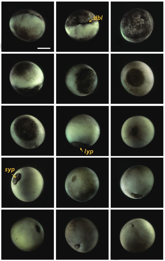
Park et al. (2013) Embryonic Development of Siberian Sturgeon

Upon fertilization, a black pigment developed at the animal                    sphere was completely divided into four similarly sized com-
pore, and the first cleavage furrow appeared at the animal pore                partments (Fig. 1I). When the 16 blastomeres were formed in
at 2 h post-fertilization (HPF) (Fig. 1A). This cleavage fur-                  the animal hemisphere (at 5 HPF; fourth cleavage), one half
row was limited to the animal hemisphere (Fig. 1B and 1C).                     (newly formed) of the 16 blastomeres in the center of the ani-
Following the first cleavage (1 h later), the second cleavage                  mal hemisphere were much smaller than the remaining half
formed typical 4-cell (four equal-sized blastomeres) embryos                   (earlier ones) (Fig. 1J-1L). The sizes and shapes of the blasto-
(Fig. 1D and 1E). Although the second cleavage furrow did                      meres that resulted from subsequent cleavages in the animal
not divide completely the vegetal hemisphere, partial infil-                   hemisphere were noticeably dissimilar (Fig. 1M and 1N), and
tration of the vegetal hemisphere was observed (Fig. 1F).                      the furrows in the vegetal hemisphere were also formed in an
At 4 HPF, 8-cell embryos (similarly sized blastomeres) were                    irregular manner (Fig. 1O). This irregular pattern of division
formed in the animal hemisphere through the third cleavage                     continued in both the animal (Fig. 1P and 1Q) and vegetal
event (Fig. 1G and 1H). At that time-point, the cleavage in-                   (Fig. 1R) hemispheres.
filtrated the entire vegetal hemisphere and the vegetal hemi-

Table 1. Staging of embryonic development in Siberian sturgeon Acipenser baerii
         Stages                               Descriptions                                         Time at 18°C (h)          Figures
            1              Fertilization                                                                   0                 -
            2              First cleavage (two cells in animal hemisphere)                                 2                 Fig. 1B
            3              Second cleavage (four-cells in animal hemisphere)                               3                 Fig. 1E
            4              Third cleavage (eight-cells in animal hemisphere; furrows                       4                 Fig. 1G and 1I
                            into vegetal hemisphere)
            5              Sixteen cells in animal hemisphere; different sizes of                          5                 Fig. 1L
                             blastomeres
            6              Fifth cleavage in animal hemisphere; irregular                                  6                 Fig. 1M and 1O
                             blastomeres; about 8-partitioned in vegetal hemisphere
            7              Irregular pattern of cleavages continues in both animal                         7                 Fig. 1P-1R
                             hemisphere and vegetal hemisphere
            8              Early phase of blastula                                                         9                 Fig. 2A-2D
            9              Late phase of blastula                                                         11                 Fig. 2E
           10              Onset of gastrulation                                                          19                 Figs. 2F and 3A
           11              Typical dorsal blastopore lip formed                                           20                 Fig. 3B
           12              Two-third embryo covered by animal materials (epiboly)                         25                 Fig. 3D
           13              Formation of large yolk plug                                                   28                 Fig. 3H
           14              Formation of small yolk plug                                                   30                 Fig. 3J
           15              Gastrulation almost complete                                                   32                 Fig. 3N and 3O
           16              Onset of neurulation (slit-like formed in neural groove)                       33                 Fig. 4B
           17              Wide neural plate formed                                                       34                 Fig. 4C and 4D
           18              Folded structure in head region                                                35                 Fig. 4E and 4F
           19              Rudimentary excretory system faintly seen                                      37                 Fig. 4G
           20              Pronephros rudiments evident and pronephros elongates                          40                 Fig. 4I-4L
           21              Tail region thickened and pronephroi perpendicular to neural tube              46                 Fig. 4O and 4P
           22              Round-shaped head; rudimentary eyes visible; pronephros                        55                 Fig. 5A-5C
                            distinct; somites formed
           23              Rudimentary heart visible and tail rod-shaped                                  59                 Fig. 5D and 5E
           24              Straightened heart elongated; each somite distinguishable;                     70                 Fig. 5G-5I
                             a pair of typical pronephros wings seen
           25              Heart s-shaped and onset of heart beating                                      73                 Fig. 5L
           26              Tail straightened; rudimentary fin bud in caudal region;                       86                 Fig. 6A-6D
                            triangular-shaped head; s-shape of heart pronounced
           27              Tail approaches s-heart and eye pigmentation evident                           94                 Fig. 6E and 6F
           28              Fully straightened tail reaches head; developed fin bud;                      101                 Fig. 7A-7C
                            embryos capable of movement
           29              First occurrence of advanced hatch                                            119                 Fig. 7D-7F
           30              Mass hatch                                                                    130                 Fig. 7G and 7H

                                                                         17                                                     http://e-fas.org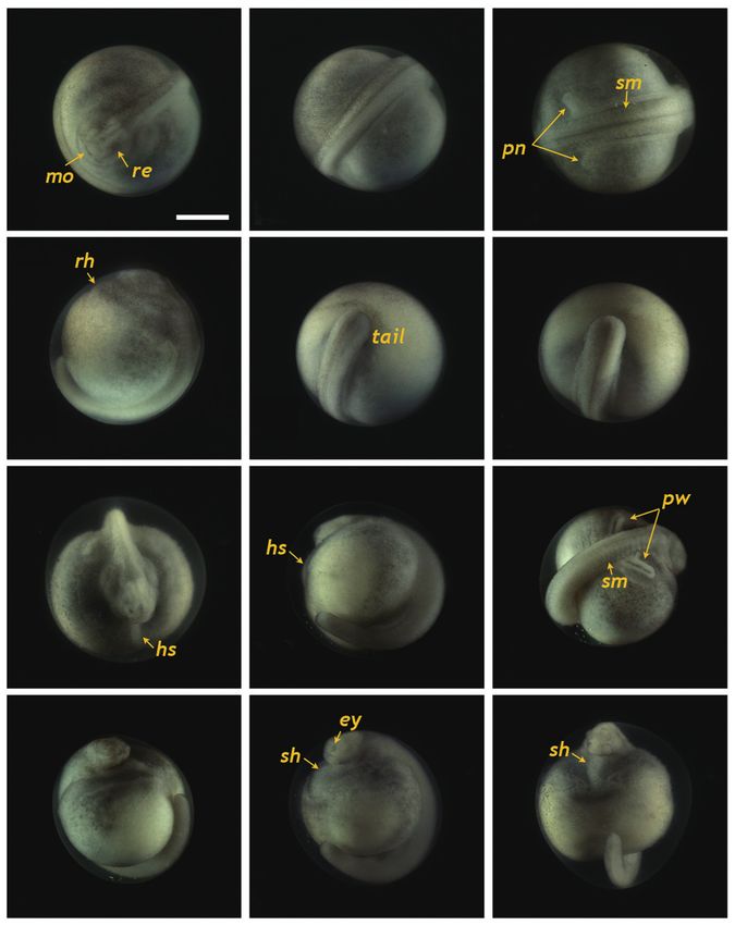
Fish Aquat Sci 16(1), 15-23, 2013

  A                        B                       C                                 A                        B                        C

  D                        E                       F                                 D                        E                        F

  G                       H                        I                               Fig. 2. Developmental progress of Siberian sturgeon Acipenser baerii
                                                                                   embryos to early blastula (A-D) and late blastula (E) with evident cleavage
                                                                                   cavity in the animal hemisphere. The embryo at the close to the onset of
                                                                                   gastrulation is shown in (F). Scale bar: A = 1 mm (A-F).

  J                        K                       L                               etal hemisphere (Fig. 2D). Cell division meant that the blas-
                                                                                   tomeres became indistinguishable from one another in the
                                                                                   animal hemisphere under the microscope at low-power mag-
                                                                                   nification, and a cleavage cavity (blastocoel) began to form at
                                                                                   the apex of the animal hemisphere (9 HPF). Meanwhile, in the
                                                                                   vegetal hemisphere, relatively small blastomeres were located
  M                       N                        O                               near the marginal zone (the equator), while larger blastomeres
                                                                                   were evident in the vegetal apex region. At 11 HPF, the pri-
                                                                                   mordial cleavage cavity became larger and more evident in
                                                                                   the animal hemisphere, whereas cell division continued to
                                                                                   generate smaller blastomeres in the vegetal hemisphere (Fig.
                                                                                   2E). Nearing the completion of blastulation (18-19 HPF), the
  P                        Q                       R                               smooth appearance of the animal hemisphere changed to a
                                                                                   milky white blastula roof (Fig. 2F).

                                                                                   Gastrulation

                                                                                      Upon the onset of gastrulation (19-20 HPF), a band was
Fig. 1. Early cleavages of Siberian sturgeon Acipenser baerii embryos.             formed close to the equator, and this was followed by the for-
(A-C) First cleavage to form two cells. (D, E) Second cleavage in animal           mation of a “dorsal blastopore lip” between the animal and
hemisphere (four cells). (F) Lateral view of four-celled embryo showing
the partial infiltration of cleavage furrow into the vegetal hemisphere.           vegetal hemispheres (Fig. 3A and 3B). At this time-point, the
(G, H) Eight cells in animal hemisphere. (I) Vegetal view of the embryos           vegetal hemisphere contained a number of divided blasto-
showing eight cells in the animal hemisphere. (J-L) Embryos showing                meres of differing sizes, whereby relatively large, countable
sixteen cells in animal hemisphere. (M-O) Irregular blastomeres formed             (distinguishable) blastomeres were present in the region close
after fifth cleavage in the animal hemisphere (animal view, lateral view
and vegetal view, respectively). (P-R) Continued cleavages in animal (P, Q)        to the apex and smaller (often uncountable) blastomeres were
and vegetal (R) hemispheres. Developmental time for each stage can be              detected in the region close to the marginal zone (Fig. 3C).
referred to Table 1. Scale bar: A = 1 mm (A-R).                                    The blastula roof of the animal hemisphere enveloped pro-
                                                                                   gressively the vegetal hemisphere. At 25 HPF, approximately
                                                                                   two-thirds of the embryo were covered by blastoderm (Fig.
Blastulation                                                                       3D and 3E), and a darkly pigmented region was formed in a
                                                                                   round shape at the surface of the animal pore in many, albeit
   As cleavage continued, small blastomeres proliferated in                        not all, of the embryos (Fig. 3F). As gastrulation continued,
the animal hemisphere (Fig. 2A), concomitant with irregu-                          epiboly covered more than two-thirds of the embryos, and
lar divisions in the vegetal hemisphere (Fig. 2B). The blas-                       the area of remaining vegetal material declined progressively
tomeres in the animal hemisphere became uncountable (Fig.                          (Fig. 3G). At 28 HPF, a large yolk plug was formed, and the
2C), although large blastomeres were still formed in the veg-                      darkly pigmented region at the animal pore decreased (Fig. 3H

http://dx.doi.org/10.5657/FAS.2013.0015                                       18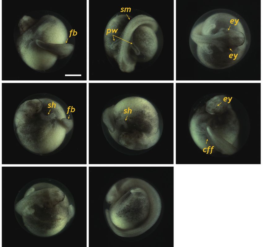
Park et al. (2013) Embryonic Development of Siberian Sturgeon

  A                       B                       C                                A                         B                        C

  D                       E                       F                                D                         E                        F

  G                       H                       I                                G                         H                        I

  J                       K                       L                                 J                        K                        L

  M                       N                       O                                M                         N                        O

Fig. 3. Gastrulation stages of Siberian sturgeon Acipenser baerii                  P
embryos. (A, B) Onset of gastrulation with the formation of dorsal
blastopore lip (dbl). (C) Vegetal view of dbl-formed embryo. (D-F) Two-
third covered epiboly in lateral, vegetal and animal views, respectively.
(G) Further covering of vegetal hemisphere by animal material in epiboly.
(H) Large yolk plug (lyp) formation at the apex of vegetal hemisphere.
(I) Animal view of the embryo with a large yolk plug. (J) Small yolk plug
(syp) formation. (K) Animal view of the embryo with a small yolk plug.
(L-N) Progressive covering to form a blastopore. (O) Completion of the           Fig. 4. Neurulation stages of Siberian sturgeon Acipenser baerii
                                                                                 embryos. (A, B) Onset of neurulation with a slit-like neural groove (ng). (C,
gastrulation. Scale bar: A = 1 mm (A-O).
                                                                                 D) Formation of neural plate. (E, F) Folded structure in head region. (G)
                                                                                 Appearance of excretory rudiments. (H) Folded structure in tail region. (I,
                                                                                 J) Pronephros rudiments (pr) running in parallel to the neural groove. (K-
and 3I). About 2 h later, the size of the yolk plug was further                  N) Elongation of evident pronephroi (pn). (O, P) Thickened tail region and
reduced to less than one-fifth the diameter of the embryo (i.e.,                 pronephroi perpendicular to the neural tube. Scale bar: A = 1 mm (A-P).
small yolk plug formation) (Fig. 3J). The pigmented region at
the surface of the animal pore was also markedly reduced in
size and coloration (Fig. 3K). The size of the yolk plug gradu-                  neural groove in the blastopore (Fig. 4A and 4B), followed
ally decreased (Fig. 3L) until the blastopore appeared only as                   by the formation of the neural plate at the dorsal surface with
a small circle at the apex of the vegetal pore (Fig. 3M and 3N).                 the folded structure in the head region (33 HPF) (Fig. 4C and
As gastrulation neared completion (32 HPF), the blastopore                       4D). With the progression of development (33-35 HPF), the
took on a slit-like appearance, which signaled the initiation of                 neural plate in the head region widened and the neural folds
neurulation (Fig. 3O).                                                           rose and thickened (Fig. 4E and 4F). In the dorsal region, the
                                                                                 neural groove was more evident and the excretory system ru-
Neurulation                                                                      diments were faintly visible parallel to the neural groove (37
                                                                                 HPF) (Fig. 4G). At that time-point, a folded shape was seen in
   Neurulation began with the appearance of a slit-shaped                        the tail region (Fig. 4H). As neurulation continued, the folding

                                                                            19                                                               http://e-fas.org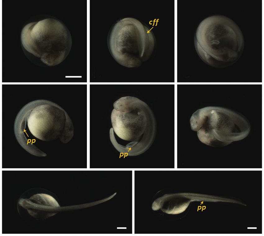
Fish Aquat Sci 16(1), 15-23, 2013

  A                       B                        C                                A                        B                       C

  D                       E                        F                                D                        E                       F

  G                       H                        I                                G                        H

   J                      K                        L
                                                                                  Fig. 6. Siberian sturgeon Acipenser baerii embryos at stage 26 (A-D) and
                                                                                  stage 27 (E-H). In stage 26, the tail was transformed to be straightened
                                                                                  structure with the rudimentary fin bud (fb) in caudal region. Somites
                                                                                  (sm), pronephros wings (pw), eye caps (ey) and s-heart (sh) were more
                                                                                  developed than previous stages. In stage 27, rudimentary fin bud was
                                                                                  further developed to caudal fin fold (cff) and tail approached the s-heart.
                                                                                  Developmental time for each stage can be referred to Table 1. Scale bar: A
                                                                                  = 1 mm (A-H).
Fig. 5. Developmental progress of Siberian sturgeon Acipenser baerii
embryos belonging to stages from 22 to 25. Developmental time for each
stage can be referred to Table 1. (A-C) Embryos at stage 22 showing the
round-shaped head, rudimentary eye (re), underdeveloped mouth (mo)                eyes and undeveloped mouth were easily visible (Fig. 5A). At
and somite (sm) formation. A pair of pronephroi (pn) became v-shaped.             this stage, the dorsal region had a rod-shaped appearance with
(D-F) Embryos at stage 23 characterized by the formation of rudimentary
                                                                                  developed somites, and the primary pronephros-wings were
heart as well as the rod-shaped tail that began to separate from yolk. (G-
I) Embryos at stage 24 displaying the heart straightened (hs) and well-           visible (Fig. 5B and 5C). A rudimentary heart formed in the
developed pronephros wing (pw). (J-L) Embryos at stage 25 showing the             embryo (59 HPF), although the head had not yet separated
s-shaped heart (sh) and evident eye caps (ey). Scale bar: A = 1 mm (A-L).         (Fig. 5D). In the caudal region, the flattened tail was trans-
                                                                                  formed to a rod-shaped structure and the tail had begun to
                                                                                  separate from the yolk sac (Fig. 5E and 5F). As development
                                                                                  continued (60-70 HPF), the head region thickened and began
of the head region was slightly reorganized and a pair of pro-                    to separate from the yolk, in which the heart was seen as a
nephros rudiments became more evident as cords running par-                       short straight tube (Fig. 5G). The tail continued to lengthen
allel to the neural groove (40 HPF) (Fig. 4I and 4J). The neural                  and pronounced somites were visible over the entire embry-
folds rose significantly and the anterior part of the pronephros                  onic body (Fig. 5H and 5I). At this point, a pair of highly dif-
was visualized as a distinct form (Fig. 4K and 4L). Thereafter,                   ferentiated pronephros-wings could be observed (Fig. 5I). At
the pronephros became elongated and thickened along with                          73 HPF, the heart became s-shaped (the so-called s-heart) and
the risen neural folds (Fig. 4M and 4N). The tail region con-                     began to beat. The eyes were more distinct in this stage (Fig.
tinued to thicken significantly as development progressed, al-                    5J-5L).
though it had not yet detached from the yolk sac membrane. At
this stage, the pronephroi were located almost perpendicular                      Progress to hatching
to the neural tube and the neural tube was mostly closed (46
HPF) (Fig. 4O and 4P).                                                               At 86 HPF, the round, rod-shaped structure of the tail
                                                                                  straightened, and fin-bud rudiments were visible in the caudal
Progress to the onset of heart beating                                            region (Fig. 6A). Other morphologic features typical of the
                                                                                  embryos at this stage were: separation of the anterior part of
   At 55 HPF, the lateral plates were fused to the prosenceph-                    the head from the yolk sac; the triangular shape of the devel-
alon (forebrain), and the head region appeared as a round-                        oped head; slightly pigmented eyes; and a more pronounced
shaped object in the dorsal view, in which the rudimentary                        s-heart (Fig. 6B-6D). At 94 HPF, the tail reached the beating

http://dx.doi.org/10.5657/FAS.2013.0015                                      20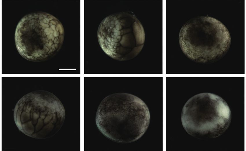
Park et al. (2013) Embryonic Development of Siberian Sturgeon

                                                                                 sturgeon embryos is different from the holoblastic cleavage
  A                       B                       C
                                                                                 seen in anuran embryos. This difference is attributable to the
                                                                                 uneven distribution of yolk constituents between the animal
                                                                                 and vegetal hemispheres in sturgeon embryos, a consequence
                                                                                 of which is a reduction of the cleavage rate in the vegetal
                                                                                 hemisphere, which contains more yolk content than the animal
                                                                                 hemisphere (Colombo et al., 2007).
  D                       E                       F
                                                                                    These differential rates of cleavages result in differently
                                                                                 sized and irregular-shaped blastomeres in the animal and
                                                                                 vegetal hemispheres at the beginning of blastulation. With
                                                                                 the progression of blastulation, a marginal zone of transition
                                                                                 between the animal and vegetal hemispheres is formed with
                                                                                 intermediate-sized blastomeres. In this zone, a “dorsal lip” ap-
  G                                   H
                                                                                 pears as a marker of the initiation of gastrulation in sturgeon
                                                                                 embryos (Bolker, 1993). Following the primary internaliza-
                                                                                 tion by involution, germ layers arise and the overall plan for
                                                                                 the body architecture is programmed during gastrulation. This
                                                                                 usually involves a complex set of morphogenetic movements
Fig. 7.    Siberian sturgeon Acipenser baerii embryos or larvae at stages
from 28 to 30. (A-C) Embryos at prehatching stage (stage 28) with                of cellular progenitors, syncytial yolk cells, and an extra-em-
well-developed caudal fin fold (cff). (D-F) Advanced hatchlings (stage           bryonic sheath (enveloping layers) (Cooper and Virta, 2007;
29) possessing pigment plug (pp). (G, H) Just hatch-out of normally              Shook and Keller, 2008). During gastrulation, the cell layers
developed embryos (stage 30). Developmental time for each stage can be
                                                                                 (i.e., the blastula roof) in the animal hemisphere progressively
referred to Table 1. Scale bars: A = 1 mm (A-F), G, H = 1 mm.
                                                                                 cover the embryos (i.e., formation of epiboly). The covered
                                                                                 embryo begins to show the “slit-like” neural groove from the
                                                                                 blastopore as a sign of neurulation, and this is soon followed
s-heart and fin folds appeared in the caudal region (Fig. 6E-                    by the formation of the neural plate at the dorsal surface, with
6H). Blood circulation was easily observable, and olfactory                      the neural folds in the head region. With the initial progress of
organs were also seen. As the tail continued to lengthen, it                     neurulation, head structuring and formation of the excretory
approached the head (101 HPF). Eye caps were evident and                         system are noticeable where the reorganized folding of the
caudal fin folds were easily distinguishable (Fig. 7A-7C). The                   head region and a pair of pronephros rudiments running paral-
embryos at this stage were capable of movement. From 119                         lel to the neural groove are visible. The pronephros is the first
HPF, several advanced embryos began to hatch, and mass                           kidney formed during development in vertebrates (so-called
hatching was observed at 130 HPF. The average hatching suc-                      embryonic kidney), and it often persists in hatchlings to play
cess based on duplicate examinations was 80.5%. Individuals                      essential roles in primary blood filtration and osmoregulation
from advanced hatches were often bent in shape (Fig. 7D-7F),                     in fish and amphibians (Drummond et al., 1998; Ichimura et
whereas most of the mass-hatched prolarvae (i.e., yolk-bear-                     al., 2012). Although functional pronephroi are found during
ing) had a straight shape (Fig. 7G and 7H). At hatching, the                     the early life stages of most fish types, the embryos of gan-
average total length of the prolarvae was 10.5 mm. They had                      oid fishes (e.g., sturgeons) usually display pronephroses that
a pigment plug (i.e., yolk plug), which is a typical feature of                  are morphologically more distinct than those of teleost fishes.
sturgeon prolarvae.                                                              With the progression of development, sturgeon embryos ac-
                                                                                 quire a pair of highly differentiated pronephros-wings. At the
                                                                                 later phase of neurulation, one of the most notable morphoge-
Discussion                                                                       netic changes is heart formation. A rudimentary heart is ini-
                                                                                 tially seen as a straight tube in Siberian sturgeon embryos, and
   Unlike many teleostean embryos that show the meroblastic                      this straightened structure becomes s-shaped (s-heart) at the
cleavages on the blastodisc exclusively in the animal hemi-                      onset of heart-beating. Thereafter, the embryos undergo fur-
sphere, sturgeon embryos display holoblastic cleavage (Conte                     ther differentiation into their morphologic structures, particu-
et al., 1988). In general, the holoblastic cleavage pattern ob-                  larly with respect to thickening of the head, lengthening of the
served in the present study is in agreement with those previ-                    caudal part, detachment of the tail from the yolk sac, appear-
ously defined for other Acipenseriform fishes (Dettlaff et al.,                  ance of the eye cap, and the formation of fin folds. Embryos
1993). In similarity with other sturgeon species, the Siberian                   at the prehatching stage are capable of movement. Hatch-out
sturgeon exhibits an uneven pattern of holoblastic cleavage in                   of the embryos is often signaled by the emergence of the tail
which the vegetal hemisphere is not completely divided with                      from the embryonic membrane. Several larvae that show ad-
each cleavage furrow. This asymmetrical cleavage pattern in                      vanced hatching remain bent in shape, unlike many normal

                                                                            21                                                     http://e-fas.org
Fish Aquat Sci 16(1), 15-23, 2013
larvae that have a straight shape. Colombo et al. (2007) have chem Physiol B Biochem Mol Biol 147, 178-190. http://dx.doi.
reported that such larvae of advanced hatchings of shovelnose org/10.1016/j.cbpb.2007.01.001.
sturgeon Scaphirhynchus platorynchus typically show no pig- Colombo RE, Garvey JE and Wills PS. 2007. A guide to the embryonic
ment in the eye cups and incomplete formation of the pigment development of the shovelnose sturgeon (Scaphirhynchus plato-
plug. However, in the present study, we found no differences rynchus), reared at a constant temperature. J Appl Ichthyol 23, 402-
in either the degree of pigmentation of the eye cups or the 410. http://dx.doi.org/10.1111/j.1439-0426.2007.00898.x.
formation of the yolk plug, which suggests that differences in Conte FS, Doroshov SI, Lutes PB and Strange EM. 1988. Hatchery
pigment accumulation patterns occur during embryonic devel- Manual for the White Sturgeon (Acipencer transmontanus Rich-
opment in the two sturgeon species. ardson) with Applications to Other North American Acipenseri-
In the present study, a complete set of images of normal dae. Publ. 3322. University of California Press, Oakland, CA, US.
embryonic development was prepared, together with embryo- Cooper MS and Virta VC. 2007. Evolution of gastrulation in the ray-
logic descriptions for 30 developmental stages of the Siberian finned (Actinopterygian) fishes. J Exp Zool B Mol Dev Evol 308,
sturgeon. Based on the results of this study, the effects of inte- 591-608. http://dx.doi.org/10.1002/jez.b.21142.
gral water temperature on the mitotic divisions and develop- Dettlaff TA and Vassetzky SG. 1991. Animal Species for Developmen-
ment of sturgeon might be examined in future studies. The tal Studies. Vol. 2. Vertebrates. Plenum Publishing, New York,
results of the present study could be useful in hatchery man- US.
agement for the reproductive control of the Siberian sturgeon, Dettlaff TA, Ginsburg AS and Schmalhausen OI. 1993. Sturgeon Fish-
and constitute a basis for future studies of developmental gene es: Developmental Biology and Aquaculture. Springer-Verlag,
expression in this sturgeon species. New York, US.
Drummond IA, Majumdar A, Hentschel H, Elger M, Solnica-Krezel L,
Schier AF, Neuhauss SC, Stemple DL, Zwartkruis F, Rangini Z,
Acknowledgements Driever W and Fishman MC. 1998. Early development of the ze-
brafish pronephros and analysis of mutations affecting pronephric
This study was supported by a research fund from the Min- function. Development 125, 4655-4667.
istry of Land, Transport and Maritime Affairs, Korea (project Ichimura K, Bubenshchikova E, Powell R, Fukuyo Y, Nakamura T,
#20088033-1). Tran U, Oda S, Tanaka M, Wessely O, Kurihara H, Sakai T and
Obara T. 2012. A comparative analysis of glomerulus development
in the pronephros of medaka and zebrafish. PLoS One 7, e45286.
References http://dx.doi.org/10.1371/journal.pone.0045286.
Karpinsky MG. 2010. Review: The Caspian Sea benthos: unique
Akbarzadeh A, Farahmand H, Mahjoubi F, Nematollahi MA, Leski- fauna and community formed under strong grazing pressure.
nen P, Rytkönen K and Nikinmaa M. 2011. The transcription of Mar Pollut Bull 61, 156-161. http://dx.doi.org/10.1016/j.marpol-
l-gulono-gamma-lactone oxidase, a key enzyme for biosynthesis bul.2010.02.009.
of ascorbate, during development of Persian sturgeon Acipenser Kim DS, Nam YK, Noh JK, Park CH and Chapman FA. 2005. Karyo-
persicus. Comp Biochem Physiol B Biochem Mol Biol 158, 282- type of North American shortnose sturgeon Acipenser brevirostrum
288. http://dx.doi.org/10.1016/j.cbpb.2010.12.005. with the highest chromosome number in the Acipenseriformes.
Bemis WE, Findeis EK and Grande L. 1997. An overview of Ichthyol Res 52, 94-97. http://dx.doi.org/10.1007/s10228-004-
Acipenseriformes. Environ Biol Fishes 48, 25-71. http://dx.doi. 0257-z.
org/10.1023/A:1007370213924. Kim KY, Lee SY, Song HY, Park CH and Nam YK. 2009. Complete
Billard R and Lecointre G. 2001. Biology and conservation of sturgeon mitogenome of the Russian sturgeon Acipenser gueldenstaedtii
and paddlefish. Rev Fish Biol Fish 10, 355-392. http://dx.doi. (Acipenseriformes; Acipenseridae). J Fish Sci Technol 12, 35-43.
org/10.1023/A:1012231526151. http://dx.doi.org/10.5657/fas.2009.12.1.035.
Birstein VJ, Hanner R and DeSalle R. 1997. Phylogeny of the Acipenseri- Pikitch EK, Doukakis P, Lauck L, Chakrabarty P and Erickson DL.
formes: cytogenetic and molecular approaches. Environ Biol Fish- 2005. Status, trends and management of sturgeon and paddlefish
es 48, 127-155. http://dx.doi.org/10.1023/A:1007366100353. fishries. Fish Fish 6, 233-265. http://dx.doi.org/10.1111/j.1467-
Blacklidge KH and Bidwell CA. 1993. Three ploidy levels indicated by 2979.2005.00190.x.
genome quantification in Acipenseriformes of North America. J Seong KB and Baik KK. 1999. The early growth of Siberian sturgeon,
Hered 84, 427-430. Acipenser baeri in the internal transplantation. Bull Natl Fish Res
Bolker JA. 1993. The mechanism of gastrulation in the white sturgeon. J Dev Inst Korea 57, 87-93.
Exp Zool 266, 132-145. http://dx.doi.org/10.1002/jez.1402660207. Shook DR and Keller R. 2008. Epithelial type, ingression, blastopore
Cho YS, Douglas SE, Gallant JW, Kim KY, Kim DS and Nam YK. architecture and the evolution of chordate mesoderm morpho-
2007. Isolation and characterization of cDNA sequences of L- genesis. J Exp Zool B Mol Dev Evol 310, 85-110. http://dx.doi.
gulono-gamma-lactone oxidase, a key enzyme for biosynthesis org/10.1002/jez.b.21198.
of ascorbic acid, from extant primitive fish groups. Comp Bio- Van Eenennaam JP, Doroshov SI, Moberg GP, Watson JG, Moore DS
http://dx.doi.org/10.5657/FAS.2013.0015 22Park et al. (2013) Embryonic Development of Siberian Sturgeon
and Linares J. 1996. Reproductive conditions of the Atlantic stur- docrinology in fisheries management and aquaculture of sturgeons.
geon (Acipenser oxyrinchus) in the Hudson River. Estuaries 19, Gen Comp Endocrinol 170, 313-321. http://dx.doi.org/10.1016/j.
769-777. http://dx.doi.org/10.2307/1352296. ygcen.2010.11.024 .
Webb MAH and Doroshov SI. 2011. Importance of environmental en-
23 http://e-fas.orgYou can also read