Temporal expression of estrogen receptor a in the hypothalamus and medulla oblongata during fasting: a role of noradrenergic neurons
←
→
Page content transcription
If your browser does not render page correctly, please read the page content below
593

Temporal expression of estrogen receptor a in the hypothalamus and
medulla oblongata during fasting: a role of noradrenergic neurons
Beverly A S Reyes1,2, Hiroko Tsukamura1, Helen I’Anson3, Maria Amelita C Estacio1, Kanjun Hirunagi1
and Kei-Ichiro Maeda1
1
    Graduate School of Bioagricultural Sciences, Nagoya University, Nagoya 464-8601, Japan
2
    Department of Neurosurgery, Farber Institute for Neurosciences, Thomas Jefferson University, 900 Walnut Street, Suite 400, Philadelphia,
      Pennsylvania 19107, USA
3
    Department of Biology, Washington and Lee University, Lexington, Virginia 24450-03, USA
(Requests for offprints should be addressed to B A S Reyes; Email: bsr103@jefferson.edu)

Abstract
Fasting-induced LH suppression is augmented by estrogen in                         and this was significantly higher than in unfasted controls
female rats. We investigated the temporal changes in the                           (8$16% DBH-ir cells expressed ERa). In the PVN, most
number of estrogen receptor a (ERa)-immunoreactive (ir)                            ERa-ir neurons were juxtaposed with DBH-ir varicosities.
cells in various brain regions in ovariectomized rats fasted for                   These results suggest that ERa is expressed in specific brain
6, 24, 30, and 48 h, commencing at 1300 h. We also                                 regions at a defined time from the onset of fasting. In
determined the anatomical relationship of ERa immuno-                              addition, the anatomical relationship of noradrenergic and
reactivity and dopamine-b-hydroxylase (DBH) neurons in                             ERa-ir neurons in the A2 region and PVN may suggest a
the A2 region of the nucleus of the solitary tract (NTS) and                       role for estrogen in increasing the activity of noradrenergic
the paraventricular nucleus (PVN). The number of ERa-ir                            neurons in the A2 region and enhancing sensitivity of the
cells significantly increased after 30 h from the onset of                         PVN to noradrenergic input arising from the lower
fasting in the PVN and NTS compared with the unfasted                              brainstem and thereby augmenting the suppression of LH
controls and was sustained until 48 h. In the A2 region                            secretion during fasting.
of 48-h fasted rats, 46$75% DBH-ir cells expressed ERa,                            Journal of Endocrinology (2006) 190, 593–600

Introduction                                                                       and parvocellular paraventricular nucleus (PVN; Panicker
                                                                                   et al. 1998). These changes in ER expression are associated
Among the environmental factors that control mammalian                             with the suppression of copulatory behavior during malnu-
reproduction, food availability plays a vital role and probably                    trition. In OVX rats, 48-h fasting significantly increases the
acts as a proximate regulator of reproductive performance                          number of ERa-ir cells in the PVN, periventricular nucleus
(Bronson 1989). Indeed, repeated investigations have shown                         (PeVN) and A2 region of nucleus of the solitary tract (NTS),
that 48-h fasting dramatically suppresses pulsatile luteinizing                    with no appreciable alteration in the VMH, POA, and arcuate
hormone (LH) secretion in female rats, and the suppression is                      nucleus (ARC; Estacio et al. 1996a). In a similar manner,
largely dependent on the estrogenic milieu (Cagampang et al.                       these increases in ERa expression could be associated with
1991, Maeda et al. 1996). As such, 48-h fasting suppressed LH                      fasting-induced LH suppression. In addition, in prepubertal
secretion only in intact and estradiol-treated rats but not in                     mice, 48-h underfeeding reduces ER-ir cells in the mPOA,
ovariectomized (OVX) rats (Cagampang et al. 1991, Maeda                            VMH and ARC (Roemmich et al. 1997), which could be
et al. 1996). Estrogen is also essential in modulating                             correlated with delayed puberty during undernutrition.
reproductive functions during seasonal shifts in reproductive                         An increase in ERa expression by 48-h fasting in the PVN
activity (Goodman et al. 1981).                                                    and A2 region of the NTS (Estacio et al. 1996a), could augment
   Several lines of evidence suggest a significant role for                        the binding potential of estrogen, and therefore, might explain
estrogen receptor (ER) in mediating reproductive events                            the estrogen-dependent suppression of LH secretion during
during energy-deficient conditions. In OVX Syrian hamsters,                        fasting. Thus, a fasting-induced increase in ERa expression
food deprivation decreases the number of ER-immuno-                                may play a key role in regulating LH secretion. 2-Deoxy-D-
reactive (ER-ir) cells in the ventromedial hypothalamus                            glucose (2DG)-induced glucoprivation suppresses LH release
(VMH) and area lateral to it, and increases the number of                          (Nagatani et al. 1996) and increases ERa-ir cells in the PVN
ER-ir cells in the medial preoptic area (mPOA; Li et al. 1994)                     and NTS within 1 h (Reyes et al. 2001), suggesting that ERa

Journal of Endocrinology (2006) 190, 593–600                                                                                       DOI: 10.1677/joe.1.06915
0022–0795/06/0190–593 q 2006 Society for Endocrinology            Printed in Great Britain           Online version via http://www.endocrinology-journals.org

                                                                                                                Downloaded from Bioscientifica.com at 09/15/2021 02:58:08PM
                                                                                                                                                              via free access
594 B A S REYES and others $ ERa expression during fasting
expression could be acutely stimulated. However, unlike Fasting
glucoprivation, fasting generates more complex physiological
Rats were randomly assigned to groups (nZ4 per group)
signals and takes longer to suppress LH secretion compared
12 days after ovariectomy and were fasted for 6, 24, 30, and
with 2DG-induced glucoprivation (Panicker et al. 1998).
48 h, starting at 1300 h. Feed was available for the control
Therefore, we determined the time course of ERa expression
groups ad libitum.
in the PVN and NTS from the onset of fasting, thereby
identifying more fully the mechanism underlying the fasting-
induced LH suppression. Immunohistochemistry
Previous studies have demonstrated noradrenergic
regulation of estrogen binding in the hypothalamus At the end of each fasting period, rats were deeply anesthetized
(Blaustein et al. 1986, Blaustein 1987, Blaustein & Turcotte with pentobarbital sodium (50 mg/kg) and perfused intracar-
1987). Noradrenergic neurons arising from the lower dially with 0$05 M PBS (pH 7$5) followed by ice-cold 4%
brainstem project to the PVN (Sawchenko & Swanson formaldehyde in 0$05 M phosphate buffer (PB). Unfasted
1982, Cunningham & Sawchenko 1988, Mezey & Palkovits controls were perfused at either 1300 or 1900 h. These periods
1991). We have demonstrated that ERa colocalized within were chosen because two of the fasted groups were perfused at
noradrenergic cell bodies is increased by pharmacological 1300 h (24- and 48-h fasting) or 1900 h, (6- and 30-h fasting)
glucoprivation (Reyes et al. 2001). In addition, lesioning and the feeding behavior was expected to occur around 1900 h,
catecholaminergic inputs to the PVN 2 weeks prior to which is the onset of darkness. Brains were postfixed for 2–3 h in
fasting and glucoprivation precluded fasting- and glucopriv- the same fixative and immersed in 30% sucrose in 0$05 M PB at
ation-induced increase in ERa-ir cells in the PVN (Estacio 4 8C. Sequential 50 mm coronal sections through the hypo-
et al. 2004). Hence, we hypothesized that during fasting thalamus and medulla oblongata were prepared using a cryostat
ERa may be colocalized in the noradrenergic neurons in the (Leica CM 1800, Leica, Nussloch, Germany) and stored at
A2 region of the NTS, and that noradrenergic terminals K20 8C in cryoprotectant until stained.
target ERa-containing neurons in the PVN. Estrogen- Every fourth and third section of the hypothalamus and
dependent activation of this pathway during fasting could medulla oblongata respectively was processed for ERa
activate the brainstem and modulate the sensitivity of the immunostaining using the avidin–biotin complex (ABC)
PVN to the noradrenergic input and thus be part of the method that has been previously described (Reyes et al. 2001,
cascade of events that leads to LH suppression. Estacio et al. 2004). The AS 409 anti-rat ERa used in this
In the present study, we investigated the time course of study was kindly supplied by Dr Hayashi (Yokohama City
ERa expression in specific brain areas of OVX rats during University, Yokohama, Japan) and its specificity has been
fasting and characterized the anatomical relationship of ERa reported elsewhere (Okamura et al. 1992).
with noradrenergic expressing neurons in the A2 region and To determine whether ERa-ir cells in the A2 region are
noradrenergic fibers in the PVN. colocalized in the neurons that produce norepinephrine (NA),
every sixth section (three sections per animal) from unfasted and
48-h fasted rats (nZ4 per group) was dual-stained with rabbit
anti-rat ERa and mouse monoclonal anti-dopamine-b-
Materials and Methods hydroxylase (DBH; Chemicon International, Temecula, CA,
USA) using an indirect immunofluorescence technique that has
Animals and treatments been described previously (Reyes et al. 2001). Tissue sections
After monitoring at least two consecutive estrous cycles, adult were incubated in a cocktail containing anti-ERa (1:20 000)
female Wistar–Imamichi rats (200–240 g) were individually and anti-DBH (1:100) for 7 days at 4 8C. ERa and DBH
caged in a controlled environment (14 h light:10 h darkness, immunoreactivities were visualized using flourescein isothio-
lights on at 0500 h and off at 1900 h, 24G2 8C). They were cyanate-conjugated donkey anti-rabbit immunoglobulin G
allowed free access to food (CE-2, Clea Japan, Inc., Tokyo, (IgG; 1:800; Jackson Laboratories, West Grove, PA, USA) and
Japan) and water unless otherwise indicated. Rats were OVX indocarbocyanine (Cy3)-conjugated donkey anti-mouse IgG
under ether anesthesia to eliminate endogenous estrogens. (1:800, Jackson Laboratories) for 2 h in the dark at room
The antibody used in this study has been reported to temperature. The sections were mounted with FluoroGuard
recognize both occupied and unoccupied ERa (Okamura anti-fade reagent (Bio-Rad) and observed under a confocal laser
et al. 1992). However, one study using the same antibody scanning microscope (MRC 1024; Bio-Rad).
shows a downregulation of ERa expression in the presence of To determine the anatomical relationship between nor-
estrogen (Okamura et al. 1994). If an increase in ERa adrenergic terminals and ERa-expressing cells, sections
expression is a key event in the enhanced LH response to containing the PVN were dual-stained with anti-ERa and
fasting observed in estradiol-treated OVX rats, then this anti-DBH using the ABC method. Sections (four sections per
increase should occur in the presence or absence of estrogen. animal) from 48-h fasted rats (nZ4) were immunostained for
We chose to use the OVX rats, so that ERa expression would ERa and DBH. Sections were incubated in a cocktail
not be masked by the presence of estrogen. of primary antibodies (anti-rat ERa at 1:20 000 and mouse
Journal of Endocrinology (2006) 190, 593–600 www.endocrinology-journals.org
Downloaded from Bioscientifica.com at 09/15/2021 02:58:08PM
via free accessERa expression during fasting $ B A S REYES and others 595
anti-DBH at 1:100) for 7 days at 4 8C. Following rinses with calculated. ERa in the medial parvocellular PVN, A2 region
0$1 M tris-buffered saline (TBS, pH 7$6), sections were of the NTS, ARC, and VMH were counted following the
incubated with biotinylated goat anti-rabbit and biotinylated anatomical levels (posterior from the bregma) represented in
horse anti-mouse IgG (1:400; Vector Laboratories, Burlin- the rat brain atlas of Paxinos & Watson (1986): PVN (2$12–
game, CA, USA), for 1 h. Subsequently, a 30-min incubation 1$8 mm), A2 (14$6–13$68 mm), ARC (2$12–4$16 mm),
of ABC (Vector Laboratories) followed. Sections were then and VMH (2$12–3$60 mm). Since our previous studies show
reacted with 0$05% 3,3 0 -diaminobenzidine and 0$05% that the medial parvocellular PVN is the area where ERa-ir
hydrogen peroxide in 0$1 M TBS for ERa and DBH cells are found during fasting (Estacio et al. 1996a, 1996b,
immunoreactivities. For all incubations and washes, sections 2004), counting was only conducted in this subnucleus of the
were continuously agitated with a rotary shaker. DBH-ir PVN. Statistical significance between groups was determined
varicosities appeared as small round structures, which were by one-way ANOVA. All statistical analyses were performed
easily distinguishable from the much larger ERa-ir cell nuclei using the StatView program (StatView-J 5$0, SAS Institute,
(Fig. 3C). Then, sections were treated with 1% osmium Inc., Cary, NC, USA) followed by post hoc Student–
tetroxide (Electron Microscopy Sciences, Fort Washington, Newman–Keuls multiple comparisons test. Values were
PA, USA) in 0$1 M PB at 4 8C for 1$5 h, rinsed for 10 min in considered significant when P!0$05.
0$1 M PB and dehydrated in an ascending series of ethanol.
Osmicated sections were flat-embedded in Araldite on glass
slides with silicon rubber (1 mm thick). The region of interest
was examined with a microscope and photographs were taken. Results
Fasting significantly increased the number of ERa-ir cells
Statistical analysis
(P!0$05) in the PVN and NTS in OVX rats 30 h after its
Slides were coded to avoid bias while counting. ERa-, DBH- onset as compared with unfasted controls and rats fasted for 6
and dual-labeled cells were counted twice and the average was and 24 h (Fig. 1). The increased expression of ERa-ir cells
Unfasted
Fasted
300 500
PVN ARC
400
b
200
b 300
200
Number of ERα-ir cells
100
a a 100
a a
0 0
300 500
NTS VMH
400
200
300
b b 200
100
a a
100
a
0 0
00
00
00
00
00
00
00
00
00
00
Clock time
13
19
13
13
19
19
13
13
13
19
Fasting
period (h) 0 6 24 30 48 0 6 24 30 48
Figure 1 Effects of fasting on the number of estrogen receptor a(ERa)-immunoreactive
(ir) cells in the paraventricular nucleus (PVN), periventricular nucleus (PeVN),
arcuate nucleus (ARC), ventromedial hypothalamus (VMH), and nucleus of the
solitary tract (NTS) in ovariectomized (OVX) rats. Values are meansGS.E.M. Values
with different letters are significantly different (P!0$05) from each other in each time
point studied (Student–Newman–Keuls multiple comparisons test after ANOVA).
www.endocrinology-journals.org Journal of Endocrinology (2006) 190, 593–600
Downloaded from Bioscientifica.com at 09/15/2021 02:58:08PM
via free access596 B A S REYES and others $ ERa expression during fasting
(P!0$05) was sustained 48 h following fasting in the same the A2 region of the NTS in 30-h (Fig. 2E) and 48-h-fasted rats
nuclei compared with unfasted controls and rats fasted for 6 (Fig. 2F) compared with unfasted rats (Fig. 2D). A large number
and 24 h. ERa expression did not change significantly at any of ERa-ir cells were found in the ARC, but there was no
time point in the ARC and VMH (Fig. 1). difference in the distribution or number of rats that were fasted
Figure 2 presents the distribution of ERa-ir cells in the PVN (Fig. 2H–I) or unfasted (Fig. 2G).
(Fig. 2A–C), NTS (Fig. 2D–F), and ARC (Fig. 2G–I) of The numbers of ir neurons for ERa, DBH, and dual-
representative animals. In 30- (Fig. 2B) and 48-h (Fig. 2C)- labeled cells in the A2 region of the NTS are presented in
fasted rats, ERa-ir cells were observed in the medial Table 1. Immunofluorescence labeling for ERa and DBH was
parvocellular PVN with more cells in the ventral and visualized through the A2 region of the NTS. Immunolabel-
periventricular portions compared with unfasted rats ing showed DBH-ir neurons in the A2 region of the NTS of
(Fig. 2A). There were many ERa-ir cells distributed throughout both unfasted and 48-h fasted rats. The number of DBH-ir
Figure 2 Estrogen receptor a(ERa)-immunoreactive (ir) cells in the PVN (A–C), NTS (D–F), and ARC (G–I) in OVX rats. ERa-ir cells in the
PVN, NTS, and ARC in OVX rats fasted for 30 h (B, E, H) and 48 h (C, F, I) respectively. Note the absence of, or few, ERa-ir cells in the PVN
(A) and NTS (D) in unfasted OVX rats. Also, there was no difference in the distribution and number of ERa-ir cells in the ARC (G–I). III, third
ventricle; cc, central canal. Scale barZ100 mm.
Journal of Endocrinology (2006) 190, 593–600 www.endocrinology-journals.org
Downloaded from Bioscientifica.com at 09/15/2021 02:58:08PM
via free accessERa expression during fasting $ B A S REYES and others 597
Table 1 Number of estrogen receptor a (ERa), dopamine-b-hydroxylase (DBH), and dual-labeled cells in the nucleus of the solitary tract of
48-h fasted and unfasted ovariectomized rats (nZ4)
Double-labeled cells
ERa DBH ERaCDBH (percent of DBH)
Group
Unfast 8$25G4$92b 73$50G8$22 6$00G3$24b 8$16b
48-h fast 40$00G6$75a 63$38G13$39 29$63G5$81a 46$75a
Superscripts with different letters indicate statistical significance.
neurons were not altered by fasting, however, more DBH-ir colleagues (2005), using ventral prostate epithelium in mice at
neurons expressed ERa after 48-h fasting compared with various postnatal periods, described the opposing actions of
unfasted rats (P!0$05, Table 1 and Fig. 3A–B). In 48-h- ERa and ERb on epithelial proliferation and differentiation
fasted rats, 46$75% DBH-ir cells also expressed ERa. This in a Yin-Yang paradigm. Using the present fasting model, it
percentage was significantly higher than in corresponding would be interesting to determine if ERb mediates estrogenic
unfasted controls (only 8$16% DBH-ir cells expressed ERa). signals in the PVN and the NTS that would describe the
Our dual-immunocytochemical studies of the PVN existence of the Yin-Yang paradigm or the opposing actions
revealed that DBH-ir varicosities, appearing as punctuate of ERa and ERb. Further studies are needed to address this
structures, were found throughout the PVN in 48-h-fasted issue.
rats. Figure 3C shows the juxtaposition of ERa-ir neurons Temporal ERa expression in the PVN and the NTS was
and DBH-ir varicosities in the PVN. evident 30 h after fasting and sustained until at least 48 h,
confirming our previous observations that fasting for 48 h
induces ERa expression in the same nuclei (Estacio et al.
Discussion 1996a, Maeda et al. 1996). Similarly, 2DG-induced
glucoprivation increases ERa expression in the same nuclei
Our present findings show that fasting induced ERa in the within an hour (Reyes et al. 2001). These data suggest that
PVN and NTS in OVX rats 30 h after its onset compared ERa expression in the PVN and A2 region of the NTS may
with unfasted rats. The significant increase in ERa expression play a role in enhancing the suppressive effect of fasting or
was sustained until at least 48 h. Additionally, ERa and DBH glucoprivation on LH secretion. It has been demonstrated
are colocalized in the A2 region of the NTS, with more that estrogen induces c-fos expression in the A2 region of the
DBH-ir neurons expressing ERa after 48-h fasting. Our NTS (Jennes et al. 1992) and upregulates receptor gene
results also provide the first anatomical evidence that in the expression in the hypothalamus (Blaustein & Turcotte 1989,
PVN, ERa-ir neurons are juxtaposed with DBH-ir Shughrue et al. 1997) and A2 region (Haywood et al. 1999).
varicosities. Thus, increased ERa expression at 30-h fasting, which was
Using immunoblot analysis, the characterization and sustained until at least 48-h fasting, may influence transcrip-
specificity of the rabbit antiserum against rat ERa used in tional gene expression that would direct the sequence
the present study have been previously described (Okamura of events culminating in intensified LH suppression by
et al. 1992). Specifically, immunocytochemistry recognizes 48-h fasting.
ERa in the pituitary and neurons in specific brain regions OVX female rats were used over intact or estradiol-treated
(Okamura et al. 1992, 1994). While it is known that ERb female rats for the purpose of eliminating the circulating
immunoreactivity is expressed in the PVN and the NTS endogenous estrogen. Although the rabbit anti-rat ERa used
(Simonian & Herbison 1997, Shughrue et al. 1997), our in this study recognizes both occupied and unoccupied ERa
previous reports have shown that 48-h fasting significantly (Okamura et al. 1992), previous investigations using
increases ERa expression in these nuclei (Estacio et al. 1996a, immunohistochemistry showed a suppressive effect of
1996b, Maeda et al. 1996, Estacio et al. 2004). To our estrogen on ER expression (Okamura et al. 1994). Therefore,
knowledge, whether ERb expression is increased during if ERa expression heralds the suppression of LH secretion in
fasting has not been elucidated. Thus, the possibility of cross- response to fasting (Maeda et al. 1996, Estacio et al. 1996a)
immunoreactivity between ERa and ERb during 30- and observed in intact (Cagampang et al. 1990) and estradiol-
48-h fasting in the PVN and the A2 regions of the NTS treated rats (Cagampang et al. 1991, Maeda et al. 1996), there
requires further investigation. ERb has a specific binding would be an increase in ERa expression in the presence or
affinity for estradiol and is capable of activating the estrogen- absence of estrogen. Using intact or estradiol-treated rats
response element reporter gene construct (Kuiper et al. 1996). would certainly mask the ERa expression.
Hence, ERb has been instrumental in understanding how In the present study, the increased ERa expression and
estrogen exerts its myriad of physiological, sometimes colocalization of ERa in DBH-ir neurons in the A2 region
opposing effects (Nillson et al. 2001). In fact, Imamov and of the NTS in fasted rats suggest that the fasting signal
www.endocrinology-journals.org Journal of Endocrinology (2006) 190, 593–600
Downloaded from Bioscientifica.com at 09/15/2021 02:58:08PM
via free access598 B A S REYES and others $ ERa expression during fasting
stimulates ERa expression in the A2 region and this may
enhance the activation of A2 noradrenergic neurons
projecting to the PVN during fasting. Estrogen activation of
brainstem noradrenergic neurons (Jennes et al. 1992) and an
increment in hypothalamic NA turnover in response to
estrogen treatment is well established (Honma & Wuttke
1980, Wise et al. 1981, Demling et al. 1985, Liaw et al. 1992).
Therefore, increase in ERa colocalized within the nor-
adrenergic neurons of the A2 region during fasting suggests
that estrogen could increase the activation of A2 nor-
adrenergic neurons, thereby increasing NA transmission
from the A2 region to the PVN and enhancing the
suppression of LH secretion. Previously, we have illustrated
that complete vagotomy restored the fasting-induced LH
suppression (Cagampang et al. 1992a) and blocked the fasting-
induced increase in ERa-ir cells in the PVN and A2 region
(Estacio et al. 1996b). These results suggest that the afferent
vagal nerve from the upper digestive tract transmits the fasting
signal to the NTS, thereby mediating increased ERa
expression in the PVN and A2 region.
We have demonstrated for the first time that noradrenergic
varicosities in the PVN are in a close anatomical relationship
with ERa-expressing cells in the rat. Anatomically, the PVN
is densely innervated by NA cell bodies originating from the
A1, A2, and A6 of the lower brainstem (Sawchenko &
Swanson 1982, Cunningham & Sawchenko 1988, Mezey &
Palkovits 1991). NA release is elevated in the PVN during
fasting (Stanley et al. 1989). Moreover, the catecholaminergic
regulation of estrogen binding in the hypothalamus has been
illustrated (Blaustein et al. 1986, Blaustein 1987, Blaustein &
Turcotte 1987). For example, the administration of a1-
noradrenergic antagonists decreases estrogen binding in
female rat hypothalamus. In addition, an interaction between
the NA system and [3H]estradiol-labeled cells have been
previously reported (Heritage et al. 1977). Recently, we have
demonstrated that lesioning catecholaminergic inputs to the
PVN by bilateral injection of saporin-conjugated anti-DBH
into the PVN 2 weeks prior to fasting and glucoprivation
precluded fasting- and glucoprivation-induced increase in
ERa-ir cells in the PVN (Estacio et al. 2004). Therefore, our
present findings support the hypothesis that A2 noradrenergic
neurons may directly influence ERa expression in the PVN
during fasting.
Estrogen enhances LH suppression induced by microinjec-
tion of NA in the PVN (Tsukamura et al. 1994). The
existence of noradrenergic synapses on corticotropin-releas-
ing hormone (CRH)-containing neurons in the PVN has
been demonstrated (Liposits et al. 1986) and this nor-
adrenergic system facilitates synthesis and/or secretion of
CRH both in vivo (Itoi et al. 1994) and in vitro (Tsagarakis et al.
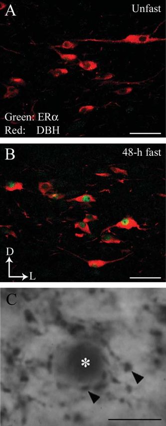
Figure 3 (A and B) Estrogen receptor a (ERa) and dopamine-b- 1988). Furthermore, noradrenergic input to the PVN induces
hydroxylase (DBH)-immunoreactivities in the A2 region in the CRH release via the activation of the a-adrenergic receptors
nucleus of the solitary tract in OVX rats. ERa (green) is colocalized (Cagampang et al. 1992b, Maeda et al. 1994). Anatomical
within DBH-ir cells (red) in 48-h fasted rats. Arrows indicate dorsal (D)
and lateral (L) orientation of the sections illustrated. Scale barZ50 mm.
studies showed that PVN neurons project to the median
(C) DBH-ir varicosities (arrowheads) juxtaposed with estrogen receptor eminence (Armstrong & Hatton 1980, Reyes et al. 2005).
a-ir soma (asterisks) in the PVN in OVX rats. Scale barZ375 mm. Since anti-CRH administered into the third ventricle reverses
Journal of Endocrinology (2006) 190, 593–600 www.endocrinology-journals.org
Downloaded from Bioscientifica.com at 09/15/2021 02:58:08PM
via free accessERa expression during fasting $ B A S REYES and others 599
fasting-induced LH suppression (Tsukamura et al. 1994), References
CRH neurons in the PVN could act at the median eminence
and/or POA to inhibit gonadotropin-releasing hormone, Armstrong WE & Hatton GI 1980 The localization of projection neurons in
which subsequently suppresses LH secretion (Maeda & the rat hypothalamic paraventricular nucleus following vascular and
neurohypophysial injections of HRP. Brain Research Bulletin 5 473–477.
Tsukamura 1996). The identity of the ERa-ir cells within Bao AM, Hestiantoro A, Van Someren EJ, Swaab DF & Zhou JN 2005
the PVN has yet to be established, but it is tempting to Colocalization of corticotrophin-releasing hormone and estrogen receptor-
speculate that the increment in ERa-ir cells in the PVN and alpha in the paraventricular nucleus of the hypothalamus in Mood
A2 region of the NTS causes an increase in the sensitivity of disorders. Brain 128 1301–1313.
the CRH-releasing system to the incoming NA signal during Blaustein JD 1987 The alpha 1-noradrenergic antagonist prazosin decreases
the concentration of estrogen receptors in female rat hypothalamus. Brain
fasting and thereby suppresses LH release. Nevertheless,
Research 404 39–50.
future studies would be useful to identify the neurochemical Blaustein JD & Turcotte J 1987 Further evidence of noradrenergic regulation
nature of ERa-containing cells expressed in the PVN during of rat hypothalamic estrogen receptor concentration: possible non-
fasting. functional increase and functional decrease. Brain Research 436 253–264.
Recently, human brain tissue sections obtained from Blaustein JD & Turcotte JC 1989 Estradiol-induced progestin receptor
autopsy specimens of patients having suffered from major immunoreactivity is found only in estrogen receptor-immunoreactive cells
in guinea pig brain. Neuroendocrinology 49 454–461.
depression/major depressive disorder or bipolar disorder Blaustein JD, Brown TJ & Swearengen ES 1986 Dopamine-beta-hydroxylase
demonstrated a colocalization of CRH and ERa in the inhibitors modulate the concentration of functional estrogen receptors
PVN (Bao et al. 2005). Impaired appetite is one of the in female rat hypothalamus and pituitary gland. Neuroendocrinology 43
common features of depressed or bipolar individuals 150–158.
(Diagnostic and Statistical Manual IV 1994, Sugahara et al. Bronson FH 1989 Mammalian Reproductive Physiology., Chicago: University of
Chicago Press.
2004, Kawa et al. 2005, Kishi & Elmquist 2005). However, a Cagampang FR, Maeda K, Yokoyama A & Ota K 1990 Effect of food
reduced appetite is observed in most depressive patients, while deprivation on the pulsatile LH release in the cycling and ovariectomized
an increase in appetite is seen in fewer patients (Diagnostic female rat. Hormone and Metabolic Research 22 269–272.
and Statistical Manual IV 1994). Together with the present Cagampang FR, Maeda KI, Tsukamura H, Ohkura S & Ota K 1991
results, it is possible that the increased ERa expression in the Involvement of ovarian steroids and endogenous opioids in the fasting-
induced suppression of pulsatile LH release in ovariectomized rats. Journal of
present experimental fasting model could somehow serve as a
Endocrinology 129 321–328.
cellular substrate related to the impairment of appetite Cagampang FR, Maeda K & Ota K 1992a Involvement of the gastric vagal
observed in depressed patients. This is an interesting concern nerve in the suppression of pulsatile luteinizing hormone release during
for further investigation. acute fasting in rats. Endocrinology 130 3003–3006.
In conclusion, our present study provides evidence that Cagampang FR, Ohkura S, Tsukamura H, Coen CW, Ota K & Maeda K
fasting induces ERa expression in the PVN, PeVN, and NTS 1992b Alpha 2-adrenergic receptors are involved in the suppression of
luteinizing hormone release during acute fasting in the ovariectomized
30 h from its onset, which is sustained until at least 48 h. estradiol-primed rats. Neuroendocrinology 56 724–728.
These results suggest that fasting creates complex physiologi- Cunningham ET Jr & Sawchenko PE 1988 Anatomical specificity of
cal signals, some of which slowly culminate in suppression of noradrenergic inputs to the paraventricular and supraoptic nuclei of the rat
LH secretion. Colocalization of ERa within the nor- hypothalamus. Journal of Comparative Neurology 274 60–76.
adrenergic neurons in the A2 region, suggests a role for Demling J, Fuchs E, Baumert M & Wuttke W 1985 Preoptic catecholamine,
GABA, and glutamate release in ovariectomized and ovariectomized
estrogen during fasting in increasing activation of the A2
estrogen-primed rats utilizing a push-pull cannula technique. Neuroendo-
noradrenergic neurons that project to the PVN. Moreover, crinology 41 212–218.
the anatomical relationship of DBH-ir varicosities and ERa- Diagnostic and Statistical Manual IV 1994 American Psychiatric Association,
ir cells in the PVN suggests that fasting enhances the Washington DC.
sensitivity of the PVN to this noradrenergic signal, Estacio MAC, Yamada S, Tsukamura H, Hirunagi K & Maeda K 1996a Effect
of fasting and immobilization stress on estrogen receptor immunoreactivity
consequently leading to the suppression of LH secretion.
in the brain in ovariectomized female rats. Brain Research 717 55–61.
Estacio MAC, Tsukamura H, Yamada S, Tsukahara S, Hirunagi K & Maeda K
1996b Vagus nerve mediates the increase in estrogen receptors in the
Acknowledgements hypothalamic paraventricular nucleus and nucleus of the solitary tract
during fasting in ovariectomized rats. Neuroscience Letters 208 25–28.
We would like to express our sincere gratitude to Dr Shinji Estacio MAC, Tsukamura H, Reyes BAS, Uenoyama Y, I’Anson H & Maeda
K-I 2004 Involvement of brainstem catecholaminergic inputs to the
Hayashi for the antibody and Dr Yoshihisa Uenoyama for his hypothalamic paraventricular nucleus in estrogen receptor a expression in
technical advice. We are also indebted to Ms Yoko Niwa and this nucleus during different stress conditions in female rats. Endocrinology
Kyoko Ohmiya for their technical assistance. This work was 145 4917–4926.
supported in part by Grants-in-Aid (Nos 10460131 and Goodman RL, Legan SJ, Ryan KD, Foster DL & Karsch FJ 1981 Importance
11660283) from the Ministry of Education, Science, Sports of variations in behavioural and feedback actions of oestradiol to the control
of seasonal breeding in the ewe. Journal of Endocrinology 89 229–240.
and Culture of Japan; the Japan Society for the Promotion of
Haywood SA, Simonian SX, van der Beek EM, Bicknell RJ & Herbison AE
Science (fellowship to H I). The authors declare that there is 1999 Fluctuating estrogen and progesterone receptor expression in
no conflict of interest that would prejudice the impartiality of brainstem norepinephrine neurons through the rat estrous cycle.
this scientific work. Endocrinology 140 3255–3263.
www.endocrinology-journals.org Journal of Endocrinology (2006) 190, 593–600
Downloaded from Bioscientifica.com at 09/15/2021 02:58:08PM
via free access600 B A S REYES and others $ ERa expression during fasting
Heritage AS, Grant LD & Stumpf WE 1977 3H estradiol in catecholamine Okamura H, Yokosuka M & Hayashi S 1994 Induction of substance
neurons of rat brain stem: combined localization by autoradiography and P-immunoreactivity by estrogen in neurons containing estrogen receptors
formaldehyde-induced fluorescence. Journal of Comparative Neurology 176 in the anteroventral periventricular nucleus of female but not male rats.
607–630. Journal of Neuroendocrinology 6 609–615.
Honma K & Wuttke W 1980 Norepinephrine and dopamine turnover rates in Panicker AK, Mangels RA, Powers JB, Wade GN & Schneider JE 1998
the medial preoptic area and the mediobasal hypothalamus of the rat brain after AP lesions block suppression of estrous behavior, but not estrous cyclicity,
various endocrinological manipulations. Endocrinology 106 1848–1853. in food-deprived Syrian hamsters. American Journal of Physiology 275
Imamov O, Shim GJ, Warner M & Gustafsson JA 2005 Estrogen receptor beta R158–R164.
in health and disease. Biology of Reproduction 73 866–871. Paxinos G & Watson C 1986 The Rat Brain in Stereotaxic Coordinates,
Itoi K, Suda T, Tozawa F, Dobashi I, Ohmori N, Sakai Y, Abe K & Demura H New York: Academic Press.
1994 Microinjection of norepinephrine into the paraventricular nucleus of Reyes BAS, Estacio MA, I’Anson H, Tsukamura H & Maeda K-I 2001
the hypothalamus stimulates corticotropin-releasing factor gene expression Glucoprivation increases estrogen receptor immunoreactivity in the brain
in conscious rats. Endocrinology 135 2177–2182. catecholaminergic neurons in ovariectomized rats. Neuroscience Letters 299
Jennes L, Jennes ME, Purvis C & Nees M 1992 c-fos expression in 109–112.
noradrenergic A2 neurons of the rat during the estrous cycle and after Reyes BAS, Valentino RJ, Xu G & Van Bockstaele EJ 2005 Hypothalamic
steroid hormone treatments. Brain Research 586 171–175. projections to locus coeruleus neurons in rat brain. European Journal of
Kawa I, Carter JD, Joyce PR, Doughty CJ, Frampton CM, Wells JE, Walsh AE Neuroscience 22 93–106.
& Olds RJ 2005 Gender differences in bipolar disorder: age of onset, course,
Roemmich JN, Li X, Rogol AD & Rissman EF 1997 Food availability affects
comorbidity, and symptom presentation. Bipolar Disorders 7 119–125.
neural estrogen receptor immunoreactivity in prepubertal mice. Endo-
Kishi T & Elmquist JK 2005 Body weight is regulated by the brain: a link
crinology 138 5366–5373.
between feeding and emotion. Molecular Psychiatry 10 132–146.
Sawchenko PE & Swanson LW 1982 The organization of noradrenergic
Kuiper GG, Enmark E, Pelto-Huikko M, Nilsson S & Gustafsson JA 1996
pathways from the brainstem to the paraventricular and supraoptic nuclei in
Cloning of a novel receptor expressed in rat prostate and ovary. PNAS 93
the rat. Brain Research 257 275–325.
5925–5930.
Shughrue PJ, Lane MV & Merchenthaler I 1997 Regulation of progesterone
Li HY, Wade GN & Blaustein JD 1994 Manipulations of metabolic fuel
availability alter estrous behavior and neural estrogen receptor immunor- receptor messenger ribonucleic acid in the rat medial preoptic nucleus by
eactivity in Syrian hamsters. Endocrinology 135 240–247. estrogenic and antiestrogenic compounds: an in situ hybridization study.
Liaw JJ, He JR, Hartman RD & Barraclough CA 1992 Changes in tyrosine Endocrinology 138 5476–5484.
hydroxylase mRNA levels in medullary A1 and A2 neurons and locus Simonian SX & Herbison AE 1997 Differential expression of estrogen
coeruleus following castration and estrogen replacement in rats. Brain receptor alpha and beta immunoreactivity by oxytocin neurons of rat
Research. Molecular Brain Research 13 231–238. paraventricular nucleus. Journal of Neuroendocrinology 9 803–806.
Liposits Z, Phelix C & Paull WK 1986 Electron microscopic analysis of Stanley BG, Schwartz DH, Hernandez L, Hoebel BG & Leibowitz SF 1989
tyrosine hydroxylase, dopamine-beta-hydroxylase and phenylethanola- Patterns of extracellular norepinephrine in the paraventricular hypo-
mine-N-methyltransferase immunoreactive innervation of the hypo- thalamus: relationship to circadian rhythm and deprivation-induced eating
thalamic paraventricular nucleus in the rat. Histochemistry 84 105–120. behavior. Life Science 45 275–282.
Maeda K & Tsukamura H 1996 Neuroendocrine mechanism mediating Sugahara H, Akamine M, Kondo T, Fujisawa K, Yoshimasu K, Tokunaga S
fasting-induced suppression of luteinizing hormone secretion in female rats. & Kudo C 2004 Somatic symptoms most often associated with depression
Acta Neurobiologiae Experimentalis (Wars) 56 787–796. in an urban hospital medical setting in Japan. Psychiatry Research 128
Maeda K, Cagampang FR, Coen CW & Tsukamura H 1994 Involvement of 305–311.
the catecholaminergic input to the paraventricular nucleus and of Tsagarakis S, Holly JM, Rees LH, Besser GM & Grossman A 1988
corticotropin-releasing hormone in the fasting-induced suppression of Acetylcholine and norepinephrine stimulate the release of corticotropin-
luteinizing hormone release in female rats. Endocrinology 134 1718–1722. releasing factor-41 from the rat hypothalamus in vitro. Endocrinology 123
Maeda K, Nagatani S, Estacio MA & Tsukamura H 1996 Novel estrogen feedback 1962–1969.
sites associated with stress-induced suppression of luteinizing hormone Tsukamura H, Nagatani S, Cagampang FR, Kawakami S & Maeda K 1994
secretion in female rats. Cellular and Molecular Neurobiology 16 311–324. Corticotropin-releasing hormone mediates suppression of pulsatile lutei-
Mezey E & Palkovits M 1991 CRF-containing neurons in the hypothalamic nizing hormone secretion induced by activation of alpha-adrenergic
paraventricular nucleus: regulation, especially by catecholamines. Frontiers receptors in the paraventricular nucleus in female rats. Endocrinology 134
in Neuroendocrinology 12 23–37. 1460–1466.
Nagatani S, Bucholtz DC, Murahashi K, Estacio MA, Tsukamura H, Foster Wise PM, Rance N & Barraclough CA 1981 Effects of estradiol and
DL & Maeda KI 1996 Reduction of glucose availability suppresses pulsatile progesterone on catecholamine turnover rates in discrete hypothalamic
luteinizing hormone release in female and male rats. Endocrinology 137 regions in ovariectomized rats. Endocrinology 108 2186–2193.
1166–1170.
Nilsson S, Makela S, Treuter E, Tujague M, Thomsen J, Andersson G, Enmark
E, Pettersson K, Warner M & Gustafsson JA 2001 Mechanisms of estrogen
action. Physiological Reviews 81 1535–1565.
Okamura H, Yamamoto K, Hayashi S, Kuroiwa A & Muramatsu M 1992 A
polyclonal antibody to the rat oestrogen receptor expressed in Escherichia
Received 11 April 2006
coli: characterization and application to immunohistochemistry. Journal of Received in final form 22 May 2006
Endocrinology 135 333–341. Accepted 6 June 2006
Journal of Endocrinology (2006) 190, 593–600 www.endocrinology-journals.org
Downloaded from Bioscientifica.com at 09/15/2021 02:58:08PM
via free accessYou can also read


