Down-regulation of BMP8A, SMADs 1/5/8 and BAX Proteins Following Methotrexate-treatment in Testicular Tissue of Swiss Albino Mice
←
→
Page content transcription
If your browser does not render page correctly, please read the page content below
Annual Research & Review in Biology

                            36(6): 1-9, 2021; Article no.ARRB.68818
                            ISSN: 2347-565X, NLM ID: 101632869

     Down-regulation of BMP8A, SMADs 1/5/8 and BAX
        Proteins Following Methotrexate-treatment in
               Testicular Tissue of Swiss Albino Mice
                             Olajumoke Omolara Ojo1*, Oluwadamilare Oluwaseun Ajayi1
                                                        and Babafemi Tosin Ogunbiyi2
         1
         Department of Biochemistry, Faculty of Science, Ekiti- State University, Ado-Ekiti, Ekiti- State,
                                                                                                 Nigeria.
      2
       Department of Biochemistry, Benjamin S. Carson (Snr) School of Medicine, Babcock University,
                                                                           Illisan, Ogun-State, Nigeria.

                                                                                             Authors’ contributions

        This work was carried out in collaboration among all authors. Author OOO designed the study,
        performed the statistical analysis, wrote the protocol and wrote the first draft of the manuscript.
       Authors OOA and BTO managed the analyses of the study. Author OOA managed the literature
                                          searches. All authors read and approved the final manuscript.

                                                                                                  Article Information

                                                                                    DOI: 10.9734/ARRB/2021/v36i630384
                                                                                                                  Editor(s):
                                                 (1) Dr. Gonzalo Emiliano Aranda Abreu, Veracruzana University, Mexico.
                                                                                                                Reviewers:
                                                              (1) Ponraj Mohanadoss, The Copperbelt University, Zambia.
                                                       (2) Ing. Hélia Némethová, Technical University of Košice, Slovakia.
                                             Complete Peer review History: http://www.sdiarticle4.com/review-history/68818

                                                                                           Received 24 March 2021
  Original Research Article                                                                 Accepted 03 June 2021
                                                                                           Published 14 June 2021

ABSTRACT

 Several studies on the adverse effects of methotrexate have been reported, especially its
 implication in the degeneration of spermatogenesis, reduced sperm count and ultimate male
 infertility. As an antagonist, methotrexate (MTX) uses folic acid to obstruct the production of some
 biomolecules involved in synthesis of DNA, RNA and protein. It is used in the treatment of cancer
 and other diseases such as psoriasis, and rheumatism. Reports have also revealed that the
 expression of Bone Morphogenetic Protein (BMP8a) promotes spermatogenesis and fertility
 through the induction and activation of signaling sets of transcription factors, SMAD1/5/8. Hence,
 the expressions of these proteins and role of apoptosis are crucial to understand the mechanism
 involved in Methotrexate-induced infertility. In view of this, albino mice (Swiss strain) were randomly
 sorted to four groups. Group I served as control while groups II, III & IV (n=5) were treated with 5,
_____________________________________________________________________________________________________

*Corresponding author: E-mail: olajumoke.ojo@eksu.edu.ng;
Ojo et al.; ARRB, 36(6): 1-9, 2021; Article no.ARRB.68818
10 and 20mg/kg/day of Methotrexate (IP) respectively. Expressions of BMP8A and SMADs 1/ 5/ 8
were done by PCR and Western blotting techniques. Germ cell apoptosis was measured by flow
cytometry and immunohistochemistry techniques. Ultrastructural changes were assessed in leydig
cells as well as sertoli cells. The results of this study reveal a down-regulation of BMP8A and
SMAD1/5/8 proteins in a dose-dependent pattern. Induction of apoptosis was also confirmed by the
expression of primary apoptotic Bax antibody. The sertoli cells which play their major roles of
nourishing and protecting the development of sperm cells were severely impaired too. These
findings suggest that the function of BMP8A and SMAD1/5/8 proteins in promoting proliferation and
differentiation of spermatogonia was severely disrupted following methotrexate exposure. Caution
should therefore be taken when administering this drug.
Keywords: Bone morphogenetic proteins (BMPs); methotrexate; apoptosis; TEM.
1. INTRODUCTION together in activating pathways which stimulate
spermatogenesis [12].
Methotrexate (MTX) is one of the many
anticancer drugs used in chemotherapy, Furthermore, programmed cell death (apoptosis)
although it is also known for its use in the has been linked with incidence of male infertility.
treatment of autoimmune diseases like Apoptosis is an indispensable biological process
rheumatoid arthritis and psoriasis [1]. MTX required for spermatogenesis in mammals.
functions as a competitive inhibitor of enzymes Asides degenerated spermatogenesis, apoptosis
which utilize folate as coenzyme. With this ability, is also relevant to the pathogenesis of male
it is able to disrupt the process of DNA replication infertility whether independently or in synergy
in cancer cells [2]. Interestingly, MTX is with genetic disorders [13,14]. Spermatogenic
reportedly used majorly by males of reproductive processes have their intricacies alongside
age for cancer treatment [3]. Male infertility, maturity of germ cells. The sertoli and leydig cells
paternal teratogenicity, hypotension and allergic are at the helm of affairs in the development,
reactions are some of the complications resulting nourishment, protection and preservation of germ
from MTX-treatment as suggested by previous cells as they are known to increase in number
studies [4]. It was reported in a recent study that [15]. Sertoli cells are activated by follicle-
MTX treatment in male mice led to reduction in stimulating hormone (FSH) to secrete androgen-
the levels of testosterone and luteinizing binding proteins. This process makes for the
hormone which are crucial hormones related to sustenance of spermatogenesis. On the other
male fertility [5]. Another study also suggested hand, leydig cells are important in that they are
that MTX causes oxidative damage to testicular responsible for the production of testosterone
tissue in male rats [6,7]. Despite these reports, and are stimulated by luteinizing hormone (LH)
there is still insufficient clarification of the [16]. Apoptosis aids the selective regulation of
mechanism responsible for MTX-induced male number of germ cells according to the ability of
infertility. the Sertoli cells [17]. The pro-apoptotic protein,
BAX, is a signaling molecule encoded by the
Bone Morphogenetic Proteins (BMPs) and BAX gene. It is a common indicator of apoptosis
SMADs are important molecules in signal in cells [18]. Similarly, Annexin V is also
transduction pathways which are pertinent to known to be another indicator of apoptotic cells
fertility in males. BMPs are proteins classified due to its affinity for phosphatidylserine which is
under a large family of growth-factor proteins [8]. an apoptotic marker on the extra-cytosolic side
In the male reproductive system, BMPs play an [19].
important role in spermatogenesis and germ cell
maintenance and development of epididymis. Evidences are yet to be considerably established
More specifically, BMP8A has been reported to with regard to the effect of MTX usage male
be a central player in the preservation of infertility and the mechanism supporting these
structure and functions of the epididymis [9,10]. observations. Hence, the purpose of this study
SMADs are known as the major signal was to investigate possible mechanisms through
transducers responsible for the regulation of cell which MTX could induce male infertility vis-à-vis
growth and development in many organs of the gene expressions of BMP8a, SMADs 1/5/8 and
body. The activity of SMADs is activated by BAX, coupled with apoptosis in testicular tissue
BMPs [11]. Thus, BMPs and SMADs function of male mice.
2Ojo et al.; ARRB, 36(6): 1-9, 2021; Article no.ARRB.68818
2. METHODOLOGY fluorescein isothiocyanate/Propidium Iodide (PI)
detects primary phases of the apoptotic process
2.1 Care of Experimental Animals by binding to phosphoserine while permitting
direct quantification. Testis tissue was
The Institutional Animal Ethics Committee of homogenized in PBS (1500xg/5mins). Cells were
College of Medicine, Ekiti State University, Ekiti- washed, and re-suspended in phosphate-buffer
State Nigeria, approved the protocols for animal saline (PBS, pH-7.0). Fluorescein isothiocyanate
handling for this study. Strict compliance with all conjugated Annexin V and PI.
the animal care and handling guidelines as
recommended by the committee was ensured. 2.6 Immunohistochemical (IHC) Staining
2.2 Animal Dosing After deparaffinising and rehydrating the tissue
sections, they were kept in a dark space (30
Twenty (20) swiss albino mice were randomly mins) with 3% hydrogen peroxide to deactivate
sorted to four experimental groups (n=5). Group I peroxidase present. Thereafter, the sections
served as control while groups II, III & IV were were rinsed and placed in 0.01% Tween 20 with
treated with 5, 10 and 20mg/kg/day of 0.01M PBS for 5 minutes (pH 7.4). The tissue
Methotrexate respectively via intraperitoneal sections were further incubated for 30 minutes
o
injection. The treatment lasted for twenty-one and then overnight both at 4 C with Bax primary
(21) days. antibodies (1:1000). After sufficient washing in
0.01% Tween 20, sections were incubated with
o
anti-immunoglobulin G for 1 hour (37 C).
2.3 Tissue Sample Preparation
Furthermore, the tissue sections were washed
and 3, 3’-diaminobenzidine was used for antigen
At autopsy, animals were killed by using over detection.
dose of anesthesia to avoid any suffering to the
animals. Testis of one side were excised and
2.7 Testicular Ultrastructure
cleaned with cold saline. Excess blood and
moisture was also discarded. Tissues were
frozen using liquid nitrogen and stored at −80 °C The ultrastructure of testis was studied [22].
(Ultra-cold Freezer, Thermo Fisher Scientific, Tiny testicular fractions were excised and fixed in
UK) until required for further analysis. Other side a mixture of 2.5% glutaraldehyde, 2%
testes were fixed in appropriate fixative for paraformaldehyde in 0.1M sodium phosphate
ultrastructural and immunohistochemistry buffer (pH 7.3) for 12 hours (4oC). After washing
examinations. with a buffer solution, tissues were fixed for 1
hour (1% osmium tetroxide and 0.1M phosphate)
o
at 4 C. Sample dehydration was carried out
2.4 Isolation Testicular Germ Cells
using increasing levels of acetone. Infiltration of
(TGCs) the samples was followed by embedding using
araldite (CY 212 /TAAB, UK). 1 µm slices were
Testicular germ cells (TGCs) were collected from cut and mounted on clean glass slides. This was
testes of experimental animals by incision using immediately followed by staining using blue
a surgical blade. The cells were subjected to toluidine stain. The slices were viewed under a
centrifugation (5 min at 800 xg). The cells were light microscope for gross scrutiny of the area
thoroughly washed with DMEM/F12 medium. and quality of the fixation process. Thin slices of
TGCs were cultured and counted in a grey-silver colour interference (70-80 nm) were
hemocytometer. Trypan blue was also used as mounted onto 300 mesh-copper grids for
staining agent for determination of cell viability. electron microscopy. Uranyl acetate (alcoholic)
Spermatocytes (>90% pachytene) and round and lead citrate (alkaline) were used as staining
spermatids were also purified [20,21]. agents. The slices were further washed and
observed under a Morgagni 268D transmission
2.5 Apoptosis Detection by Flow electron microscope (FEI Company,
Cytometry Netherlands).
FITC Annexin V Apoptosis Detection Kit protocol 2.8 Total RNA Extraction
was used for detecting apoptosis via flow
cytometry. The analysis investigated induction of Total RNA extraction from testicular germ cells
apoptosis in testicular germ cells. Annexin V- was carried out using Trizol reagent.
3Ojo et al.; ARRB, 36(6): 1-9, 2021; Article no.ARRB.68818
Spectrophotometry was used to quantify isolated 3.3 Immunohistochemical Analysis of
total RNA as well as its level of purity by BAX in Testes of Mice
measuring the optical density (OD) (260 and
280nm). Optical density ratios (260nm/280nm) Positive staining of BAX antibody was observed
for all the samples ranged from 1.8 to 2.0. The in the testes of mice treated with 5, 10 and
amplification and quantification of target genes 20mg/kg/day (Fig. 3a). BAX positive staining in
were performed in a reaction of 20 mL for 40 5mg/kg was significantly intense in the
cycles (SYBR Green Kit Germany) [23]. spermatocytes and spermatogonia cells,
indicating increased rate of apoptosis among the
2.9 Expression of BMP8A protein by Methotrexate treated groups. Positive staining of
Western Blotting BAX antibody was also seen in the nucleus of
elongated spermatids (Est), spermatogonia (Sg),
Total protein was extracted from testicular tissue round spermatids (RSt) and pachytene
with urea lysis buffer according to modified spermatocytes (PSc) (Fig. 3b).
protocol of [24]. Protein separation and transfer
was probed using BMP8B protein and β - actin 3.4 Effect of MTX on Expression of
antibodies. Bound antibody was detected using BMP8A and Bax Genes in Testis
HRP conjugated secondary antibody. Blot
development was done via enhanced A significant difference was observed in RT-PCR
chemiluminescent detection reagent which was results (p < 0.05) in gene expression levels of
further used for densitometric analysis and BMP8a and Bax when compared with the control
normalization. Data from western blotting group (Fig. 4).
(quantitative) will be calculated from
densitometric analysis of bands with the NIH
3.5 Effect of MTX on Testicular
image J software. The values were normalized
with β -actin which served as loading control. Ultrastructure
2.10 Statistical Analysis Leydig cell ultrastructure in MTX-treated mice
indicated elevation in hypertrophy with shrunken
nuclei. There was also a corresponding rise in
Analysis of variance (ANOVA) was used to
the number of Iipid droplets in comparison to the
analyze statistical comparisons between the
control group. Condensed mitochondria were
experimental groups (GraphPad Prism software).
detected in mice administered 10 and 20 mg/kg
Results were expressed as mean ± Standard
MTX (Fig. 5a, b, ‘c and d’).
Error Mean (SEM). Statistically significant values
were considered at p < 0.05.
Series of abnormal and irregular morphological
characteristics were seen in Sertoli cell
3. RESULTS ultrastructure following MTX-exposure. These
include increased vacuole number in the
3.1 MTX and Viability of Spermatogenic cytoplasm and expanded endoplasmic reticulum.
Cells Shrunken mitochondria and intact cell structures
were also visible (VI ‘b, c, d’). Control group
The viability determined by trypan blue staining
sertoli cells showed intact mitochondria with their
showed significant decrease of germ cells in all
cristae in the cytoplasm. The cell nucleoli were
the MTX- treated samples when compared with
also intact as well as the chromatin structures
the control group in a dose-dependent manner
(Fig. 6b).
(Fig. 1).
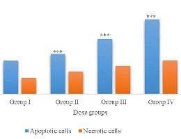
3.2 Methotrexate Effect on Sperma- 3.6 Effect of Methotrexate on BMP8a and
togenic Cells SMAD1/5/8 Proteins
Increased concentrations of Methotrexate Methotrexate differentially altered the levels of
resulted in an increased percentage of Annexin BMP8a and SMAD1/5/8 protein expressions
V-positive spermatogenic cells in dose in a concentration-dependent manner in total
dependent pattern (Fig. 2). Annexin V positivity is spermatogenic cells. Significant downregulation
an attribute of apoptotic cells; hence, an of the protein was seen across the groups (Fig. 6
appropriate method to quantify apoptosis. a and b).
4Ojo et al.; ARRB, 36(6): 1-9,
9, 2021;
2021 Article no.ARRB.68818
Fig. 1. Graph showing Effect MTX on viability
iability of spermatogenic cells
All the values are expressed as mean ± SEM, (n=5), ***POjo et al.; ARRB, 36(6): 1-9,
9, 2021;
2021 Article no.ARRB.68818
Fig. 4. Bar chat represents the fold change of BMP8a and BAX genes in testes of mice treated
with MTX for 21days
The data are expressed as mean ± SD (n=5). *, **, and *** indicate significant difference as compared to controls
at (pOjo et al.; ARRB, 36(6): 1-9, 2021; Article no.ARRB.68818
concerns. One of such side effects is the with MTX (5, 10 and 20mg/kg/day). These
induction of infertility in male mice. From observations are further supported by the clear
previous studies, it has been proposed that positive staining of BAX antibody observed in
treatment with MTX causes male infertility nuclei of spermatogonia, spermatocytes and
through increased oxidative stress and reduction spermatids in MTX-treated mice. Annexin V, a
of DNA integrity in sperm [5,25]. Our findings signaling protein which detects the presence of
from this study revealed a significant (p ˂ 0.05) apoptotic cells by signaling expression of surface
downregulation of BMP8A gene in mice testes, phosphatidylserine, was also determined in mice
with the lowest expression of BMP8A observed spermatogenic cells. Increasing Annexin V
at 20m/kg/day dose. This is quite profound positivity from increased doses of MTX in this
because BMP8A is important in the sustenance study confirms the presence of apoptotic cells in
and maintenance of spermatogenesis via the MTX-treated mice. These observations further
activation of SMADs 1/5/8 as well as SMADs 2/3 confirm that MTX treatment may also induce
in spermatogonia [12]. Hence, BMP8A activates male infertility by inducing apoptosis in
promotes spermatogenesis via proliferation and spermatogonia.
differentiation of spermatogonia [12]. Thus, any
impairment in the expression of BMP8A will In addition to the aforementioned parameters, we
negatively regulate SMAD 1/5/8 signaling, which also assessed the ultrastructures of sertoli and
in turn causes reduced spermatogonial leydig cells in testes of MTX-treated rats to check
proliferation and differentiation culminating in for structural irregularities. Results showed that
infertility. Hereafter, downregulated expression of the structure of the sertoli cells was
BMP8A observed from our findings show a compromised in MTX-treated mice. The
relationship between MTX treatment in mice compromise of sertoli cell structure by MTX will
testis and infertility. To further confirm this, result in failure to secrete androgen-binding
SMADs 1/5/8 expression result from this study proteins necessary for spermatogenesis. Also, it
also showed a significantly dose-dependent was revealed from this study that the number of
downregulation (p ˂ 0.05). SMADs 1/5/8 lipid droplets increased as MTX dose increases
activation by BMP8A is responsible for when compared to the control rats. This
spermatogonial diversification [26]. The observed abnormality can be attributed to the cytotoxic
downregulation of SMADs 1/5/8 can be linked effect of MTX on the structure of the leydig cells,
with the downregulation of BMP8A observed in especially in mice treated with 10 and 20mg/kg.
mice spermatogonia. Based on these The observed increase in lipid droplets in the
observations, we propose that downregulated MTX-treated rats indicates impairment in
expressions of BMP8A and SMADs 1/5/8 are lipophagy. Lipophagy is an autophagic process
likely signaling mechanisms that are associated of breaking down lipid droplets; a process used
with MTX-induced male infertility in mice. by most eukaryotic cells to produce energy.
Lipophagy modulates breaking down of
To obtain a closer perspective of the involvement cholesteryl esters to free cholesterol which is the
of apoptosis in MTX-induced infertility, this study precursor for the production of testosterone
also assessed the expression of BAX gene, a [29,26]. Previous research also reported the
pro-apoptotic molecule, in the testes of mice. importance of lipophagy in stimulation of
BAX is a regulator of apoptosis involved in a spermatogenesis [30,31]. It is possible that
series of signaling reactions which culminate in lipophagy was compromised in MTX-treated rats
the release of cytochrome C, a potent death due to decreased production of Luteinizing
signal, as well as other pro-apoptotic factors hormone (LH) which positively regulates
[27,28]. Result from expression of BAX gene in lipophagy. In a study by [6], it was reported that
MTX-treated mice showed a dose-dependent MTX-treated mice showed significantly
increase in BAX expression. This indicates that decreased LH and testosterone levels. Thus,
MTX treatment is capable of inducing production impaired lipophagy induced by MTX could be
of BAX antibodies, thus increasing the rate of linked with reduced testosterone production from
apoptosis in mice spermatogonia. This is spermatogenesis, ultimately causing male
attributed mainly to increased level of caspases infertility.
released from the mitochondria causing
proliferation of apoptotic cells and ultimately 5. CONCLUSION
programmed cell death. In addition to this,
immunohistochemical analysis showed presence This study suggests that apoptosis via
of BAX in mice testes of all the groups treated downregulation of BMP8A and BAX genes
7Ojo et al.; ARRB, 36(6): 1-9, 2021; Article no.ARRB.68818
occurs in spermatogonia following methotrexate- Methotrexate, blood pressure and markers
treatment in testicular tissue of swiss albino of arterial function in patients with
mice. This might be the likely mechanism rheumatoid arthritis: a repeated cross-
through which MTX induces male infertility. sectional study. Ther. Adv. Musculoskelet.
Dis. 2017;9(9):213-229.
DISCLAIMER 5. Ojo OO, Adesua OO, Titiloye O.
Therapeutic effect of quercetin against
The products used for this research are methotrexate-induced male infertility. J.
commonly and predominantly use products in our Toxicol. Pharmacol. 2019;1:023.
area of research and country. There is absolutely 6. Ojo O. O. Induction of Oxidative Stress by
no conflict of interest between the authors and Methotrexate in the testicular tissue of
producers of the products because we do not swiss albino mice. Annals of Clinical
intend to use these products as an avenue for Toxicology Ann Clin Toxicol. 2019;2(3):
any litigation but for the advancement of 1022.
knowledge. Also, the research was not funded by 7. Yapca OE, Borecki B, Turan MI, Gulapoglu
the producing company rather it was funded by M, Saman S. The effect of mirtazapine on
personal efforts of the authors. methotrexate-induced oxidative damage
and infertility in rats. Science Asia. 2014;
ETHICAL APRROVAL 40:152-156.
8. Semet M, Paci M, Saïas-Magnan J,
The Institutional Animal Ethics Committee of Metzler-Guillemain C, Boissier R,
College of Medicine, Ekiti State University, Ekiti- Lejeune H, Perrin J.The impact of drugs on
State Nigeria, approved the protocols for animal male fertility: a review Andrology. 2017;
handling for this study. Strict compliance with all 5(4).
the animal care and handling guidelines as DOI.org/10.1111/andr.1236:640-663.
recommended by the committee was ensured. 9. Itman C, Loveland KL. SMAD expression
in the testis: an insight into BMP regulation
ACKNOWLEDGEMENTS of spermatogenesis. Dev Dyn. 2008;237:
97–111.
The authors acknowledge Mr. Oyewumi Titiloye
10. Lochab AK, Extavour CG. Bone
John and all the staff of Chemical Pathology
morphogenetic factor (BMP) signaling in
Department, College of Medicine, University of
animal reproductive system development
Ibadan Oyo-State Nigeria.
and function. Dev Biol. 2017;427(2):258-
COMPETING INTERESTS 269.
11. Zhao GQ, Liaw L, Hogan BL. Bone
Authors have declared that no competing morphogenetic protein 8A plays a role in
interests exist. the maintenance of spermatogenesis and
the integrity of the epididymis. Dev.
REFERENCES 1998;125:1103-1112.
12. Lasorella A, Benezra R, Iavarone A. The
1. Cronstein BN. Low-dose methotrexate: a ID proteins: master regulators of cancer
mainstay in the treatment of rheumatoid stem cells and tumour aggressiveness.
arthritis. Pharm. Rev. 2005;57(2):163–72. Nat. Rev. Cancer. 2014;14(2):77–91.
2. Rajagopalan, PT, Zhang Z, McCourt L, 13. Wu FJ, Lin TY, Sung LY, Chang WF, Wu
Dwyer M, Benkovic SJ, Hammes GG PC, Luo CW. BMP8A sustains
Interaction of dihydrofolate reductase with spermatogenesis by activating both SMAD
methotrexate: Ensemble and single- 1/5/8 and SMAD 2/3 in spermatogonia.
molecule kinetics. Proceedings of the Sci. Sig. 2017;10:eaal1910.
National Academy of Sciences of the 14. Weng C, Li Y, Xu D, Shi Y, Tang
United States of America. 2002;99(21): H. Specific cleavage of Mcl-1 by caspase-3
13481–6. in tumor necrosis factor-related apoptosis-
3. Gutierrez JC, Hwang K. The toxicity of inducing ligand (TRAIL)-induced apoptosis
methotrexate in male fertility and paternal in Jurkat leukemia T cells. J. Biol.
teratogenicity. Expert Opin Drug Metab Chem. 2005;280(11):10491–500.
Toxicol. 2017;13(1):51-58. 15. Vermes I, Haanen C, Steffens-Nakken H,
4. Mangoni AA, Baghdadi LR, Shanahan Reutelingsperger C. A novel assay for
EM, Wiese MD, Tommasi S, Elliot D. apoptosis. Flow cytometric detection of
8Ojo et al.; ARRB, 36(6): 1-9, 2021; Article no.ARRB.68818
phosphatidylserine expression in early 24. Saraiva KLA, Silva Junior VA, Dias ESF,
apoptotic cells using fluorescein-labelled Peixoto CA. Morphological changes in the
Annexin V. Journal of Immunological testis induced by diethylcarbamazine.
Methods. 1995;184(1):39-51. Reprod Toxicol. 2006;22:754-759.
16. Vaithinathan S, D’Cruz, SC, Mathur PP. DOI: 10.1016/j.reprotox.2006.07.008.
Apoptosis and male infertility. In: S.J. 25. Livak KJ, Schmittgen TD. Analysis of
Parekattil and A. Agarwal (eds.), Male relative gene expression data using real –
infertility: Contemporary Clinical time quantitative PCR and the 2(-Delta
Approaches, Andrology, ART & Delta C (T)) Method. Methods. 2001;25:
Antioxidants. 2012;329-335 402-408.
17. Iammarrone E, Balet R, Lower AM, Gillott 26. Ojo OO. Expression of Bax and Bcl-2
C, Grudzinskas JG. Male infertility. Best Apoptotic Regulatory Proteins in
Pract Res Clin Obstet Gynaecol. 2003; Melphalan- induced Spermatogenic
17(2):211–29. Dysfunction. International Research
18. Al-Agha O, Axiotis C. An in-depth look at Journal of Biological Sciences Asian Pac.
Leydig cell tumor of the testis. Arch Pathol J. Health Sci. 2020;7(2):7-11.
Lab Med. 2007;131(2):311–7. 27. Ley D, Jones J, Parrish J, Salih S, Caldera
19. McLachlan RI, Wreford NG, Meachem SJ, F, Tirado E, Leader B, and Sumona S.
De Kretser DM, Robertson DM. Effects of Methotrexate reduces DNA integrity in
testosterone on spermatogenic cell sperm from men with inflammatory bowel
populations in the adult rat. Biol Reprod. disease. Gastroenterology. 2018;154:
1994;51(5):945–55. 2064-2067.
20. Pierrat B, Simonen M, Cueto M, Mestan J, 28. Ojo OO, Bhadauria S, Rath SK. Dose-
Ferrigno P, Heim J. SH3GLB, a new Dependent Adverse Effects of Salinomycin
endophilin-related protein family featuring on Male Reproductive Organs and Fertility
an SH3 domain. Genomics. 2001;71(2): in Mice. PLOS One. 2013;2(6):133-140,
222–34. e69086.
21. Sofiadis A, Becker S, Hellman U, Hultin- DOI: 10.1371/journal.pone.0069086
Rosenberg L, Dinets A, Hulchiy M, 29. Ghribi O, Dewitt DA, Forbes MS, Herman
Zedenius J, Wallin G, Foukakis T, Höög A, Savory J. Co-involvement of mitochondria
Auer G, Lehtiö J, Larsson C. Proteomic and endoplasmic reticulum in regulation of
profiling of follicular and papillary apoptosis: Changes in cytochrome c Bcl-2
thyroid tumors. European Journal of and Bax in the hippocampus of aluminum
Endocrinology. 2012;166(4):657–67. –treated rabbits. Brain Res. 2001;903:
22. Picton HM, Wyns C, Anderson RA, 66-73.
Goossens E, Jahnukainen K, Kliesch S, 30. Ma Y, Zhou Y, Zhu YC, Wang SQ, Ping P,
Mitchell RT, Pennings G, Rives N, Chen XF. Lipophagy contributes to
Tournaye H. et al. A European perspective testosterone biosynthesis in male rat
on testicular tissue cryopreservation for leydig cells. Endocrinology. 2018;159(2):
fertility preservation in prepubertal and 1119-1129.
adolescent boys. Human Rep. 2015;30: 31. Michael Garland, Mitra Geier Sean Bugel,
2463–2475. Prarthana Shankar Aryl Hydrocarbon
DOI: 10.1093/humrep/dev190 Receptor Mediates Larval Zebrafish Fin
23. Romrell LI, BeIlve AR, Fawcett DW. Duplication Following Exposure to
Separation of mouse spermatogenic Cells Benzofluoranthenes. Toxicological
of sedimentation velocity. Dev Biol. 1976; Sciences. 2020;176(1).
119-131. DOI: 10.1093/toxsci/kfaa063
_________________________________________________________________________________
© 2021 Ojo et al.; This is an Open Access article distributed under the terms of the Creative Commons Attribution License
(http://creativecommons.org/licenses/by/4.0), which permits unrestricted use, distribution, and reproduction in any medium,
provided the original work is properly cited.
Peer-review history:
The peer review history for this paper can be accessed here:
http://www.sdiarticle4.com/review-history/68818
9You can also read