Upregulation of microRNA 140 3p mediates dachshund nephropathy through cell cycle dependent mechanisms
←
→
Page content transcription
If your browser does not render page correctly, please read the page content below
MOLECULAR MEDICINE REPORTS 23: 134, 2021

       Upregulation of microRNA‑140‑3p mediates dachshund
     family transcription factor 1 expression in immunoglobulin A
        nephropathy through cell cycle‑dependent mechanisms
                          XIAOBIN ZHOU1, YAO LU2, PENGFEI GUO3 and CHENGLIN ZHOU1

                   1
                  Department of Clinical Laboratory, Taizhou People's Hospital, Taizhou, Jiangsu 225300;
      2
       Department of Teaching Research of Medical Technology, Gannan Medical University, Ganzhou, Jiangxi 341000;
            3
              Department of Clinical Laboratory, Shanghai Tenth People's Hospital, Shanghai 200072, P.R. China

                                      Received May 29, 2020; Accepted November 11, 2020

                                                  DOI: 10.3892/mmr.2020.11773

Abstract. Immunoglobulin A nephropathy (IgAN) is a kidney           release from HMCs cultured with pIgA‑IgAN. The findings
disease and one of the commonest forms of glomerulonephritis        of the present study demonstrated that DACH1 decreased
worldwide. The present study investigated the role of               HMC growth and the release of inflammatory cytokines from
dachshund family transcription factor 1 (DACH1) in IgAN             HMCs may be targeted by miR‑140‑3p. The results suggested
and identified one of its binding microRNAs (miRNAs). The           that DACH1 could be associated with the progression of IgAN
expression of DACH1 in human mesangial cells (HMCs)                 and provide a potential target for further studies related to the
incubated with polymeric IgA (pIgA) isolated and purified           mechanism of IgAN.
from the serum of patients with IgAN or healthy individuals
was evaluated by reverse transcription‑quantitative (RT‑q)          Introduction
PCR and western blotting. Cell proliferation and cell cycle
assays were performed in DACH1‑overexpressing HMCs                  Immunoglobulin A nephropathy (IgAN) is an immune
to identify the role of DACH1 in IgAN and enzyme‑linked             complex glomerulonephritis characterized by IgA deposition
immunosorbent assay was carried out to verify the release           in the mesangial area, abnormal proliferation of mesangial
of inflammatory factors from HMCs. The target miRNAs of             cells and excessive expansion of the mesangial matrix (1,2).
DACH1 were predicted using bioinformatics software and              It is one of the commonest causes of end‑stage kidney failure
miR‑140‑3p was identified as a target of DACH1 by luciferase        and there is at present no specific treatment targeting IgAN (3).
report assay, RT‑qPCR and western blotting. The results             Thus, research has focused on exploring the pathogenesis
demonstrated that DACH1 was downregulated in HMCs                   of IgAN and developing effective treatment against it. As
cultured with pIgA‑IgAN at both mRNA and protein levels.            IgA deposition in the mesangial region is a key step in the
Overexpression of DACH1 suppressed HMC growth and                   development of IgAN (4) and the etiology and pathogenesis
inhibited inflammatory cytokine release from HMCs cultured          of this disease are unclear, the pathogenesis of IgAN can
with pIgA‑IgAN. The expression of DACH1 was negatively              be discussed from the perspective of IgA deposition in the
regulated by miR‑140‑3p in IgAN and miR‑140‑3p inhibition           mesangial region.
suppressed HMC growth and inhibited inflammatory cytokine                Dachshund family transcription factor 1 (DACH1) is a key
                                                                    component of the retinal determination gene network family
                                                                    that has been shown to be closely associated with organo‑
                                                                    genesis and tumorigenesis (5,6). DACH1 participates in cell
Correspondence to: Dr Chenglin Zhou, Department of Clinical         differentiation and proliferation in renal development (7) and
Laboratory, Taizhou People's Hospital, 366 Taihu Road, Taizhou,     decreased expression of DACH1 is associated with the progres‑
Jiangsu 225300, P.R. China                                          sion and severity of glomerulopathy (8). DACH1 disorder has
E‑mail: jszhoucl@126.com                                            been reported in a variety of human malignancies such as
                                                                    breast cancer (9), ovarian cancer (10), renal carcinoma (11) and
Abbreviations: IgAN, immunoglobulin A nephropathy; DACH1,
                                                                    gastric cancer (12). It acts as a cell‑fate determination factor
dachshund family transcription factor 1; HMCs, human mesangial
cells; pIgA, polymeric IgA; 3'UTR, 3'‑untranslated region; CXCL1,   that regulates cell growth and development (13,14), but the
chemokine cc‑motif ligand 1; NC, negative control.                  mechanism and function of DACH1 dysregulation in IgAN
                                                                    has yet to be explored.
Key words: dachshund family transcription factor 1, human                Abnormal cell proliferation is a sign of cancer transforma‑
mesangial cells, cell differentiation, cell cycle, miR‑140‑3p,      tion (15) and abnormal expression of genes in cancer cells can
immunoglobulin A nephropathy                                        be directly involved in the regulation of cell growth and cell
                                                                    cycle progression (16,17). Malfunctions in the cell cycle usually
                                                                    result in uncontrolled cell growth characteristics, allowing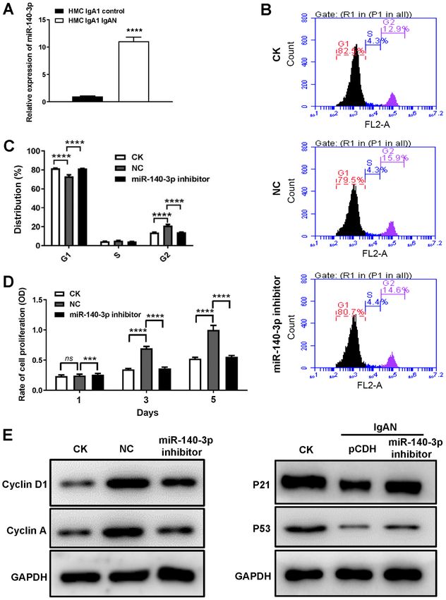
2 ZHOU et al: miR-140-3p MEDIATES DACH1 EXPRESSION IN IgAN VIA CELL CYCLE-DEPENDENT MECHANISMS
cancer cells to proliferate excessively and eventually leading 0.1 mg/ml streptomycin (Beyotime Institute of Biotechnology).
to the tumorigenesis (18). DACH1 inhibits cell proliferation HMCs and 293T were cultured at 37˚C in an atmosphere of 5%
and enhance cell cycle arrest. For example, Chen et al (19) CO2.
demonstrated that DACH1 participates in p53‑mediated p21
induction and cell cycle arrest in non‑small‑cell lung cancer. Plasmids. Lentiviral vector plasmid pCDH‑CMV‑MCS‑puro
Wu et al (10) revealed that low expression of DACH1 in breast (pCDH) and recombinant plasmid pCDH‑CMV‑DACH1‑puro
cancer cells promotes tumor growth by downregulating Nanog (pDACH1) were purchased from GenScript for the transfection
and Sox2. Kalousova et al (20) confirmed that DACH1 can and production of lentiviruses. The pGL3‑Control plasmid
bind to the promoter of the cell cycle inhibitor p27 to inhibit the and pGL3‑DACH1 3'UTR luciferase reporter plasmid were
proliferation and cell cycle progression of insulin‑producing purchased from GenScript for use in detection of luciferase
cells. The present study explored the role of DACH1 in IgAN activity following transfection.
in terms of cell proliferation and cell cycle progression.
MicroRNAs (miRNAs) are small non‑coding RNAs Lentivirus transduction and production. Lentivirus packaging
that consist of ~22 nucleotides (21). miRNAs can degrade vector psPAX2 (5 µg) and envelope vector pMD2.G (10 µg)
target mRNAs or inhibit their translation by binding to the were co‑transfected with lentivirus plasmid pCDH (5 µg)
3'‑untranslated region (3'UTR) of the target mRNAs to or pDACH1 (5 µg) into 293T cells using Lipofectamine
negatively regulate gene expression (22). Studies have shown 2000 (50 µl) (Thermo Fisher Scientific, Inc.) to produce the
that miRNAs are involved in the regulation of cell proliferation, corresponding lentivirus solution. The culture supernatant was
differentiation, cell cycle, apoptosis and inflammation (22‑25). collected 48‑72 h after transfection. The supernatant contained
The present study evaluated the expression and function of the corresponding virus and was used for subsequent cell
DACH1 in human mesangial cells (HMCs) cultured with infection experiments.
polymeric IgA (pIgA) that was isolated and purified from
the serum of patients with IgAN or healthy individuals. Reverse transcription‑quantitative (RT‑q) PCR. Total RNA was
Bioinformatics analysis was used to screen for candidate extracted from 5x10⁶ cells using TRIzol® reagent kit according
miRNAs associated with DACH1 expression. Among the to the manufacturer's protocol (Thermo Fisher Scientific, Inc.)
predicted miRNAs, miRNA (miR)‑140‑3p was selected and and reverse transcribed into single‑stranded cDNA according
its expression and effect on cell proliferation and cell cycle to the instructions of the Reverse Transcription kit (Vazyme
progression in IgAN were investigated. It was found that Biotech Co., Ltd.). The expression of mRNA was detected by
miR‑140‑3p directly suppressed the expression of DACH1 RT‑qPCR using the Rotor‑Gene Q detection system (Qiagen
and promoted the proliferation and cell cycle progression of GmbH) and the reaction volume was 20 µl, with the following
HMCs in IgAN. The findings of the present study may provide cycling conditions: 95˚C for 5 min, then 45 cycles of 95˚C
a theoretical basis for the study of the mechanism of IgAN. for 10 sec and 60˚C for 30 sec. The expression of miRNA
or mRNA was calculated using the 2‑ΔΔCq method (28). The
Materials and methods mRNA expression level was normalized to that of GAPDH
and the expression level of miRNA was normalized to U6
Human IgAN samples. The serum of 30 patients with IgAN RNA. The primer sequences used were: IL‑6 forward, 5'‑AAC
(men=23; women=7; age, 20‑30 years n=19 and >30 years TCCTTCTCCACAAGCGCCTT‑3' and reverse, 5'‑GTCAAT
n=11) and 30 normal individuals was collected. Inclusion TCG T TC T GA AGAG GT G ‑3'; IL‑8 forward, 5'‑GGTC TC
criteria included cases of lgA nephropathy and exclusion ACCTCCCAACTGC‑3' and reverse, 5'‑TCAG CTCGAACA
criteria included other nephropathies. The recruitment of CTTTGAATAT‑3'; IL‑13 forward, 5'‑TGAG GAG CTG GT
patients was conducted at Shanghai Tenth People's Hospital CAACATCA‑3' and reverse, 5'‑CCACCTCGATTTTGGTGT
between January 2020 and April 2020. The serum collec‑ CT‑3'; chemokine cc‑motif ligand 1 (CXCL1) forward, 5'‑CTC
tion was approved by the patients and all provided written CTG CGAGTG G CACTG CTG CTC‑3' and reverse, 5'‑GAG
informed consent. All blood samples (each sample ~5 ml) were GCA AGC T TT C CG C CC ATT C TT‑3'; GAPDH forward,
anonymized. The present study was approved by the clinical 5'‑GAGTCAACGGATT TGGTCGT‑3' and reverse, 5'‑GAT
research ethics committee of Shanghai Tenth People's Hospital CTCGCTCCTGGAAGATG‑3'; and U6 forward, 5'‑CTTCGG
(approval no. SHSY‑IEC‑4.1/20‑117‑01). CAGCACATATAC‑3' and reverse, 5'‑TTCACGAATTTGCGT
GTCAT‑3'. The experiment was repeated three times.
Extraction of polymeric IgA (pIgA). The pIgA was isolated
and purified from the serum of patients with IgAN or healthy Western blot analysis. The total protein of each group was
individuals using a jacalin‑agarose column as previously harvested and collected using RIPA lysis buffer (Thermo
described (26,27). The extracted pIgA was stored at ‑80˚C Fisher Scientific, Inc.). Protein concentration was measured
until use. The final concentration of pIgA was 0.5 mg/ml. with a BCA Protein Assay kit (Beyotime Institute of
Biotechnology). Equal amounts of protein (30 µg/lane) were
Cell lines and culture. Human mesangial cells (HMCs) and separated by 10% sodium dodecyl sulfate‑polyacrylamide
293T (cat. no. KCB200744YJ) cells were purchased from gel electrophoresis and transferred to polyvinylidene fluoride
the Conservation Genetics CAS Kunming Cell Bank. HMCs membranes (EMD Millipore). The membrane was blocked by
were cultured in RPMI‑1640 medium (Invitrogen; Thermo incubation with 5% non‑fat milk for 1 h at 24˚C and then with
Fisher Scientific, Inc.) containing 10% fetal calf serum (Gibco; primary antibodies overnight at 4˚C. The primary antibodies
Thermo Fisher Scientific, Inc.) and 80 U/ml penicillin and used were: Anti‑DACH1 (cat. no. ab176718; Abcam; 1:1,000),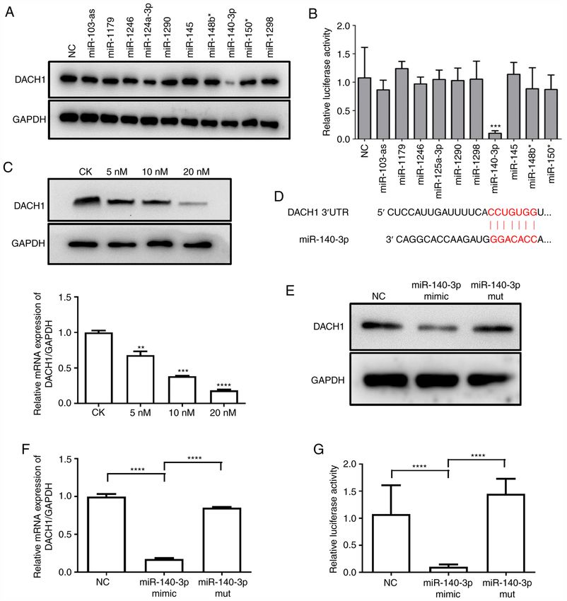
MOLECULAR MEDICINE REPORTS 23: 134, 2021 3
anti‑Cyclin D1 (cat. no. 55506; Cell Signaling Technology, cells were harvested using Dual‑Lumi™ II Luciferase Reporter
Inc.; 1:1,000), anti‑Cyclin A (cat. no. 91500; Cell Signaling Assay kit (Beyotime Institute of Biotechnology) according to
Technology, Inc.; 1:1,000) and anti‑P21 (cat. no. 2947; Cell the manufacturer's instructions and the luciferase activity was
Signaling Technology, Inc.; 1:1,000), anti‑P53 (cat. no. 2527; detected using a multimode reader (BioTek Instruments, Inc.).
Cell Signaling Technology, Inc.; 1:1,000). After washing three The Firefly luciferase activity was normalized to the Renilla
times with Tris buffered saline‑Tween (TBS‑T; 50 mM Tris, luciferase activity.
150 mM NaCl, 0.05% Tween‑20) the membranes were incu‑
bated with goat anti‑rabbit (cat. no. ab6721; Abcam; 1:5,000) Target prediction. miRTar (miRTar, developed by Dr.
or anti‑mouse (cat. no. ab205719; Abcam; 1:5,000) IgG/horse‑ Hsien‑Da Huang, http://mirtar.mbc.nctu.edu.tw/human/) was
radish peroxidase secondary antibody at room temperature for used to predict binding sequences of miR‑140‑3p in the 3'UTR
1 h. Protein expression was detected using a chemilumines‑ of DACH1.
cence detection system (Tanon 4600SF; Tanon Science and
Technology Co., Ltd.). miRNA mimics, mutation and inhibition. Synthetic miR‑140‑3p
mimic, mutation, inhibitor, other miRNAs and negative control
Cell proliferation assay. HMCs of each group were seeded were obtained from GenScript. The miR‑140‑3p mutant
into 96 well plates at 1x10 4 cells/well one day prior to cell sequence was 5'‑UACCACAGGGUAGAACCACGG‑3', the
viability measurement at day 1, with additional measurements miR‑140‑3p mutant sequence was 5'‑AUGCACAGGGUAGAA
taken on days 3 and 5. Cell proliferation assay was determined CCACGG‑3' and the negative control sequence was 5'‑UUC
using the CCK‑8 cell count kit (cat. no. E606335, Sangon UCCGAACGUGUCACGUTT‑3'. RT‑qPCR results confirmed
Biotech Co., Ltd.). According to the manufacturer's instruc‑ that miR‑140‑3p mimic, mutation and inhibitor were success‑
tions, CCK‑8 was mixed with DMEM at 1:10 v/v, then added fully transfected into HMCs or 293 (Fig. S1).
to 96 well plates and incubated at 37˚C for 2 h. The optical
density (OD) values were measured at a wavelength of 562 nm Statistical analysis. Quantitative data are presented as the
using a multimode reader (BioTek Instruments, Inc.) mean ± standard deviation. The Tukey multiple comparisons
test was used for statistical analysis, a one‑way ANOVA was
Flow cytometry. Cell cycle distribution was detected performed before Tukey's test. P>0.05 was considered no
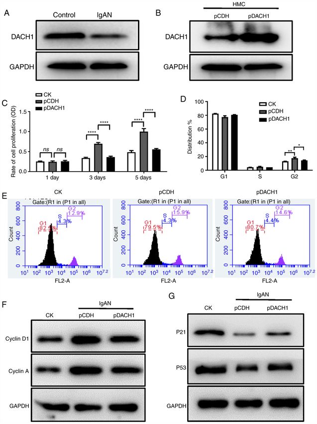
by Annexin /propidium iodide (PI) single staining statistical significance (ns), P4 ZHOU et al: miR-140-3p MEDIATES DACH1 EXPRESSION IN IgAN VIA CELL CYCLE-DEPENDENT MECHANISMS Figure 1. Overexpression of DACH1 reduces HMCs proliferation and inhibits cell cycle progression. (A) The expression of DACH1 in HMCs cultured with pIgA‑IgAN or pIgA‑control was detected by western blotting. (B) Western blotting for DACH1 protein in HMCs infected with DACH1 or control lentivirus (pCDH). (C) HMCs stably transduced with lentiviral DACH1, as well as the control empty vector (pCDH) were seeded and examined for cell proliferation with CCK‑8 test on days 1, 3 and 5 (****P
MOLECULAR MEDICINE REPORTS 23: 134, 2021                                                            5

Figure 2. Overexpression of DACH1 decreases the release of inflammatory cytokines. (A) Secretion of IL‑6, IL‑8, IL‑13 and CXCL1 (****P
6              ZHOU et al: miR-140-3p MEDIATES DACH1 EXPRESSION IN IgAN VIA CELL CYCLE-DEPENDENT MECHANISMS

Figure 3. miR‑140‑3p directly targets DACH1. (A) The effect of miRNAs on endogenous DACH1 expression. HMCs were transfected with 10 miRNA mimics
(20 nM) and NC, western blotting was performed at 48 h after transfection. (B) Inhibition of DACH1 3'UTR reporter activity by miRNAs. Luciferase activity
was detected in 293T cells co‑transfected with 10 miRNA mimics (20 nM) or NC together with the pGL3‑DACH1 3'UTR luciferase reporter (***P
MOLECULAR MEDICINE REPORTS 23: 134, 2021                                                                 7

Figure 4. DACH1 decrease IgAN cell proliferation and cell cycle progression by regulating miR‑140‑3p expression. (A) Expression of miR‑140‑3p in HMCs
incubated with pIgA or control was detected by reverse transcription‑quantitative PCR (****P
8              ZHOU et al: miR-140-3p MEDIATES DACH1 EXPRESSION IN IgAN VIA CELL CYCLE-DEPENDENT MECHANISMS

Figure 5. DACH1 decreases the release of inflammatory cytokines by positively regulating miRNA‑140‑3p expression. (A) Secretion of IL‑6, IL‑8, IL‑13 and
CXCL1 in the supernatant of HMCs transfected with the miR‑140‑3p inhibitor or NC when pIgA‑IgAN was added or not. (B) Reverse transcription‑quantitative
PCR analyses of the mRNA levels of IL‑6, IL‑8, IL‑13 and CXCL1 in HMCs transfected with the miR‑140‑3p inhibitor or NC when pIgA‑IgAN was added or
not. Data are expressed as mean ± standard deviation. *P
MOLECULAR MEDICINE REPORTS 23: 134, 2021                                                       9

    According to previous study, abnormal miRNA expression         References
is present in most patients with IgAN and is closely associated
with the extent of the disease (40). miRNAs participate             1. Lai KN: Pathogenesis of IgA nephropathy. Nat Rev Nephrol 8:
                                                                       275‑283, 2012.
in almost every process involved in cancer occurrence,              2. McGrogan A, Franssen CF and de Vries CS: The incidence of
development and progression (22,41‑43). The present study              primary glomerulonephritis worldwide: A systematic review of
confirmed that miR‑140‑3p expression was upregulated in                the literature. Nephrol Dial Transplant 26: 414‑430, 2011.
                                                                    3. Mestecky J, Novak J, Moldoveanu Z and Raska M: IgA nephrop‑
HMCs treated with pIgA‑IgAN and that the upregulation of               athy enigma. Clin Immunol 172: 72‑77, 2016.
miR‑140‑3p meditated the expression of DACH1. The present           4. Boyd JK, Cheung CK, Molyneux K, Feehally J and Barratt J: An
study also demonstrated that miR‑140‑3p regulated cell                 update on the pathogenesis and treatment of IgA nephropathy.
                                                                       Kidney Int 81: 833‑843, 2012.
growth, cell cycle progression and the release of inflammatory      5. Zhu H, Wu K, Yan W, Hu L, Yuan J, Dong Y, Li Y, Jing K, Yang Y
cytokines in HMCs. Although the findings of the present study          and Guo M: Epigenetic silencing of DACH1 induces loss of trans‑
indicated that DACH1 was probably involved in the regulation           forming growth factor‑β1 antiproliferative response in human
                                                                       hepatocellular carcinoma. Hepatology 58: 2012‑2022, 2013.
of IgAN and was regulated by miR‑140‑3p, a variety of               6. Popov VM, Wu K, Zhou J, Powell MJ, Mardon G, Wang C
other factors are involved in regulating the occurrence and            and Pestell RG: The Dachshund gene in development and
development of IgAN and further studies on the pathogenesis            hormone‑responsive tumorigenesis. Trends Endocrinol Metab 21:
                                                                       41‑49, 2010.
of IgAN are warranted.                                              7. Ikeda K, Watanabe Y, Ohto H and Kawakami K: Molecular
    In summary, the present study demonstrated that DACH1              interaction and synergistic activation of a promoter by Six, Eya,
was downregulated in HMCs in the presence of pIgA‑IgAN.                and Dach proteins mediated through CREB binding protein. Mol
                                                                       Cell Biol 22: 6759‑6766, 2002.
Moreover, DACH1 regulated cell proliferation, cell cycle            8. Liu QQ, Zhou YQ, Liu HQ, Qiu WH, Liu H, Hu TY, Xu Q, Lv YM
progression and the release of inflammatory cytokines in               and Wu KM: Decreased DACH1 expression in glomerulopathy
HMCs and these phenomena may be targeted by miR‑140‑3p.                is associated with disease progression and severity. Oncotarget 7:
                                                                       86547‑86560, 2016.
This novel pathway is therefore expected to provide a weak          9. Sunde JS, Donninger H, Wu K, Johnson ME, Pestell RG,
theoretical basis for IgAN research.                                   Rose GS, Mok SC, Brady J, Bonome T and Birrer MJ: Expression
                                                                       profiling identifies altered expression of genes that contribute to
                                                                       the inhibition of transforming growth factor‑beta signaling in
Acknowledgements                                                       ovarian cancer. Cancer Res 66: 8404‑8412, 2006.
                                                                   10. Wu K, Li A, Rao M, Liu M, Dailey V, Yang Y, Di Vizio D,
Not applicable.                                                        Wang C, Lisanti MP, Sauter G, et al: DACH1 is a cell fate deter‑
                                                                       mination factor that inhibits cyclin D1 and breast tumor growth.
                                                                       Mol Cell Biol 26: 7116‑7129, 2006.
Funding                                                            11. Dalgin GS, Drever M, Williams T, King T, DeLisi C and Liou LS:
                                                                       Identification of novel epigenetic markers for clear cell renal cell
                                                                       carcinoma. J Urol 180: 1126‑1130, 2008.
No funding was received.                                           12. Yamada Y, Arao T, Gotoda T, Taniguchi H, Oda I, Shirao K,
                                                                       Shimada Y, Hamaguchi T, Kato K, Hamano T, et al: Identification
Availability of data and materials                                     of prognostic biomarkers in gastric cancer using endoscopic
                                                                       biopsy samples. Cancer Sci 99: 2193‑2199, 2008.
                                                                   13. Chen R, Amoui M, Zhang Z and Mardon G: Dachshund and eyes
All data generated or analyzed during this study are included          absent proteins form a complex and function synergistically to
in this published article.                                             induce ectopic eye development in Drosophila. Cell 91: 893‑903,
                                                                       1997.
                                                                   14. Shen W and Mardon G: Ectopic eye development in Drosophila
Authors' contributions                                                 induced by directed dachshund expression. Development 124:
                                                                       45‑52, 1997.
                                                                   15. Fan GK, Imanaka M, Yang B and Takenaka H: Characteristics
XZ and YL designed the study and performed the experiments.            of nasal inverted papilloma and its malignant transformation:
XZ drafted the manuscript. PG and CZ were major contribu‑              A study of cell proliferation and programmed cell death. Am
                                                                       J Rhinol 20: 360‑363, 2006.
tors in the conception, design and reviewing of the manuscript.    16. Liu M, Zhang H, Li Y, Wang R, Li Y, Zhang H, Ren D, Liu H,
Based on their contributions, XZ was listed as the first author,       Kang C and Chen J: HOTAIR, a long noncoding RNA, is a
while CZ was the author for correspondence. All authors read           marker of abnormal cell cycle regulation in lung cancer. Cancer
                                                                       Sci 109: 2717‑2733, 2018.
and approved the final manuscript.                                 17. Wang W, Dong M, Cui J, Xu F, Yan C, Ma C, Yi L, Tang W,
                                                                       Dong J and Wei Y: NME4 may enhance nonsmall cell lung
                                                                       cancer progression by overcoming cell cycle arrest and promoting
Ethics approval and consent to participate                             cellular proliferation. Mol Med Rep 20: 1629‑1636, 2019.
                                                                   18. Hanahan D and Weinberg RA: Hallmarks of cancer: The next
All patients provided written informed consent and all serum           generation. Cell 144: 646‑674, 2011.
                                                                   19. Chen K, Wu K, Cai S, Zhang W, Zhou J, Wang J, Ertel A,
samples were anonymized. The present study was approved                Li Z, Rui H, Quong A, et al: Dachshund binds p53 to block
by the clinical research ethics committee of Shanghai Tenth            the growth of lung adenocarcinoma cells. Cancer Res 73:
People's Hospital (approval no. SHSY‑IEC‑4.1/20‑117‑01).               3262‑3274, 2013.
                                                                   20. Kalousova A, Mavropoulos A, Adams BA, Nekrep N, Li Z,
                                                                       Krauss S, Stainier DY and German MS: Dachshund homologues
Patient consent for publication                                        play a conserved role in islet cell development. Dev Biol 348:
                                                                       143‑152, 2010.
                                                                   21. He L and Hannon GJ: MicroRNAs: Small RNAs with a big role
Not applicable.                                                        in gene regulation. Nat Rev Genet 5: 522‑531, 2004.
                                                                   22. Bartel DP: MicroRNAs: Genomics, biogenesis, mechanism, and
Competing interests                                                    function. Cell 116: 281‑297, 2004.
                                                                   23. Kloosterman WP and Plasterk RH: The diverse functions of
                                                                       microRNAs in animal development and disease. Dev Cell 11:
The authors declare that they have no competing interests.             441‑450, 2006.
10 ZHOU et al: miR-140-3p MEDIATES DACH1 EXPRESSION IN IgAN VIA CELL CYCLE-DEPENDENT MECHANISMS
24. Li K, Du Y, Jiang BL and He JF: Increased microRNA‑155 and 34. Morgan DO: Principles of CDK regulation. Nature 374: 131‑134,
decreased microRNA‑146a may promote ocular inflammation 1995.
and proliferation in Graves' ophthalmopathy. Med Sci Monit 20: 35. Kim JK and Diehl JA: Nuclear cyclin D1: An oncogenic driver in
639‑643, 2014. human cancer. J Cell Physiol 220: 292‑296, 2009.
25. Wang W, Zhao LJ, Tan YX, Ren H and Qi ZT: miR‑138 induces 36. Wolf G and Shankland SJ: Cell cycle control in glomerular
cell cycle arrest by targeting cyclin D3 in hepatocellular carci‑ disease. Prog Cell Cycle Res 5: 71‑79, 2003.
noma. Carcinogenesis 33: 1113‑1120, 2012. 37. Shankland SJ and Wolf G: Cell cycle regulatory proteins in renal
26. Tamouza H, Vende F, Tiwari M, Arcos‑Fajardo M, Vrtovsnik F, disease: Role in hypertrophy, proliferation, and apoptosis. Am
Benhamou M, Monteiro RC and Moura IC: Transferrin receptor J Physiol Renal Physiol 278: F515‑F529, 2000.
engagement by polymeric IgA1 induces receptor expression and 38. Li Y, Liu Z, Guo X, Shu J, Chen Z and Li L: Aristolochic acid
mesangial cell proliferation: Role in IgA nephropathy. Contrib I‑induced DNA damage and cell cycle arrest in renal tubular
Nephrol 157: 144‑147, 2007. epithelial cells in vitro. Arch Toxicol 80: 524‑532, 2006.
27. Lai KN, To WY, Li PK and Leung JC: Increased binding of 39. Marshall CB and Shankland SJ: Cell cycle and glomerular
polymeric lambda‑IgA to cultured human mesangial cells in IgA disease: A minireview. Nephron Exp Nephrol 102: e39‑48, 2006.
nephropathy. Kidney Int 49: 839‑845, 1996. 40. Dai Y, Sui W, Lan H, Yan Q, Huang H and Huang Y: Microarray
28. Livak KJ and Schmittgen TD: Analysis of relative gene expres‑ analysis of micro‑ribonucleic acid expression in primary immu‑
sion data using real‑time quantitative PCR and the 2(‑Delta Delta noglobulin A nephropathy. Saudi Med J 29: 1388‑1393, 2008.
C(T)) method. Methods 25: 402‑408, 2001. 41. Silveri L, Tilly G, Vilotte JL and Le Provost F: MicroRNA
29. Schena FP and Nistor I: Epidemiology of IgA nephropathy: A involvement in mammary gland development and breast cancer.
global perspective. Semin Nephrol 38: 435‑442, 2018. Reprod Nutr Dev 46: 549‑556, 2006.
30. Tian J, Wang Y, Liu X, Zhou X and Li R: Rapamycin ameliorates 42. Zhang B, Pan X, Cobb GP and Anderson TA: microRNAs as
IgA nephropathy via cell cycle‑dependent mechanisms. Exp Biol oncogenes and tumor suppressors. Dev Biol 302: 1‑12, 2007.
Med (Maywood) 240: 936‑945, 2015. 43. Khoshnaw SM, Green AR, Powe DG and Ellis IO: MicroRNA
31. Kurogi Y: Mesangial cell proliferation inhibitors for the treat‑ involvement in the pathogenesis and management of breast
ment of proliferative glomerular disease. Med Res Rev 23: 15‑31, cancer. J Clin Pathol 62: 422‑428, 2009.
2003.
32. Rodrigues JC, Haas M and Reich HN: IgA nephropathy. Clin This work is licensed under a Creative Commons
J Am Soc Nephrol 12: 677‑686, 2017. Attribution-NonCommercial-NoDerivatives 4.0
33. Leung JC, Chan LY, Tang SC, Lam MF, Chow CW, Lim AI International (CC BY-NC-ND 4.0) License.
and Lai KN: Oxidative damages in tubular epithelial cells in
IgA nephropathy: Role of crosstalk between angiotensin II and
aldosterone. J Transl Med 9: 169, 2011.You can also read


