SNORA72 Activates the Notch1/c-Myc Pathway to Promote Stemness Transformation of Ovarian Cancer Cells - Frontiers
←
→
Page content transcription
If your browser does not render page correctly, please read the page content below
ORIGINAL RESEARCH
                                                                                                                                      published: 03 November 2020
                                                                                                                                     doi: 10.3389/fcell.2020.583087

                                            SNORA72 Activates the
                                            Notch1/c-Myc Pathway to Promote
                                            Stemness Transformation of Ovarian
                                            Cancer Cells
                                            Liwen Zhang 1,2,3,4† , Rong Ma 1,2,3,4† , Mengcong Gao 1,2,3,4 , Yanyun Zhao 1,2,3,4 , Xuemei Lv 1,2,3,4 ,
                                            Wenjing Zhu 1,2,3,4 , Li Han 1,2,3,4 , Panpan Su 1,2,3,4 , Yue Fan 1,2,3,4 , Yuanyuan Yan 1,2,3,4 ,
                                            Lin Zhao 1,2,3,4 , Heyao Ma 1,2,3,4 , Minjie Wei 1,2,3,4* and Miao He 1,2,3,4*
                                            1
                                              Department of Pharmacology, School of Pharmacy, China Medical University, Shenyang, China, 2 Liaoning Key Laboratory
                                            of Molecular Targeted Anti-Tumor Drug Development and Evaluation, China Medical University, Shenyang, China, 3 Liaoning
                                            Cancer Immune Peptide Drug Engineering Technology Research Center, China Medical University, Shenyang, China,
                                            4
                                              Key Laboratory of Precision Diagnosis and Treatment of Gastrointestinal Tumors, Ministry of Education, China Medical
                                            University, Shenyang, China

                         Edited by:         Cancer stem cells (CSCs) are responsible for the migration and recurrence of
                  Richard G. Pestell,
        Baruch S. Blumberg Institute,
                                            cancer progression. Small nucleolar RNAs (snoRNAs) play important roles in tumor
                      United States         development. However, how snoRNAs contribute to the regulation of the stemness of
                      Reviewed by:          ovarian CSCs (OCSCs) remains unclear. In the present study, we found that SNORA72
                        Chunliang Li,
                                            was significantly upregulated in OVCAR-3 spheroids (OS) and CAOV-3 spheroids (CS)
        St. Jude Children’s Research
              Hospital, United States       with the OCSC characteristics attained by serum-free culture in a suspension of
                  Shreya Raghavan,          OVCAR-3 (OV) and CAOV-3 (CA) cells. The overexpression of SNORA72 increased
 Texas A&M University, United States
                                            self-renewal abilities and migration abilities in OV and CA cells and upregulated the
                  *Correspondence:
                            Miao He
                                            expressions of the stemness markers Nanog, Oct4, and CD133. In addition, the
               hemiao_cmu@126.com           ectopic expression of SNORA72 can elevate the messenger RNA (mRNA) and protein
                           Minjie Wei
                                            expression levels of Notch1 and c-Myc in parental cells. The opposite results were
               weiminjiecmu@163.com
     † These                                observed in SNORA72-silenced OCSCs. Moreover, we found that Notch1 knockdown
               authors have contributed
                    equally to this work    inversed the migration abilities and self-renewal abilities raised by overexpressing
                                            SNORA72. In summary, stemness transformation of ovarian cancer cells can be
                    Specialty section:
         This article was submitted to
                                            activated by SNORA72 through the Notch1/c-Myc pathway. This study introduces a
                  Stem Cell Research,       novel therapeutic strategy for improving the treatment efficiency of ovarian cancer.
                a section of the journal
  Frontiers in Cell and Developmental       Keywords: SNORA72, ovarian cancer stem cells (OCSCs), Notch1, c-Myc, stemness
                                Biology
            Received: 14 July 2020
        Accepted: 05 October 2020          INTRODUCTION
      Published: 03 November 2020
                              Citation:
                                           Ovarian cancer has the highest mortality rate among all gynecological cancers (Siegel et al.,
      Zhang L, Ma R, Gao M, Zhao Y,        2020). More than 80% of patients with ovarian cancer present with advanced-stage disease when
     Lv X, Zhu W, Han L, Su P, Fan Y,      diagnosed (Narod, 2016; Li et al., 2018). It has been reported that approximately 15–25% of
     Yan Y, Zhao L, Ma H, Wei M and        patients are resistant to initial platinum-based chemotherapy; even patients with an initial complete
    He M (2020) SNORA72 Activates
           the Notch1/c-Myc Pathway
                                           Abbreviations: CA, CAOV-3 cells; cDNA, complementary DNA; CRC, colorectal cancer; CS, CAOV-3 spheroid cells;
to Promote Stemness Transformation         CSCs, cancer stem cells; ncRNA, non-coding RNA; OCSCs, ovarian cancer stem cells; OV, OVCAR-3 cells; OS, OVCAR-
              of Ovarian Cancer Cells.     3 spheroid cells; PDAC, pancreatic ductal adenocarcinoma; PFS, progression-free survival; qRT-PCR, quantitative real-time
      Front. Cell Dev. Biol. 8:583087.     PCR; shRNA, short hairpin RNA; snoRNAs, small nucleolar RNAs; SNORDs, C/D box snoRNAs; SNORAs, H/ACA box
      doi: 10.3389/fcell.2020.583087       snoRNAs.

Frontiers in Cell and Developmental Biology | www.frontiersin.org                 1                                      November 2020 | Volume 8 | Article 583087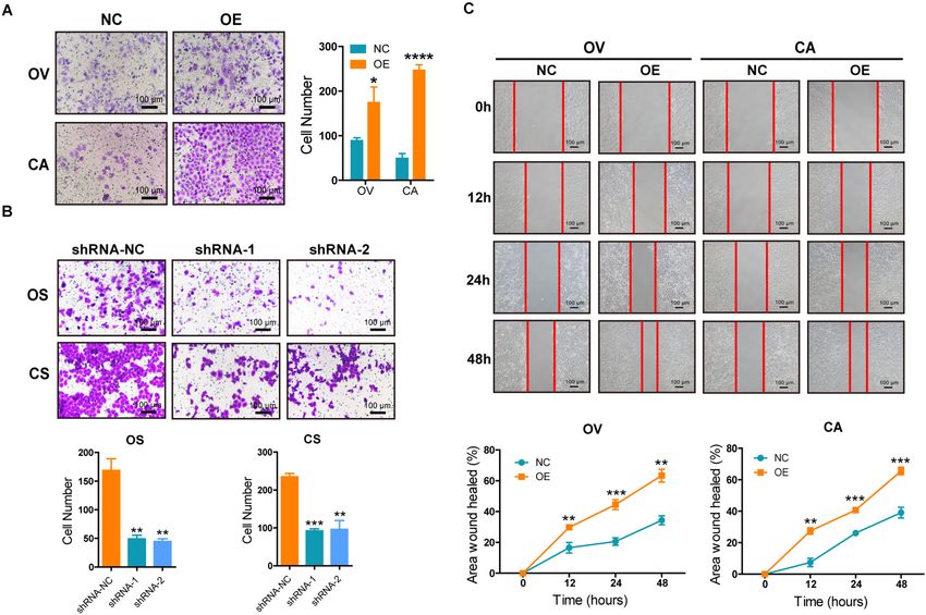
Zhang et al.                                                                                           SNORA72 Promotes the Stemness of OCCs

response will relapse and develop resistance (Agarwal and Kaye,          at a density of 1 × 10⁵ cell/ml from monolayers of OV and
2003; Bapat et al., 2005; Zhang et al., 2014; Ottevanger, 2017).         CA into a serum-free Dulbecco’s modified Eagle’s medium
Targeting the roots of metastasis and resistance of ovarian cancer       (DMEM)/F12 medium (Hyclone) with 10 ng/ml basic fibroblast
cells is the key to treating ovarian cancer. Cancer stem cells           growth factor (bFGF; Peprotech Corporation, United States),
(CSCs) are rare cells in cancers with indefinite potential for           20 ng/ml epidermal growth factor (EGF; Peprotech Corporation),
self-renewal that drive tumorigenesis and recreate heterogeneity.        and 2% B27 (Invitrogen Corporation, United States). After
Growing evidence shows that CSCs not only influence cancer               several generations, the cells grew into nonadherent spherical
initiation but also drive tumor progression, therapy resistance,         clusters termed OVCAR-3 spheres (OS) and CAOV3 spheres
and metastatic growth (Reya et al., 2001; Chen et al., 2017; Lytle       (CS). The cell lines used in this study did not suffer
et al., 2018). Therefore, exploring the targets and mechanisms of        from mycoplasma infection. All cell lines were authenticated
maintaining the stemness of ovarian CSCs (OCSCs) will provide            using STR profiling.
better insights into the treatment of ovarian cancer.
    Small nucleolar RNAs (snoRNAs) are one of the best-                  Limiting Gradient Dilution
characterized classes of non-coding RNAs (ncRNAs) in the                 Single cell suspensions of OV, OS, CA, and CS cells were
nucleus and are 60–300 nucleotides in length. They are classified        seeded into 96-well plates at a density of 200, 100, 50, or
as C/D box snoRNAs (SNORDs), serving as guides for 20 -                  25 cells/well, and each gradient has 20 wells. The number of
O-ribose methylation of ribosomal RNAs (rRNAs) or small                  wells with colonies in each cell gradient (Wells with Colonies)
nuclear RNAs (snRNAs), and H/ACA box snoRNAs (SNORAs),                   and the total number of monoclonal cell spheres in each cell
guiding the isomerization of uridine residues into pseudouridine         gradient (Total colonies) were counted, and the percentage of
(Jorjani et al., 2016). Accumulating evidence suggests that              the total number of monoclonal cell spheres to the total cells
the dysregulation of snoRNAs can function in controlling                 of each cell gradient (Total colonies/Total cells × 100%) was
cell fate and regulating tumorigenesis and development                   calculated after 9 days.
(Gong et al., 2017; Romano et al., 2017). Cui et al. found
increased SNORA23 expression in human pancreatic ductal
adenocarcinoma (PDAC) tissues, and SNORA23 promoted                      Flow Cytometry
the survival and invasion of PDAC cells (Cui et al., 2017).              CD133 expression in OV and OS cells was measured by flow
SNORA21 was systematically identified as a key oncogenic                 cytometry. The cells were collected and washed several times with
snoRNA in colorectal cancer (CRC), which might serve as                  phosphate-buffered saline (PBS). The cells were incubated with
a prognostic biomarker (Yoshida et al., 2017). In recent                 CD133 antibody (1:20, BD Pharmingen, United States) at 4◦ C for
years, the impact of snoRNAs on the characteristics of CSCs              15–20 min in the dark. After that, the cells were washed with cold
has gained more and more attention. SNORA42 has been                     PBS three times. Finally, we tested single cell suspensions on a
reported to contribute to the self-renewal and proliferation             flow cytometer (ACEA Biosciences Inc., China).
abilities of lung tumor-initiating cells (Mannoor et al., 2014).
However, abnormal snoRNAs in OCSCs and their functions have              Quantitative Reverse Transcription PCR
rarely been reported.                                                    Total RNA was extracted using the TRIzol reagent, and snoRNAs
    In this study, we found that SNORA72 was highly expressed            were transcribed into complementary DNA (cDNA) in a 10-µl
in ovarian sphere cells with OCSC-like characteristics. The              reaction volume, which contains 2 µl 5× M-MLV buffer, 0.3 µl
ectopic expression of SNORA72 increased the self-renewal                 M-MLV (200 U/µl), 0.25 µl RNase inhibitor (40 U/µl), 1 µl
and migration abilities of ovarian cancer cells. Conversely,             dNTP (1 U/µl), 1 µl RT-primer (5 µM), and 5.45 µl RNA
the knockdown of SNORA72 inhibited the self-renewal and                  (200 ng/µl) at 42◦ C for 60 min, 70◦ C for 10 min, and then kept
migration abilities of OCSCs. Furthermore, we demonstrated that          at 4◦ C. The messenger RNAs (mRNAs) of other genes reacted
SNORA72 might maintain the stemness of OCSCs by activating               in 10 µl volume with 2 µl 5× RT, 0.5 µl enzyme mix, 0.5 µl
the Notch1/c-Myc pathway.                                                primer mix, and 7 µl RNA at 37◦ C for 15 min, 9◦ C for 5 min,
                                                                         and then kept at 4◦ C. Specific SNORA72 primer sequences were
                                                                         designed by RiboBio (Guangzhou, China) and small nuclear
MATERIALS AND METHODS                                                    RNA U6 was used as the internal control. The mRNA expression
                                                                         levels were normalized to that of β-actin. The primer sequences
Induction of Ovarian Cancer Spheroid                                     were: β-actin forward 50 -TCCTCCCTGGAGAAGAGCTA-30 ,
Cells                                                                    reverse 50 -TCCTGCTTGCTGATCCACAT-30 ; Oct4 forward
A method for inducing cancer cells to CSCs was reported                  50 -CTTGCTGCAGAAGTGGGTGGAGGAA-30 , reverse 50 -CTG
previously (Zhong et al., 2010; Wang et al., 2012; He et al.,            CAGTGTGGGTTTCGGGCA-30 ; Nanog forward 50 AATACCT
2019). In brief, OVCAR-3 (OV) (RRID:CVCL_0465) and                       CAGCCTCCAGCAGATG-30 , reverse 50 -TGCGTCACACCATT
CAOV3 (CA) (RRID:CVCL_0201) cells (Cell Bank of the                      GCTATTCTTC-30 ; c-Myc forward 50 -CGACGAGACCTTC
Chinese Academy of Sciences, Shanghai, China) were cultured              ATCAAAAAC-30 , reverse 50 -CTTCTCTGAGACGAGCTTGG-
in RPMI-1640 medium (Hyclone, United States) with 10%                    30 ; and CD133 forward 50 -GTGGCGTGTGCGGCTATGAC-30 ,
fetal bovine serum (FBS) (Tian Jin Hao Yang Biological                   reverse 50 -CCAACTCCAACCATGAGGAAGACG-30 . The
Manufacture CL., LTD, China). Tumor spheres were derived                 2−11Ct method was used to calculate the relative fold.

Frontiers in Cell and Developmental Biology | www.frontiersin.org    2                                 November 2020 | Volume 8 | Article 583087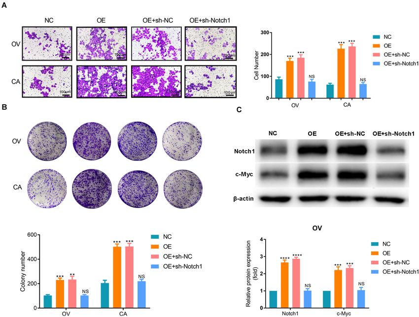
Zhang et al.                                                                                                                  SNORA72 Promotes the Stemness of OCCs

Reverse Transcription Polymerase Chain                                                   Plate Clone Formation Assay
Reaction                                                                                 The cells transfected with the indicated plasmids for 48 h
The reverse transcription process was consistent with the                                were digested using 0.25% trypsin and pipetted into single
quantitative reverse transcription PCR (qRT-PCR) process. After                          cells. The resuspended single cells were seeded in a six-well
the reverse transcription product was diluted 10 times, it was                           plate (1,000 cells per well) and placed in an incubator at
diluted with 5 µl Taq enzyme, 0.5 µl upstream primer, 0.5 µl                             37◦ C, 5% CO2 atmosphere, and saturated humidity for 2–3
downstream primer, and 4 µl cDNA to form a 10-µl reaction                                weeks. Afterward, the cells were washed with PBS two to three
volume in the following procedure: 98◦ C for 10 s; 60◦ C for 30 s;                       times, and the cells were fixed with 1 ml 4% paraformaldehyde
72◦ C for 60 s, 25 cycles for snoRNA reaction, and 30 cycles                             in each well. The cells were stained with 0.5% crystal violet
for mRNA reaction.                                                                       (Sigma-Aldrich 46364) for 30 min and then washed until
                                                                                         the background color was clean. Pictures were taken and the
Transfection                                                                             number of colonies were counted using a Nikon eclipse TE2000-
SNORA72-cDNA plasmids (Shanghai Genechem Co., LTD,                                       U microscope.
China) were used to transfect the OV and CA cells in
a six-well plate to overexpress SNORA72. The shRNA-                                      Sphere Formation Assays
SNORA72 plasmids (Shanghai Genechem Co., LTD) were                                       After transfection with the indicated plasmids for 48 h, OS
transfected into the OS and CS cells seeded in six-well                                  and CS cells were cultured in ultra-low adherent six-well plates
plates to inhibit SNORA72 expression. shRNA-Notch1 plasmids                              with a density of 2,000 cells/well in DMEM-F12 medium
(Shanghai Genechem Co., LTD) were used to transfect OV and                               (HyClone) containing 10 ng/ml bFGF (Peprotech Corporation),
CA cells to inhibit the expression of Notch1. Lipofectamine                              20 ng/ml EGF (Peprotech Corporation), and 2% B27 (Invitrogen
3000 (Invitrogen) was used for transfection according to the                             Corporation). After culturing for 14 days, spheres with a diameter
manufacturer’s instructions.                                                             >150 µm were counted.

  FIGURE 1 | SNORA72 is overexpressed in ovarian cancer stems cells (OCSCs). (A) Morphology of OVCAR-3 (OV), OVCAR-3 spheroid (OS), CAOV-3 (CA), and
  CAOV-3 spheroid (CS) cells shown under a microscope (×10). (B) Expression of CD133 detected by flow cytometry in OV vs OS and in CA vs CS cells.
  (C) Migration abilities of OV, OS, CA, and CS cells by Transwell assay. (D) Differentiation morphology of OS and CS cells at 0, 12, 24, 48, and 72 h. (E) Hierarchical
  clustering analysis of small nucleolar RNA (snoRNA) expression from non-coding RNA-ChIP data in OV and OS cells. Red, higher expression levels; green, lower
  expression levels. (F) Relative SNORA72 expression to U6, as an endogenous control, analyzed by qRT-PCR in OV and OS cells. The SNORA72 expression in OV
  cells was set as 1. Data are shown as the mean ± SD from three independent experiments. **P < 0.01, ****P < 0.0001.

Frontiers in Cell and Developmental Biology | www.frontiersin.org                    3                                        November 2020 | Volume 8 | Article 583087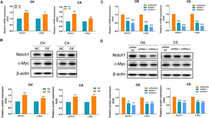
Zhang et al.                                                                                                                            SNORA72 Promotes the Stemness of OCCs

Bioinformatics Analysis                                                                     analysis. We performed gene correlation analysis using the
Kaplan–Meier survival curves of progression-free survival (PFS)                             cBioPortal2 and the online tool R2: Genomics Analysis and
for SNORA72 were produced using an online tool called                                       Visualization Platform3 .
Kaplan–Meier Plotter1 , and 1,435 patients were included for
                                                                                            2
                                                                                                http://www.cbioportal.org/
1                                                                                           3
    http://kmplot.com/analysis/index.php?p=service&cancer=ovar                                  https://hgserver1.amc.nl/cgi-bin/r2/main.cgi

TABLE 1 | Limited gradient dilution analysis experiment for OV, OS, CA and CS.

                                            OV                                  OS                                          CA                                     CS

Dilution ratio               1/1      1/2         1/4    1/8     1/1      1/2         1/4           1/8     1/1       1/2         1/4       1/8     1/1      1/2         1/4    1/8
Cells/well                   200      100         50     25     200       100         50            25      200      100          50        25     200      100          50     25
Wells with colonies          12        8          7       3      20       20          18            16       14        9          7          4      20       20          18     15
Total colonies               163       49         21      9    1,215      559        235           107      176       54          25        11     1,179    535         233     113
Total cells                 4,000    2,000       1,000   500   4,000     2,000       1,000         500     4,000     2,000       1,000      500    4,000   2,000        1,000   500
Colonies/total cells (%)    4.07      2.45        2.1    1.8   30.37     27.95       23.5          21.4     4.4       2.7         2.5       2.2    29.48   26.75        23.3    22.6

OV, OVCAR-3 cells; OS, OVCAR-3 spheroids cells; CA, CAOV-3 cells; CS, CAOV-3 spheroids cells.

    FIGURE 2 | SNORA72 overexpression increases the self-renewal of OVCAR-3 (OV) and CAOV-3 (CA) cells. Expression of SNORA72 determined in OV and CA
    ovarian cells at 24, 48, and 72 h after transfection of the SNORA72 overexpression (OE) or negative control (NC) plasmids by (A) quantitative reverse transcription
    PCR (qRT-PCR) and (B) RT-PCR analysis. (C) Changes of the clone performing ability detected by the plate clone formation assays in OV and CA cells transfected
    with the OE or NC plasmids. (D) Relative mRNA expressions of Nanog, Oct4, and CD133 to β-actin, as an endogenous control, analyzed by qRT-PCR in OV and CA
    cells after transfection of the OE or NC plasmids for 48 h. The expression level in OV and CA cells after transfection of NC plasmids was set as 1. *P < 0.05,
    **P < 0.01, ***P < 0.001, and ****P < 0.0001.

Frontiers in Cell and Developmental Biology | www.frontiersin.org                     4                                                  November 2020 | Volume 8 | Article 583087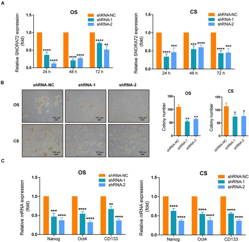
Zhang et al.                                                                                                           SNORA72 Promotes the Stemness of OCCs

Transwell Assay                                                                     membrane were counted under a light microscope and analyzed
The upper chambers of Transwell plates precoated with Matrigel                      using ImageJ software.
(Corning, United States) were plated with 1 × 10⁴ OV or CA
cells in 500 µl serum-free RPMI-1640 medium or 1 × 10⁴                              Wound-Healing Assays
OS or CS cells in 500 µl serum-free DMEM/F12 medium. The                            Cells were cultured in six-well plates until 70% confluency. OV
lower chambers were filled with 500 µl medium containing                            and CA cells were transfected with SNORA72-NC, SNORA72-
10% FBS. Then, the cells were allowed to migrate toward the                         OE, SNORA72-OE+sh-NC, and SNORA72-OE+sh-Notch1.
lower chambers for 48 h in an atmosphere of 5% CO2 at 37◦ C.                        Linear “scratches” were created on the monolayer cells in straight
The chambers were then fixed with methanol and stained with                         lines with sterile tips. The cells were washed three times with PBS
0.1% crystal violet. The number of cells migrating across the                       and serum-free medium was added. The cells were photographed

  FIGURE 3 | SNORA72 knockdown decreases the self-renewal of OVCAR-3 spheroid (OS) and CAOV-3 spheroid (CS) cells. (A) Expression of SNORA72 determined
  in OS and CS cells at 24, 48, and 72 h after transfection of the SNORA72 silencing (shRNA-1 and shRNA-2) or control (shRNA-NC) plasmids by quantitative reverse
  transcription PCR (qRT-PCR) analysis. (B) Self-renewal abilities were measured by colony formation assays in OS and CS cells transfected with the shRNA-1,
  shRNA-2, or shRNA-NC plasmids. (C) Relative mRNA expressions of Nanog, Oct4, and CD133 to β-actin, as an endogenous control, analyzed by qRT-PCR in OS
  and CS cells after transfection of the shRNA-1, shRNA-2, or shRNA-NC plasmids for 48 h. The expression level in OS and CS cells after transfection of shRNA-NC
  plasmids was set as 1. *P < 0.05, **P < 0.01, ***P < 0.001, and ****P < 0.0001.

Frontiers in Cell and Developmental Biology | www.frontiersin.org               5                                       November 2020 | Volume 8 | Article 583087
Zhang et al.                                                                                                             SNORA72 Promotes the Stemness of OCCs

after 0, 24, and 48 h of incubation under a microscope (Nikon                         RESULTS
Eclipse TE2000-U, Japan). Wound closure was quantified using
ImageJ software.                                                                      SNORA72 Is Highly Expressed in OCSCs
                                                                                      We induced and successfully enriched OCSCs according to
Western Blot                                                                          section “Induction of Ovarian Cancer Spheroids Cells” and our
The cells were inoculated into RIPA lysis buffer containing                           previous reports (He et al., 2019; Zhu et al., 2019). As shown in
1% proteinase inhibitor cocktail solution and 1% phosphatase                          Figure 1A, the OS and CS had a dense spherical morphology. The
inhibitor cocktail solution. Extracted protein samples were                           expression of CD133, an OCSC marker, was found to be higher in
loaded on SDS-polyacrylamide separating gel and transferred                           OS and CS than in its parental OV and CA cells by flow cytometry
onto PVDF membranes (Millipore). The primary antibodies used                          (Figure 1B). In addition, we also compared the monoclonal cell
were c-Myc (1:1,000, Cell Signaling Technology, United States),                       sphere formation ability of OV vs OS as well as CA vs CS cells
Notch1 (1:1,000, Cell Signaling Technology), and β-actin                              through limiting gradient dilution analysis. As shown in Table 1,
(1:1,000, Absin Bioscience Inc., China). Protein expression was                       under each cell dilution gradient, the proportion of monoclonal
quantitatively analyzed using Scion Image Software (Scion Corp.,                      spheres in OS and CS cells is higher than in that OV and CA
Frederick, MA, United States).                                                        cells, indicating that spheroid cells have a strong ability to form
                                                                                      monoclonal spheres. The Transwell assay showed that OS and
                                                                                      CS had significantly higher number of cells migrating across the
Statistical Analysis                                                                  membrane than did OV and CA (Figure 1C). Moreover, in order
Statistical analyses were performed using the GraphPad Prism                          to identify the differentiation potential of OS and CS cells, we
7.0 software. Differences between two groups were statistically                       transferred serum-free non-adherent cultured OS and CS cells
analyzed using Student’s t test, which was considered statistically                   into 1640 medium with 10% FBS. OS and CS cells began to grow
significant when the P value was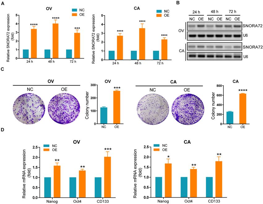
Zhang et al. SNORA72 Promotes the Stemness of OCCs
spheroids at 72 h (Figure 1D). Therefore, these results suggested plasmids (OE) after 24, 48, and 72 h were upregulated 3. 38-, 4.
that OS and CS cells had stem cell properties. 01-, and 2.93-fold, respectively, by qRT-PCR analysis compared
In order to explore the factors that affect the maintenance to the control (NC). In CA cells, SNORA72 expression levels in
of the stemness of OCSCs, we performed non-coding RNA the OE groups were 2. 69-, 3. 55-, and 2.29-fold, respectively,
chromatin immunoprecipitation (ChIP) analysis of OV and OS compared to those in the NC groups after 24, 48, and 72 h
cells. Interestingly, we found that a large number of snoRNAs transfection (Figure 2A). Similarly, agarose electrophoresis after
were abnormally expressed between OV and OS. We speculated RT-PCR also showed an increased expression of SNORA72 in OE
that snoRNAs might play major roles in the stemness phenotype ovarian cancer cells (Figure 2B). Next, we detected changes in
transformation of ovarian cancer cells. Further analysis revealed the clone performing ability after SNORA72 overexpression by
that SNORA72 was highly expressed in OS cells compared to plate clone formation assays. We found that the overexpression
OV cells (Figure 1E). Consistent with this, we observed that of SNORA72 markedly increased the number of clones and the
SNORA72 expression was 10.06 ± 0.392-fold in OS cells relative size of OV and CA cells (Figure 2C). In addition, we observed
to OV cells by qRT-PCR analysis (Figure 1F). Thus, the data that the mRNA expressions of the stemness biomarkers CD133,
indicate that it is necessary to explore the effects of SNORA72 Nanog, and Oct4 (Zhu et al., 2019) were significantly increased
on stemness maintenance in ovarian cancer cells. in OV and CA cells after the overexpression of SNORA72
(Figure 2D). The expression levels of CD133 at different times
are shown in Supplementary Figures S1A,B. The results show
SNORA72 Maintains Self-Renewal of that the expression levels of CD133 can be increased by the
OCSCs overexpression of SNORA72.
In order to investigate the effects of SNORA72 on the stemness We also successfully constructed SNORA72 knockdown
phenotype transformation of ovarian cancer cells, we first ovarian cancer cells. Transfection of sh-SNORA72 plasmids after
transfected SNORA72-cDNA plasmids into OV and CA cells 24, 48, and 72 h notably reduced SNORA72 expression in both OS
to evaluate changes in their self-renewal abilities. SNORA72 and CS cells (Figure 3A). Additionally, sphere formation assays
expression levels in OV cells transfected with SNORA72-cDNA showed that the knockdown of SNORA72 markedly decreased
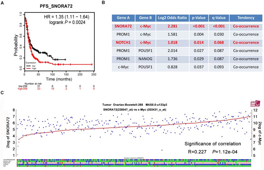
FIGURE 5 | SNORA72 expression is positively associated with the c-Myc expression of ovarian cancer patients. (A) Online tool Kaplan–Meier Plotter used to analyze
the relationship between SNORA72 expression and the progression-free survival (PFS) of ovarian cancer patients. (B) The relationship between SNORA72
expression and stemness markers was analyzed by the online analysis website cBioPortal. (C) R2: Genomics Analysis and Visualization Platform used to analyze the
expression correlation between SNORA72 and c-Myc.
Frontiers in Cell and Developmental Biology | www.frontiersin.org 7 November 2020 | Volume 8 | Article 583087Zhang et al. SNORA72 Promotes the Stemness of OCCs
the number of spheres of OS and CS cells (Figure 3B). We also cells compared to the control cells. All the data suggest that
found that the expressions of CD133, Nanog, and Oct4 mRNAs SNORA72 can promote the migration abilities of ovarian cancer
were significantly downregulated in sh-SNORA72-transfected cells (Figure 4C).
OS and CS cells than in sh-NC-transfected cells (Figure 3C).
The expression levels of CD133 at different times are shown SNORA72 Expression Is Positively
in Supplementary Figures S1C,D. The results show that the
expression levels of CD133 can be decreased by the knockdown
Correlated With c-Myc Expression in
of SNORA72. All these findings suggest that SNORA72 can Ovarian Cancer Patients
promote the self-renewal ability of ovarian cancer cells. In order to assess the effects of SNORA72 expression on the
progression of ovarian cancer patients, we used the online
analysis website Kaplan–Meier Plotter to analyze the association
SNORA72 Maintains the Migration of SNORA72 expression with PFS in ovarian cancer patients.
Abilities of OCSCs We found that patients with a high SNORA72 expression had
Increased migration ability is one of the features of CSCs. a shorter PFS time (HR = 1.35, P = 0.0024; Figure 5A).
We explored the effects of SNORA72 on the migration These findings suggest that SNORA72 has a higher degree
ability of OCSCs. The Transwell assays showed that the of correlation with poor prognosis in ovarian cancer patients.
overexpression of SNORA72 significantly increased the number Next, we used the online analysis website cBioPortal to analyze
of cells migrating across the membrane in OV and CA cells whether SNORA72 was related to certain stemness markers
(Figure 4A), while the number of cells migrating across the in ovarian cancer patients. The analysis results showed that
membrane in OS and CS cells after knocking down SNORA72 the expressions of SNORA72 and c-Myc had co-expression
markedly decreased (Figure 4B). In addition, the wound- characteristics (P < 0.001; Figure 5B). We further used the R2:
healing assays showed that the overexpression of SNORA72 Genomics Analysis and Visualization Platform to analyze the
significantly increased the wound-healed areas in OV and CA expression correlation between SNORA72 and c-Myc and also
FIGURE 6 | SNORA72 induces the stemness of ovarian cancer by activating the Notch1/c-Myc pathway. (A) Relative mRNA expressions of Notch1 and c-Myc to
β-actin, as an endogenous control, analyzed by quantitative reverse transcription PCR (qRT-PCR) in OVCAR-3 (OV) and CAOV-3 (CA) cells after transfection of the
transfected (OE) or control (NC) plasmids for 48 h. The expression level in OV and CA cells after transfection of NC plasmids was set as 1. (B) Relative protein
expressions of Notch1 and c-Myc to β-actin analyzed by Western blot in OV and CA cells after transfection of the OE or NC plasmids for 48 h. The expression level
in OV and CA cells after transfection of NC plasmids was set as 1. (C) Relative mRNA expressions of Notch1 and c-Myc to β-actin analyzed by qRT-PCR in
OVCAR-3 spheroid (OS) and CAOV-3 spheroid (CS) cells after transfection of the shRNA-1, shRNA-2, or shRNA-NC plasmids for 48 h. The expression level in OS
and CS cells after transfection of shRNA-NC plasmids were set as 1. (D) Relative protein expressions of Notch1 and c-Myc to β-actin analyzed by Western blot in
OS and CS cells after transfection of the shRNA-1, shRNA-2, or shRNA-NC plasmids for 48 h. The expression level in OS and CS cells after transfection of
shRNA-NC plasmids was set as 1. *P < 0.05, **P < 0.01, ***P < 0.001, and ****P < 0.0001.
Frontiers in Cell and Developmental Biology | www.frontiersin.org 8 November 2020 | Volume 8 | Article 583087Zhang et al. SNORA72 Promotes the Stemness of OCCs
found a significantly positive relationship between SNORA72 and self-renewal abilities, and protein expressions of Notch1 and
c-Myc expressions (R = 0.227, P = 1.12e−04 ; Figure 5C). c-Myc. We found that silencing Notch1 could inverse the effects
of SNORA72 overexpression on the migration, self-renewal
SNORA72 Promotes Stemness of OCSCs abilities, and protein expressions of Notch1 and c-Myc in OV
and CA cells (Figures 7A–C). Thus, these findings suggest
by Regulating the Notch1/c-Myc
that SNORA72 can induce the stemness of ovarian cancer by
Pathway activating the Notch1/c-Myc pathway.
Based on the results of the bioinformatics analyses, we assume
that SNORA72 may regulate c-Myc expression to maintain the
stemness of OCSCs. c-Myc is a major target gene of the Notch
pathway. Therefore, we measured the expression changes of DISCUSSION
c-Myc and Notch1 after inferring SNORA72 expression. We
found that the mRNA and protein levels of Notch1 and c-Myc Ovarian cancer is a malignant cancer with a high migration and
expressions were both markedly elevated in OV and CA cells invasive potential and a low survival rate since many patients
transfected with SNORA72-OE plasmids compared with the cells are at an advanced stage when diagnosed (Vaughan et al., 2011).
transfected with NC plasmids (Figures 6A,B). Conversely, the CSCs are considered the source of cancer cells that arise and
knockdown of SNORA72 in OS and CS cells notably decreased are responsible for the metastasis and relapse of cancer (Peiris-
the mRNA and protein expressions of Notch1 and c-Myc Pages et al., 2016). Therefore, exploring the mechanisms of cancer
(Figures 6C,D). Next, we respectively transfected SNORA72-NC, development and searching for therapeutic strategies for the
SNORA72-OE, SNORA72-OE+sh-NC, or SNORA72-OE+sh- effective eradication of cancer by targeting CSCs have attracted
Notch1 plasmids in OV and CA cells and detected the migration, much attention (Eun et al., 2017; Shibata and Hoque, 2019).
FIGURE 7 | SNORA72-NC, SNORA72-OE, SNORA72-OE+sh-NC, or SNORA72-OE+sh-Notch1 plasmids were, respectively, transfected in OVCAR-3 (OV) or
CAOV-3 (CA) cells. (A) Migration abilities of OV and CA cells by the Transwell assay. (B) The self-renewal abilities of OV and CA cells were measured by colony
formation assays. (C) Relative protein expressions of Notch1 and c-Myc to β-actin analyzed by Western blot in OV cells. **P < 0.01, ***P < 0.001, and
****P < 0.0001.
Frontiers in Cell and Developmental Biology | www.frontiersin.org 9 November 2020 | Volume 8 | Article 583087Zhang et al. SNORA72 Promotes the Stemness of OCCs
A growing body of evidence has shown that snoRNAs have In summary, we showed that SNORA72 was markedly
oncogenic or tumor-suppressive functions in various cancers upregulated in OCSCs. SNORA72 promotes the self-renewal and
(Stepanov et al., 2015; Liang et al., 2019). It has been reported that migration of ovarian cancer cells and is vital for the maintenance
SNORA42 could have an oncogenic role in lung tumorigenesis of stemness in OCSCs. Additionally, we also demonstrated
(Mei et al., 2012) and could enhance prostate cancer cell viability that SNORA72 induced the stemness transformation of ovarian
and migration (Yi et al., 2018). On the other hand, Zheng et al. cancer cells by activating the Notch1/c-Myc pathway. Our
demonstrated that SNORD78 was upregulated in cancer stem- study provides crucial evidence that SNORA72 contributes to
like non-small-cell lung carcinoma (NSCLC) cells, which showed ovarian cancer development and will be helpful for developing
that snoRNAs were required for the self-renewal of CSCs (Zheng a novel therapeutic strategy for improving ovarian cancer
et al., 2015). In the present study, we found a higher expression of treatment efficiencies.
SNORA72 in OS cells with OCSC-like characteristics compared
to the parental OV cells by non-coding RNA-ChIP analysis.
Further qRT-PCR analysis also demonstrated the abnormally DATA AVAILABILITY STATEMENT
high expression of SNORA72 in OS and CS cells, suggesting that
SNORA72 may promote the stemness of ovarian cancer cells. The datasets presented in this study can be found in
Cancer stem cells have self-renewal abilities and increased online repositories. The names of the repository/repositories
migration abilities (Lytle et al., 2018). We found that the and accession number(s) can be found in the article/
overexpression of SNORA72 promoted the self-renewal and Supplementary Material.
migration abilities of OV and CA ovarian cancer cells by a plate
clone formation assay, wound-healing assay, and Transwell assay.
Conversely, silencing SNORA72 reduced the self-renewal and
migration abilities of OS and CS cells. CD133 is one of the OCSC AUTHOR CONTRIBUTIONS
markers (Curley et al., 2009; Liou, 2019), and Nanog and Oct4 are
MH, MW, LwZ, and RM contributed to the conception and
also major CSC markers (Zhang et al., 2013; Iv Santaliz-Ruiz et al.,
design. LwZ, RM, MG, YZ, XL, WZ, LH, PS, and YF conducted
2014; He et al., 2019). In this study, SNORA72 overexpression
the experiments and acquired data. LwZ, RM, MG, MH, and
elevated the expressions of CD133, Nanog, and Oct4 in OV and
MW analyzed the data. LwZ, MH, MW, RM, YY, HM, and LnZ
CA cells, while SNORA72 knockdown decreased the expressions
wrote, reviewed, and/or revised the manuscript. MH and MW
of these stemness markers in OCSCs. Thus, SNORA72 plays an
supervised the study. All authors contributed to the article and
important role in the stemness transformation and maintenance
approved the submitted version.
of ovarian cancer cells.
NOTCH signaling is a major cell communication system
during organ development and plays important roles in
oncogenesis and stem regulation of cells (Andersson et al., FUNDING
2011). The activation of NOTCH1, one of four main NOTCH
receptors, drives the metastasis of ovarian carcinoma cells This work was supported by the National Natural
(Wieland et al., 2017) and the resistance of OCSCs (Islam and Science Foundation of China (NSFC, nos. 81972794 and
Aboussekhra, 2019). C-Myc, a CSC survival factor, controls the 81902708), Major Special S&T Projects in Liaoning Province
balance between the self-renewal and differentiation of stem cells (2019JH1/10300005), Key R&D Guidance Plan Projects in
(Wilson et al., 2004). C-Myc is also an important transcriptional Liaoning Province (2019JH8/10300011), Liaoning Revitalization
target of Notch1 signaling in various cancers (Klinakis et al., Talents Program (no. XLYC1807201), Shenyang S&T Projects
2006). As in our previous study, Notch1 and c-Myc were (19-109-4-09), and Liaoning Provincial Department of Education
highly expressed in OS cells (Zhu et al., 2019). Interestingly, Scientific Research Project (QN2019034).
we identified a positive relationship between SNORA72 and
c-Myc mRNA expressions in patients with ovarian cancer by
R2: Genomics Analysis and Visualization Platform analysis. We SUPPLEMENTARY MATERIAL
further demonstrated that SNORA72 significantly increased the
protein expressions of Notch1 and c-Myc in OV and CA cells. The Supplementary Material for this article can be found
In contrast, knocking down SNORA72 notably reduced their online at: https://www.frontiersin.org/articles/10.3389/fcell.2020.
expressions in OS and CS cells. 583087/full#supplementary-material
REFERENCES Bapat, S. A., Mali, A. M., Koppikar, C. B., and Kurrey, N. K. (2005). Stem and
progenitor-like cells contribute to the aggressive behavior of human epithelial
Agarwal, R., and Kaye, S. B. (2003). Ovarian cancer: strategies for overcoming ovarian cancer. Cancer Res. 65, 3025–3029. doi: 10.1158/0008-5472.CAN-04-
resistance to chemotherapy. Nat. Rev. Cancer 3, 502–516. doi: 10.1038/ 3931
nrc1123 Chen, D., Wu, M., Li, Y., Chang, I., Yuan, Q., Ekimyan-Salvo, M., et al. (2017).
Andersson, E. R., Sandberg, R., and Lendahl, U. (2011). Notch signaling: simplicity Targeting BMI1(+) cancer stem cells overcomes chemoresistance and inhibits
in design, versatility in function. Development 138, 3593–3612. doi: 10.1242/ metastases in squamous cell carcinoma. Cell Stem Cell 20, 621–634.e626. doi:
dev.063610 10.1016/j.stem.2017.02.003
Frontiers in Cell and Developmental Biology | www.frontiersin.org 10 November 2020 | Volume 8 | Article 583087Zhang et al. SNORA72 Promotes the Stemness of OCCs
Cui, L., Nakano, K., Obchoei, S., Setoguchi, K., Matsumoto, M., Yamamoto, T., et al. Romano, G., Veneziano, D., Acunzo, M., and Croce, C. M. (2017). Small non-
(2017). Small nucleolar noncoding RNA SNORA23, Up-regulated in human coding RNA and cancer. Carcinogenesis 38, 485–491. doi: 10.1093/carcin/
pancreatic ductal adenocarcinoma, regulates expression of spectrin repeat- bgx026
containing nuclear envelope 2 to promote growth and metastasis of Xenograft Shibata, M., and Hoque, M. O. (2019). Targeting cancer stem cells: a strategy for
tumors in mice. Gastroenterology 153, 292–306.e292. doi: 10.1053/j.gastro.2017. effective eradication of cancer. Cancers 11:732. doi: 10.3390/cancers11050732
03.050 Siegel, R. L., Miller, K. D., and Jemal, A. (2020). Cancer statistics, 2020. CA Cancer
Curley, M. D., Therrien, V. A., Cummings, C. L., Sergent, P. A., Koulouris, C. R., J. Clin. 70, 7–30. doi: 10.3322/caac.21590
Friel, A. M., et al. (2009). CD133 expression defines a tumor initiating cell Stepanov, G. A., Filippova, J. A., Komissarov, A. B., Kuligina, E. V., Richter, V. A.,
population in primary human ovarian cancer. Stem Cells 27, 2875–2883. doi: and Semenov, D. V. (2015). Regulatory role of small nucleolar RNAs in human
10.1002/stem.236 diseases. Biomed. Res. Int. 2015:206849. doi: 10.1155/2015/206849
Eun, K., Ham, S. W., and Kim, H. (2017). Cancer stem cell heterogeneity: origin Vaughan, S., Coward, J. I., Bast, R. C. Jr., Berchuck, A., and Berek, J. S. (2011).
and new perspectives on CSC targeting. BMB Rep. 50, 117–125. doi: 10.5483/ Rethinking ovarian cancer: recommendations for improving outcomes. Nat.
bmbrep.2017.50.3.222 Rev. Cancer 11, 719–725. doi: 10.1038/nrc3144
Gong, J., Li, Y., Liu, C. J., Xiang, Y., Li, C., Ye, Y., et al. (2017). A Pan-cancer Wang, L., Mezencev, R., Bowen, N. J., Matyunina, L. V., and McDonald, J. F. (2012).
analysis of the expression and clinical relevance of small nucleolar RNAs in Isolation and characterization of stem-like cells from a human ovarian cancer
human cancer. Cell Rep. 21, 1968–1981. doi: 10.1016/j.celrep.2017.10.070 cell line. Mol. Cell Biochem. 363, 257–268. doi: 10.1007/s11010-011-1178-6
He, M., Wu, H., Jiang, Q., Liu, Y., Han, L., Yan, Y., et al. (2019). Hypoxia- Wieland, E., Rodriguez-Vita, J., Liebler, S. S., Mogler, C., Moll, I., Herberich, S. E.,
inducible factor-2alpha directly promotes BCRP expression and mediates the et al. (2017). Endothelial Notch1 activity facilitates metastasis. Cancer Cell 31,
resistance of ovarian cancer stem cells to adriamycin. Mol. Oncol. 13, 403–421. 355–367. doi: 10.1016/j.ccell.2017.01.007
doi: 10.1002/1878-0261.12419 Wilson, A., Murphy, M. J., Oskarsson, T., Kaloulis, K., Bettess, M. D., Oser, G. M.,
Islam, S. S., and Aboussekhra, A. (2019). Sequential combination of cisplatin with et al. (2004). c-Myc controls the balance between hematopoietic stem cell
eugenol targets ovarian cancer stem cells through the Notch-Hes1 signalling self-renewal and differentiation. Genes Dev. 18, 2747–2763. doi: 10.1101/gad.
pathway. J. Exp. Clin. Cancer Res. 38:382. doi: 10.1186/s13046-019-1360-3 313104
Iv Santaliz-Ruiz, L. E., Xie, X., Old, M., Teknos, T. N., and Pan, Q. (2014). Yi, C., Wan, X., Zhang, Y., Fu, F., Zhao, C., Qin, R., et al. (2018). SNORA42
Emerging role of nanog in tumorigenesis and cancer stem cells. Int. J. Cancer enhances prostate cancer cell viability, migration and EMT and is correlated
135, 2741–2748. doi: 10.1002/ijc.28690 with prostate cancer poor prognosis. Int. J. Biochem. Cell Biol. 102, 138–150.
Jorjani, H., Kehr, S., Jedlinski, D. J., Gumienny, R., Hertel, J., Stadler, P. F., et al. doi: 10.1016/j.biocel.2018.07.009
(2016). An updated human snoRNAome. Nucleic Acids Res. 44, 5068–5082. Yoshida, K., Toden, S., Weng, W., Shigeyasu, K., Miyoshi, J., Turner, J., et al.
doi: 10.1093/nar/gkw386 (2017). SNORA21 - an oncogenic small nucleolar RNA, with a prognostic
Klinakis, A., Szabolcs, M., Politi, K., Kiaris, H., Artavanis-Tsakonas, S., and biomarker potential in human colorectal cancer. Ebiomedicine 22, 68–77. doi:
Efstratiadis, A. (2006). Myc is a Notch1 transcriptional target and a requisite for 10.1016/j.ebiom.2017.07.009
Notch1-induced mammary tumorigenesis in mice. Proc. Natl. Acad. Sci. U.S.A. Zhang, S., Cui, B., Lai, H., Liu, G., Ghia, E. M., Widhopf, G. F., et al. (2014). Ovarian
103, 9262–9267. doi: 10.1073/pnas.0603371103 cancer stem cells express ROR1, which can be targeted for anti-cancer-stem-
Li, S. S., Ma, J., and Wong, A. S. T. (2018). Chemoresistance in ovarian cancer: cell therapy. Proc. Natl. Acad. Sci. U.S.A. 111, 17266–17271. doi: 10.1073/pnas.
exploiting cancer stem cell metabolism. J. Gynecol. Oncol. 29:e32. doi: 10.3802/ 1419599111
jgo.2018.29.e32 Zhang, Z., Zhu, Y., Lai, Y., Wu, X., Feng, Z., Yu, Y., et al. (2013). Follicle-
Liang, J., Wen, J., Huang, Z., Chen, X. P., Zhang, B. X., and Chu, L. (2019). stimulating hormone inhibits apoptosis in ovarian cancer cells by regulating the
Small nucleolar RNAs: insight into their function in cancer. Front. Oncol. 9:587. OCT4 stem cell signaling pathway. Int. J. Oncol. 43, 1194–1204. doi: 10.3892/ijo.
doi: 10.3389/fonc.2019.00587 2013.2054
Liou, G. Y. (2019). CD133 as a regulator of cancer metastasis through the cancer Zheng, D., Zhang, J., Ni, J., Luo, J., Wang, J., Tang, L., et al. (2015). Small nucleolar
stem cells. Int. J. Biochem. Cell Biol. 106, 1–7. doi: 10.1016/j.biocel.2018. RNA 78 promotes the tumorigenesis in non-small cell lung cancer. J. Exp. Clin.
10.013 Cancer Res. 34:49. doi: 10.1186/s13046-015-0170-5
Lytle, N. K., Barber, A. G., and Reya, T. (2018). Stem cell fate in cancer growth, Zhong, Y., Guan, K., Guo, S., Zhou, C., Wang, D., Ma, W., et al. (2010). Spheres
progression and therapy resistance. Nat. Rev. Cancer 18, 669–680. doi: 10.1038/ derived from the human SK-RC-42 renal cell carcinoma cell line are enriched in
s41568-018-0056-x cancer stem cells. Cancer Lett. 299, 150–160. doi: 10.1016/j.canlet.2010.08.013
Mannoor, K., Shen, J., Liao, J., Liu, Z., and Jiang, F. (2014). Small nucleolar RNA Zhu, W., Niu, J., He, M., Zhang, L., Lv, X., Liu, F., et al. (2019). SNORD89
signatures of lung tumor-initiating cells. Mol. Cancer 13:104. doi: 10.1186/1476- promotes stemness phenotype of ovarian cancer cells by regulating Notch1-c-
4598-13-104 Myc pathway. J. Transl. Med. 17:259. doi: 10.1186/s12967-019-2005-1
Mei, Y. P., Liao, J. P., Shen, J., Yu, L., Liu, B. L., Liu, L., et al. (2012). Small nucleolar
RNA 42 acts as an oncogene in lung tumorigenesis. Oncogene 31, 2794–2804. Conflict of Interest: The authors declare that the research was conducted in the
doi: 10.1038/onc.2011.449 absence of any commercial or financial relationships that could be construed as a
Narod, S. (2016). Can advanced-stage ovarian cancer be cured? Nat. Rev. Clin. potential conflict of interest.
Oncol. 13, 255–261. doi: 10.1038/nrclinonc.2015.224
Ottevanger, P. B. (2017). Ovarian cancer stem cells more questions than answers. Copyright © 2020 Zhang, Ma, Gao, Zhao, Lv, Zhu, Han, Su, Fan, Yan, Zhao, Ma, Wei
Semin. Cancer Biol. 44, 67–71. doi: 10.1016/j.semcancer.2017.04.009 and He. This is an open-access article distributed under the terms of the Creative
Peiris-Pages, M., Martinez-Outschoorn, U. E., Pestell, R. G., Sotgia, F., and Lisanti, Commons Attribution License (CC BY). The use, distribution or reproduction in
M. P. (2016). Cancer stem cell metabolism. Breast Cancer Res. 18:55. doi: 10. other forums is permitted, provided the original author(s) and the copyright owner(s)
1186/s13058-016-0712-6 are credited and that the original publication in this journal is cited, in accordance
Reya, T., Morrison, S. J., Clarke, M. F., and Weissman, I. L. (2001). Stem cells, with accepted academic practice. No use, distribution or reproduction is permitted
cancer, and cancer stem cells. Nature 414, 105–111. doi: 10.1038/35102167 which does not comply with these terms.
Frontiers in Cell and Developmental Biology | www.frontiersin.org 11 November 2020 | Volume 8 | Article 583087You can also read