Slug promoted vasculogenic mimicry in hepatocellular carcinoma
←
→
Page content transcription
If your browser does not render page correctly, please read the page content below
J. Cell. Mol. Med. Vol 17, No 8, 2013 pp. 1038-1047

                              Slug promoted vasculogenic mimicry in
                                               hepatocellular carcinoma

               a, #
  Dan Sun             , Baocun Sun a, b, c, #, *, Tieju Liu a, c, #, Xiulan Zhao a, c, #, Na Che a, c, Qiang Gu                                     a, c
                                                                                                                                                           ,
                         Xueyi Dong a, Zhi Yao a, Rui Li a, Jing Li a, Jiadong Chi a, Ran Sun a

                                    a
                                   Department of Pathology, Tianjin Medical University, Tianjin, China
                      b
                        Department of Pathology, Tianjin Cancer Hospital, Tianjin Medical University, Tianjin, China
                      c
                        Department of Pathology, Tianjin General Hospital, Tianjin Medical University, Tianjin, China

                                             Received: February 23, 2013; Accepted: May 13, 2013

Abstract
Vasculogenic mimicry (VM) refers to the unique capability of aggressive tumour cells to mimic the pattern of embryonic vasculogenic networks.
Epithelial–mesenchymal transition (EMT) regulator slug have been implicated in the tumour invasion and metastasis of human hepatocellular
carcinoma (HCC). However, the relationship between slug and VM formation is not clear. In the study, we demonstrated that slug expression
was associated with EMT and cancer stem cell (CSCs) phenotype in HCC patients. Importantly, slug showed statistically correlation with VM
formation. We consistently demonstrated that an overexpression of slug in HCC cells significantly increased CSCs subpopulation that was
obvious by the increased clone forming efficiency in soft agar and by flowcytometry analysis. Meantime, the VM formation and VM mediator
overexpression were also induced by slug induction. Finally, slug overexpression lead to the maintenance of CSCs phenotype and VM formation
was demonstrated in vivo. Therefore, the results of this study indicate that slug induced the increase and maintenance of CSCs subpopulation
and contributed to VM formation eventually. The related molecular pathways may be used as novel therapeutic targets for the inhibition of HCC
angiogenesis and metastasis.

                          Keywords: Slug  cancer stem cells  vasculogenic mimicry  hepatocellular carcinoma

Introduction
A non-angiogenesis-dependent pathway, in which tumours can                           Slug (SNAI2), belonging to zinc-finger transcription factors, was
feed themselves, has been reported [1]. The process by which                    reported to be an essential mediator of Twist1-induced EMT and metas-
a vessel is formed from tumour cells is called vasculogenic mimicry             tasis [10]. CSCs have been shown to not only promote tumour angio-
(VM). The presence of VM was associated with a high tumour                      genesis [11] but also have the ability of transdifferentiation into
grade, invasion and metastasis, and short survival [2–5].                       endothelial cells. In recent research, slug overexpression was associated
    Our previous studies have implicated transcriptional factors                with CSC ‘stemness’ behaviour [12, 13]. Slug not only can regulate the
Twist1 and epithelial–mesenchymal transition (EMT)in the formation              cancer stem cell immunophenotype but also can mediate radioresistance
of VM by human hepatocellular carcinoma (HCC) cells in vivo and                 and chemoresistance by inducing cancer stem-like properties [14].
in vitro [6]. It has been suggested that there is a direct link between              However, the relationship of slug, CSCs phenotype and VM in
the EMT and the gain of epithelial stem cell properties. The recent             HCC is currently unknown. In this study, we try to identify the poten-
study found that EMT could promote the property of stemness in nor-             tial contribution of slug to tumour VM formation and thus provide
mal cells as well as cancer cells [7–9].                                        novel therapeutic strategies for HCC.

#These authors equally contribute to this study.                                Materials and methods
*Correspondence to: Prof. Baocun SUN,
Department of Pathology and Cancer Hospital and General Hospital of
Tianjin Medical University, Tianjin 300070, China.                              Patient samples
Tel: +86-13602111192
Fax: +86-22-23542527                                                            Through the Tumor Tissue Bank of Tianjin Cancer Hospital, tissue speci-
E-mail: baocunsun2012@yahoo.com.cn                                              mens were obtained from 113 patients who underwent hepatectomy for

doi: 10.1111/jcmm.12087
                                                                                                                                      ª 2013 The Authors.
                           Journal of Cellular and Molecular Medicine Published by Foundation for Cellular and Molecular Medicine/Blackwell Publishing Ltd
                                            This is an open access article under the terms of the Creative Commons Attribution License, which permits use,
                                                                  distribution and reproduction in any medium, provided the original work is properly cited.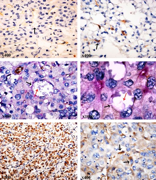
J. Cell. Mol. Med. Vol 17, No 8, 2013

HCC between 2001 and 2010. The diagnoses of these HCC samples were            (Santa Cruz, Dallas, TX, USA), CD90 (Gene Tex, Irvine, CA, USA) at
verified by pathologists. Detailed pathological and clinical data were        37°C for 1 hr. Subsequently, the cells were stained with fluorescein iso-
collected for all samples including Edmondson tumour grade, metastasis        thiocyanate (FITC) or tetramethyl rhodamine isothiocyanate–conjugated
and survival duration. Tissue collection and analysis in this study were      mouse and rabbit immunoglobulin G antibody. All flow cytometric data
approved by the Ethical Committee of Tianjin Medical University, China.       were analysed with CFlow software.

Immunohistochemical and histochemical                                         Invasion
double-staining methods
                                                                              Cell migration assay was performed with Transwell cell culture inserts
The assay was performed as previously described [5, 6].                       (Invitrogen, Carlsbad, CA, USA) according to the manufactory’s instruction.

Quantitation of slug, CD90, E-cadherin, vimentin,                             Soft agar colony formation assay
VEGF and VE-cadherin staining                                                 The assay was performed as previously described [16].

At least 10 power fields were chosen per case and >500 cells were
counted for each power field. Scoring system was modified and used            3D cultures
according to evaluation standard [15]. The percentage of the staining
cells (P) was scored as follows: 0 (negative staining), 1 (≤10% of            The assays were performed as previously described [5, 6].
cells), 2 (10–50%) and 3 (≥50%) for slug quantitation. 0 (negative
staining), 1 (≤25% of cells), 2 (≤50%) and 3 (>50%) for CD90, E-cadh-
erin, vimentin, VEGF and vascular endothelial (VE)-cadherin quantitation
respectively. Staining intensity (I) was graded as follows: 0 (no stain-      Xenograft
ing), 1 (weak staining), 2 (moderate staining), 3 (intense staining).
Samples in each power field were evaluated for both factors, i.e. P plus      All animal work had been conducted according to the guidelines of
I. The scoring of each case was a mean value of chosen power fields.          Tianjin Medical University, China. Male BALB/c nude mice, 5 weeks of
The cases with scoring ≥3 were identified as positive expression.             age, were purchased from Beijing, China. 5 9 106 viable cells/0.1 ml of
                                                                              PBS were injected into the armpit of 20 mice with a 26-gauge needle.
                                                                              For 30 days, the mice were monitored and tumour sizes were measured
                                                                              weekly using a calliper. The tumour volume (TV) was calculated by the
Cell culture and stable cell lines
                                                                              following formula: TV = 1/2 9 a 9 b2 (in which a is the length and b
                                                                              is the width of tumour).
Human liver cancer cell lines (HepG2, SMMC7221, Bel7402, Huh-7)
were obtained from American Type Culture Collection (ATCC, USA), and
the Cell Bank of the Chinese Academy of Medical Sciences (Beijing,
China). Transfection in HepG2 and Huh-7 cells was performed with              Statistical analysis
lipofectamine 2000 reagent, and clones selected by G418.
                                                                              The data analysis was performed with the SPSS16.0 (SPSS, Chicago, IL,
                                                                              USA) software package. All P values were two-sided, and statistical signif-
                                                                              icance was set at P = 0.05.
Expression plasimids

Full-length Slug complementary DNA (cDNA) was generated by normal
human embryo total cDNA, and digested with XhoI/EcoRI and subcl-              Results
oned into pcDNA3.1 vectors. The resulting constructs were confirmed
by DNA sequencing.
                                                                              Expression of slug in correlation with cancer
                                                                              stem cell phenotype in human HCC tissue
Western blot analysis and immunofluorescence
staining                                                                      Based on the criteria Hotz et al. [17] established with minor modifica-
                                                                              tion, slug in primary HCC tissue was identified in the cytoplasm as
See supplementary data.                                                       well as in the nucleus of cancer cells (Fig. 1A–C). The percentage of
                                                                              the positive cells ≥10% was considered as slug-positive case. Thirty
                                                                              nine (35.5%) of 113 cases displayed slug overexpression. By quanti-
Flow cytometry                                                                tation of slug expression, the scoring of slug staining was
                                                                              3.35  0.09 and 1.43  0.07 in slug-positive group and -negative
Indirect labelling method was performed. After trypsinized, cells were        group respectively (P = 0.000). The scoring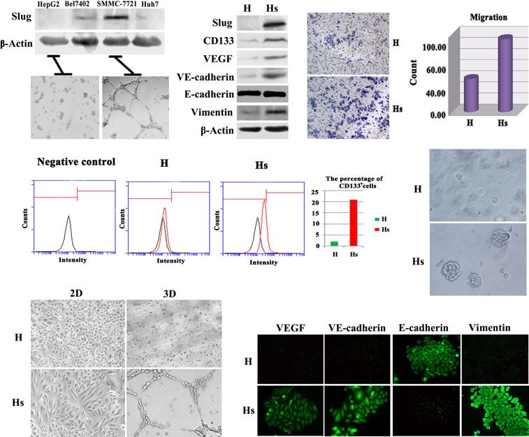
A                                                 B                                                    C

Fig. 1 Expression of slug in human hepatocellular carcinoma (HCC) tissue. (A–C) Slug-positive expression in primary HCC tissue was identified in
the cytoplasm as well as in the nucleus of cancer cells. Black arrows showed slug-positive tumour cells either could form vascular vessels or
involved in mosaic vessels with endothelial cells including red blood cells.

HCC cells. Interestingly, we observed that slug-positive tumour cells       brane positivity of CD90 staining (Fig. 2B). The percentage of CD90+
had close relationship with vascular vessel formation. Slug-positive        cells in the total tumour cells ranged from 5% to 30% in the positive
tumour cells either could form vascular vessels or involved in mosaic       case. Immunohistochemistry of 113 HCC tissues showed a moder-
vessels with endothelial cells (Fig. 1A–C arrow), suggesting that slug      ate-to-strong CD90 expression in 22 (19.5%) cases. More patients
played an important role in tumour vasculature. Slug had been shown         with high slug expression displayed high CD90 expression (30.8%,
to induce EMT, a fundamental mechanism of embryogenesis and pro-            12/39), whereas low slug expression showed 1.4% (10/74) cases
gressive disease. Then, we next examined EMT makers E-cadherin              with CD90-positive expression. By the quantitation of slug and CD90
and vimentin expression (Figure S1A–D). 74.4% (29/39) cases of              expression, slug staining showed a higher scoring in CD90-positive
slug overexpression showed a reduced E-cadherin expression pattern          group (2.59  0.23) than -negative group (1.98  0.11; P = 0.019),
(Figure S1B), whereas 41.9% (31/74) cases of low slug expression            and the scoring of CD90 was 3.15  0.16 in slug-positive group and
had a reduced pattern, with a statistically significant difference          2.26  0.11 in slug-negative group (P = 0.000). By statistical analysis,
(v2 = 10.810, P = 0.001). The scoring of E-cadherin was                     there is correlation between slug and CD90 expression (r = 0.207,
2.28  0.25 in slug-positive group and 3.19  0.24 in slug-negative         P = 0.028).
group (P = 0.019). Similarly, more patients with slug overexpression
displayed vimentin expression (28.2%, 11/29; Figure S1D), whereas
low slug expression show vimentin expression in only 12.2% (9/74)           Expression of slug in correlation with VM in
cases (v2 = 4.513, P = 0.034). The scoring of vimentin was                  human HCC tissue
2.77  0.19 in slug-positive group and 2.11  0.14 in slug-negative
group (P = 0.006). Statistically significant correlations were found        By CD31 and periodic acid-Schiff (PAS) histochemical and immuno-
among E-cadherin, vimentin and slug expression (r = 0.309,                  histochemical double staining, typical microvessels showed positive
P = 0.001 for slug and E-cadhern; r = 0.200, P = 0.034 for slug and         reaction for CD31 on their luminal surface and PAS-positive reaction
vimentin). Slug overexpression significantly correlated with reduced        in their wall. Based on our previous studies [5, 6], CD31/PAS double
E-cadherin expression and increased vimentin expression. Similar            staining was used to identify VM in HCC tissue. CD31-negative, PAS-
results were obtained when slug, E-cadherin and vimentin expression         positive vascular-like patterns containing red blood cells, which
was quantified. Slug expression showed a lower scoring in E-cadher-         formed by HCC cells, were deemed VM. HCC cells formed extracellu-
in-positive group (1.81  0.14) than -negative group (2.35  0.14)          lar matrix–rich channels (PAS-positive), with the absence of necrosis
(P = 0.009), and a higher scoring in vimentin-positive group                and inflammatory cells infiltrating around the channels. VM channels
(2.30  0.29) than -negative group (2.05  0.11), although no               were not lined by endothelial cells as demonstrated by the lack of
significance was found (P = 0.367).                                         CD31 (brown) staining (Fig. 2C–D). The highest VM area was identi-
    Recent reports [18–20] indicate that the emergence of CSCs              fied and individual VM channel counts were made on a 2009 field. At
occurs, in part, as a result of EMT. There is a direct link between the     least 40 power fields were chosen per case. By VM channel counting,
EMT program and the gain of epithelial stem cell properties. We next        the median value showed 4.20  0.80 in slug-positive expression
examined the existence of CSCs in HCC tissues. CD90 is identified as        and 0.18  0.12 in slug-negative expression. There were significant
specific antigenic markers for HCC stem cells. Two distinct CD90            differences between the two groups (t = 6.662, P = 0.000). There-
staining patterns were observed. The first pattern was observed in the      fore, the presence of VM was closely associated with slug-positive
cancer cells that formed tubular structures. Hepatocellular cancer          expression.
cells arranged in tubular structure showed apical/endoluminal cell              VEGF is one of the most potent and specific angiogenic factors of
surface CD90 staining (Fig. 2A). We observed that CD90+ tumour              tumour-induced angiogenesis. Cancer stem cell-like cells expressed
cells of the pattern were involved in vessels formation (Fig. 2A            much higher levels of VEGF and formed more tumours with more blood
arrow). The second pattern was the abundant cytoplasmic and mem-            vessels than cancer cells that did not have stem cell characteristics.

1040                                                                                                                              ª 2013 The Authors.
                        Journal of Cellular and Molecular Medicine Published by Foundation for Cellular and Molecular Medicine/Blackwell Publishing Ltd
J. Cell. Mol. Med. Vol 17, No 8, 2013
A B
Fig. 2 Expression of slug in correlation
with cancer stem cell phenotype and vas-
culogenic mimicry (VM) in human hepato-
cellular carcinoma (HCC) tissue. (A)
Hepatocellular cancer cells arranged in C D
tubular structure showed apical/endolumi-
nal cell surface CD90 staining. Black
arrow showed that CD90+ tumour cells
were involved in blood vessel formation.
(B) CD90 positivity was observed as
abundant cytoplasmic and membrane
staining in HCC specimen. (C–D) Vasculo-
genic mimicry present in HCC tissue by
CD31/PAS double staining. (E) VEGF posi-
tivity present in the cytoplasm in HCC tis-
sue with slug-positive expression. (F)
Vascular endothelial (VE)-cadherin positiv- E F
ity present in the cytoplasm in HCC tissue
and tumour cells with spindle morphology
displayed stronger VE-cadherin expres-
sion.
Accordingly, VEGF creates a perivascular niche for CSCs and study, 48 (42.5%) of 113 cases showed VE-cadherin expression in
stimulates cancer stemness and renewal [21]. By IHC, we found that HCC specimen. By statistical analysis, VE-cadherin was associated
VEGF expression was present in 61.1% (69/113) HCC samples with VM formation (r = 0.399, P = 0.000), suggesting that it could
(Fig. 2E). VEGF expression displayed a close relationship with CSCs be a VM mediator in HCC. We observed that tumour cells with spindle
phenotype (CD90-positive expression) by statistical analysis morphology displayed stronger VE-cadherin expression (Fig. 2F),
(r = 0.255, P = 0.006). Meantime, VEGF showed a higher expression suggesting that epithelial–endothelial transition (EET) existed in HCC.
in slug positive than in slug negative (v2 = 4.429, P = 0.035) and Importantly, VE-cadherin expression was positively related with slug
displayed significant correlation with slug overexpression by statisti- expression (r = 0.205, P = 0.030). By the quantitation of slug and
cal analysis (r = 0.198, P = 0.036). In addition, by the quantitation VE-cadherin expression, the scoring of VE-cadherin was 3.28 0.26
of slug and VEGF expression, the scoring of VEGF was 4.26 0.18 in slug-positive group and 2.12 0.22 in slug-negative group
in slug-positive group and 3.55 0.16 in slug-negative (P = 0.002), and slug staining showed a higher scoring in VE-cadher-
group (P = 0.008), and slug staining showed a higher scoring in in-positive group (2.35 0.16) than -negative group (1.91 0.13;
VEGF-positive group (2.26 0.13) than -negative group P = 0.033).
(1.84 0.17; P = 0.048).
Vascular endothelial-cadherin was transmembrane glycoprotein
that was expressed in the adherens junctions between vascular endo- Constitutive activation of slug-induced EMT,
thelial cells (EC). Vascular endothelial-cadherin was essential during developed CSCs phenotype and VM in HCC cells
embryonic angiogenesis. Vascular endothelial-cadherin expression by in vitro
EC played an important role in regulating vascular morphology and
stability. In addition, VE-cadherin exclusively expressed by highly We compared the level of protein of slug expression and found that
aggressive melanoma cells was critical in melanoma VM. In our slug was differentially expressed in various HCC cell lines by western
ª 2013 The Authors. 1041
Journal of Cellular and Molecular Medicine Published by Foundation for Cellular and Molecular Medicine/Blackwell Publishing Ltdblotting. We found that HepG2 and huh7 had a low-level slug expres- In this study, HepG2 and huh7 cells transfected with slug cDNA
sion in contrast with the SMMC-7721, which presented a high level expressed higher levels of slug protein than the untransfected cells as
(Fig. 3A). We then used a well-established Matrigel culture for investi- analysed by western blot (Fig. 3B). To determine the endogenous
gating VM formation. Interestingly, HepG2 and huh7 cells with low slug expression in HepG2 cells, the band intensities of slug or b-actin
slug expression could not form typical pipe-like structures; in was quantified by Image J software. The protein ratios were calcu-
contrast, SMMC-7721 with high slug expression could form VM lated by dividing the intensity of slug band by the intensity of b-actin
(Fig. 3A). band. The slug/b-actin ratio was 0.49 in HepG2 cells, which
A B C
E
D
F
G
Fig. 3 Constitutive activation of slug-induced epithelial–mesenchymal transition, developed CSCs phenotype and vasculogenic mimicry (VM) in
human hepatocellular carcinoma (HCC) cells in vitro. (A) HepG2 and huh7 had a low-level slug expression in contrast with the SMMC-7721, which
presented a high level. HepG2 and huh7 cells with slug low expression could not form typical pipe-like structures; in contrast, SMMC-7721 with
high slug expression could form VM. (B) HepG2 cells transfected with slug cDNA expressed higher levels of slug protein, CD133+ expression, VEGF,
vascular endothelial (VE)-cadherin and vimentin expression. (C) An increase in cell invasion was observed in the HepG2-slug cells (HS) when com-
pared with HepG2-control (H) cells. (D) Flowcytometry analysis showed that HepG2-slug developed a subpopulation (~21.4%) of CD133+ or CD90+
CSCs phenotype, whereas parental HepG2 cells displayed a CD133 or CD90 phenotype. (E) HepG2-slug exhibited higher colony-forming effi-
ciency and formed more colonies than HepG2-control cells. (F) HepG2-slug formed typical pipe-like structures within the 3D Matrigel medium with
exogenous slug expression. (G) By immunofluorescence, VEGF and VE-cadherin expression was detected in the cytoplasm of HepG2-slug (Hs).
E-cadherin expression was identified in the cell membrane and less intensive in the cytoplasm in Hs. Vimentin expression was displayed in the
cytoplasm in Hs.
1042 ª 2013 The Authors.
Journal of Cellular and Molecular Medicine Published by Foundation for Cellular and Molecular Medicine/Blackwell Publishing LtdJ. Cell. Mol. Med. Vol 17, No 8, 2013
suggested that there was endogenous slug expression in HepG2 cells. HepG2-slug cells into nude mice, xenografts showed a higher rate of
In contrast, the slug/b-actin ratio was 1.14 in HepG2 cells with slug tumour growth as compared with the parental HepG2 cells (Fig. 4A).
transfectant, suggesting that the exogenous slug expression was Although the HepG2-slug xenografts did not give rise to spontaneous
much higher than the endogenous slug expression. In the invasion distant organ metastases by 28 days of tumour growth, there was
assay presented in Figure 3C, an increase in cell invasion was cancer embolus present in blood vessels (Fig. 4D). In contrast,
observed in the HepG2-slug cells (HS) when compared with HepG2- HepG2 cancer xenografts that typically fail to grow vigorously in nude
control cells (H). HepG2-slugs showed spindle morphology in 2D mice displayed low propensity to invasive to vessels. Remarkably,
culture (Fig. 3F) compared with control cells, suggesting that an EMT after in vivo growth, HepG2-slug xenografts maintained CSCs pheno-
phenotype might be induced by slug introduction. We then analysed type with the presence of subpopulations of cancer cells harbouring a
the effect of slug overexpression on EMT phenotype in HepG2 CD133+ or CD90+ phenotype (Fig. 4B and C) that was not seen in the
and huh7 cells. Western blot analysis and immunofluorescence HepG2 xenografts where the predominant phenotype was CD133 or
showed that the expression of epithelial marker E-cadherin was CD90 . Meantime, EMT phenotype was also persisted in HepG2-slug
downregulated and mesenchymal marker vimentin was upregulated xenograft (Fig. 4B).
when compared with the negative vector controls (Fig. 3B, G and Vasculogenic mimicry was identified by endomucin/PAS double
Figure S2A). staining (Fig. 4E). Monoclonal Rabbit anti-Human HLA-DR antibody
Meantime, Flowcytometry analysis showed that HepG2 and huh7 was used to identify human cell and we found that HLA-DR-positive
cells with slug overexpression developed a subpopulation (~21.4%) cells had been incorporated into VM channels (Fig. 4F black arrow).
of CD133+ or CD90+ CSCs phenotype, while parental HepG2 and huh7 In contrast, the mouse liver tissue showed HLA-DR negative
cells displayed a CD133 or CD90 phenotype (Fig. 3D and Fig- (Fig. 4G).
ure S2B). To further demonstrate HepG2-slug developed the CSCs- By VM channel counting, the median value showed 7.00 0.37
like subpopulation that was not seen in the cultured HepG2 cells, soft in HepG2-slug xenograft and 1.10 0.35 in HepG2 xenograft. There
agar colony formation assays were performed. The colonies were were significant differences between the two groups (t = 11.696,
scored to determine the colony-forming efficiency (CFE) after culture. P = 0.000). Therefore, the results demonstrated that slug over-
Our studies showed that HepG2-slug exhibited higher CFE and formed expression contributed to VM formation in vivo.
more colonies than HepG2-control cells (Fig. 3E). HepG2-slug cells Likewise, VEGF and VE-cadherin showed higher expression
had a CFE of 12.8 1.46%, whereas control HepG2 cells had a lower (Fig. 4H and I) in HepG2-slug compared with HepG2 xenograft, sug-
CFE of 3.00 1.71% (t = 6.031, P = 0.000). The HepG2-slug cells gesting that the increased CSCs subpopulation induced by slug over-
showed ~fourfold increase in CFE when compared with that of expression constitutively expressed VEGF and VE-cadherin, played an
HepG2-control cells, suggesting that HepG2-slug cells have a higher important role in contributing VM formation in vivo.
proliferative potential and might developed more CSCs sub- To evaluate whether endogenous slug plays any role in HCC
population. cells with high slug expression, we knocked down slug expression
Remarkably, HepG2-slug and huh7-slug cells displayed a higher in the SMMC-7721 cells with VM formation ability using slug siRNA.
vasculogenic capacity than control cells. HepG2 and Huh7 cells with- The concomitant decrease in the slug protein level in the slug siR-
out VM formation ability formed typical pipe-like structures within the NA-treated cells was evident from the Western blot data. Remark-
3D Matrigel medium with exogenous slug expression (Fig. 3F and ably, with slug knock down, SMMC-7721 cells could not form pipe-
Figure S2C). Our results provided further support for the possible like structure on Matrigel (VM, Figure S3), suggesting that slug
role of slug in promoting VM formation. In addition, HepG2-slug and played an important role in VM formation. Then, SMMC-7221 and
huh7-slug acquired higher endothelial cell marker VE-cadherin SMMC-7721 cells with slug silencing were injected into the armpit
and VEGF expression than HepG2-control cells (Fig. 3B, G and of nude mice. SMMC-7721 cells with slug silencing showed the
Figure S2A). The expression of VE-cadherin and VEGF in HepG2-slug reduced VE-cadherin, VEGF and mesenchymal marker vimentin
and huh7-slug cells suggested that HepG2-slug and huh7-slug cells expression and the increased E-cadherin expression, the restored
with more CSCs subpopulation might have the capacity of transdiffer- CD90- non-CSCs phenotype and the inhibition of VM formation
entiation to differentiate into endothelial cells and acquire endothelial (Figure S3).
cell phenotype. Therefore, HepG2-slug and huh7-slug cells were more
potent in vascular channel formation.
Discussion
HepG2-slug xenografts maintained CSCs and Tumour growth and invasion are dependent on a persistent blood
EMT phenotype, augmented vasculogenic supply; therefore, the capability of generating neovessels through
mimicry diverse mechanisms is associated with its malignant potential in
tumour [22–27]. Vasculogenic mimicry means that tumour cells
To characterize the molecular mechanisms linking slug activation with have a larger plasticity and can make space for blood inflow when
tumour progression in vivo, we developed a xenograft model of the more aggressive tumour need more blood during the process of
human HCC progression employing the HepG2-slug cells and parental tumour growth and invasion. Vasculogenic mimicry is easy to be
HepG2 cells as control. Following subcutaneously transplant of found in the more aggressive tumour. After the blood supply for
ª 2013 The Authors. 1043
Journal of Cellular and Molecular Medicine Published by Foundation for Cellular and Molecular Medicine/Blackwell Publishing LtdA
B
D E
C
F G
H I
1044 ª 2013 The Authors.
Journal of Cellular and Molecular Medicine Published by Foundation for Cellular and Molecular Medicine/Blackwell Publishing LtdJ. Cell. Mol. Med. Vol 17, No 8, 2013
tumour growth and invasion had been satisfied by VM, the endothe- the pattern of embryonic vascular networks [34, 35]. Our results indi-
lial cells can grow into the space made by tumour cells and then cated that the mechanism of VM formation induced by slug overex-
angiogenesis and vasculogenesis are induced consequently. This pression was closely related to an increase in CSCs subpopulation
study demonstrates a novel role of transcriptional repressor slug in generated from EMT. Then, the increased CSCs can transdifferentiate
the development of VM in HCC. The formation of fluid-conducting into different phenotype, they express angiogenic and vasculogenic
networks by non-endothelial cells has been described for melano- markers such as VEGF and VE-cadherin and they are able to organize
mas, hepatocellular, breast, colon and prostate carcinomas as a pseudovascular network.
result of VM, which is a feature associated with a pluripotent gene The development of EMT, stemness, a CSCs phenotype and VM
expression pattern in aggressive tumour cells. In our study, the formation in vivo was also associated with an increase in slug
vigorous VM was present in slug overexpression patients. Slug over- expression indicating that slug were responsible for the maintaining
expression could promote VM, suggesting that HCC cells with slug of CSCs phenotype during in vivo growth. Meantime, VE-cadherin
overexpression have a more aggressive phenotype and a bigger and VEGF expression was also increased in HepG2-slug xenograft
capability of growth and invasion. and it suggested that the process of EET we named before
Our data showed that a correlation between expression levels of occurred with the non-CSCs/CSCs switch in vivo. The EET pro-
slug and decreased E-cadherin expression and increased vimentin moted by slug overexpression can be used for vascular structure.
expression was obvious in these human specimens, thus indicating CSCs in HepG2-slug in vivo can further differentiate into endothelial
that slug is sufficient to induce EMT. The induction of EMT can cell-like tumour cells to participate in the construction of tumour
generate a population with stem cell characteristics from well-differ- microcirculation. In addition, cancer stem–like cells might also
entiated epithelial cells and cancer cells [28–30]. In our study, we directly contribute to the tumour angiogenesis by converting to
found that slug overexpression not only related to EMT but also endothelial cell [36, 37]. Our study showed that the tumours in
related to CSCs phenotype. Our study suggested that slug overex- HepG2-slug xenograft presented more vascular vessels of human
pression might lead to poor prognosis through promoting VM that tumour cell origin than HepG2 xenograft. It demonstrated that slug
could be induced by an EMT-like conversion and an increased popu- overexpression could contribute to tumour angiogenesis in vivo,
lation of cells displaying CSC markers demonstrating the plasticity of especially contribute to vascular vessels formation of tumour cell
epithelial cells. origin.
Ectopic slug overexpression in vitro also showed that an EMT Therefore, in a word, this study demonstrated that slug promote
phenotype was induced. Meantime, our data showed that EMT and VM in HCC by the induction of EMT, pluripotency and CSCs-like phe-
CSCs phenotype induced by slug overexpression could be linked to notype in vitro, in vivo and in HCC patients. Vasculogenic mimicry
each other. By overexpressing the major EMT regulator slug in HepG2 represents an important survival mechanism contributing to the fail-
cells, we induced an EMT-like state (decreased E-cadherin and ure of currently available angiogenesis inhibitors to fully effect
increased expression of mesenchymal markers) and showed that tumour eradication. Thus, our study suggested that the molecular
these cells had an increased ability to self-renew and a higher CFE, a target of slug might act on specific CSCs subpopulation and opened
property normally associated with epithelial cancer stem cells. of course interesting new therapeutic perspectives for the treatment
Remarkably, there was significant difference for the formation of vas- of HCC.
cular network on Matrigel between HepG2-slug and parental HepG2.
With the ectopic introduction of slug with up-regulation in HepG2
dells, the cells with slug transfectant formed typical pipe-like struc- Acknowledgements
tures, although there was a lack of tube formation in the parental
This study was partly supported by a grant from a key project of the National
cells. Our study provided further support for the role of slug in pro-
Natural Science Foundation of China (no. 81230050), the National Natural
moting vascular channels formation. Science Foundation of China (nos. 81172046, 81173091), the Cooperation
Recent studies have suggested that tumour cells might be the project of China-Sweden (no. 09ZCZDSF04400), the Research Fund for the
progenitor for tumour vasculature [31, 32]. Most likely, tumour cells Doctoral Program of Higher Education (no. 20111202110010), the 973
that display stem cell–like characteristics can undergo asymmetric Program from the Ministry of Science and Technology of China (no.
cell division giving rise to tumour cells that trigger angiogenic pro- 2009CB918903), “211 Project” Graduate Innovation Grant of Tianjin Medical
grammes [33]. CSCs might have the unique ability to express an University (no. 2010GSI08), key project of the Tianjin Natural Science
endothelial phenotype and to form vessel-like networks, ‘mimicking’ Foundation (no. 12JCZDJC23600).
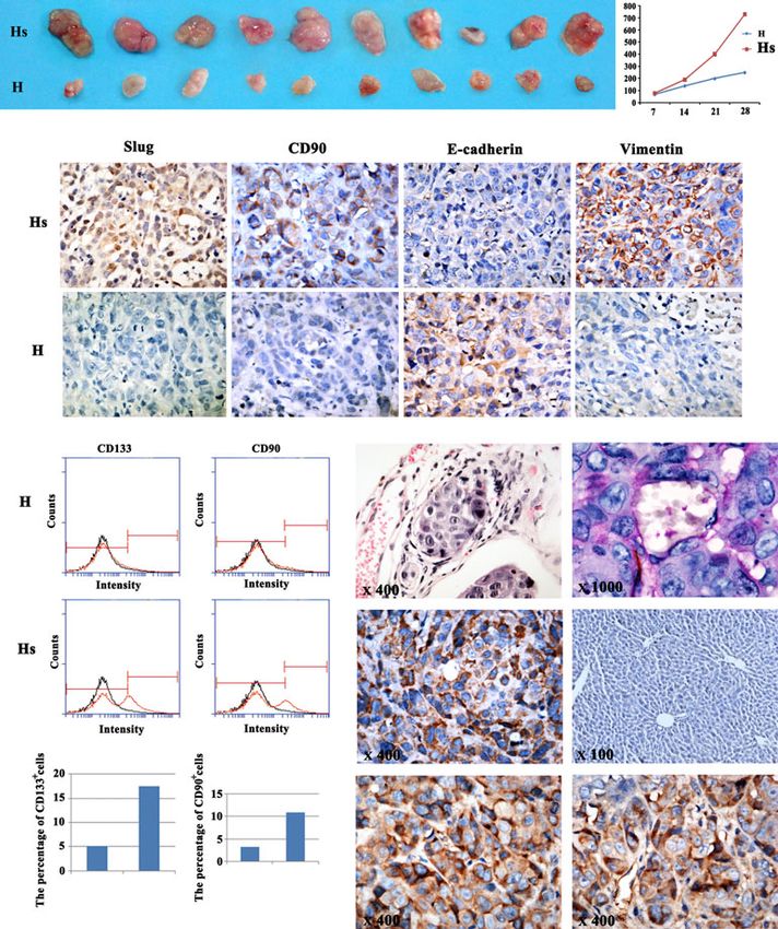
Fig. 4 HepG2-slug (Hs) xenografts maintained CSCs and epithelial–mesenchymal transition phenotype, augmented vasculogenic mimicry. (A) Xeno-
grafts showed a higher rate of tumour growth in Hs as compared with the parental HepG2 (H) cells. (B) Positive slug and CD90 expression, the
downregulated E-cadherin expression and upregulated vimentin expression present in Hs xengraft. (C) Flowcytometry analysis showed that CD133+
and CD90+ phenotype were present in Hs xenograft. (D) There was cancer embolus present in blood vessels in Hs xenograft. (E) Vasculogenic
mimicry (VM) was present in Hs xenograft. (F) Monoclonal Rabbit anti-Human HLA-DR antibody was used to identify human cell and positive stain-
ing was in the cytoplasm. Black arrow showed that HLA-DR-positive cells had been incorporated into VM channels. (G) The mouse liver tissue
showed HLA-DR negative. (H and I) VEGF and VE-cadherin showed higher expression in Hs xenograft.
ª 2013 The Authors. 1045
Journal of Cellular and Molecular Medicine Published by Foundation for Cellular and Molecular Medicine/Blackwell Publishing LtdDisclosure Figure S3 (A) The decreased expression of slug protein in the slug
siRNA-treated SMMC-7721 cells (S: SMMC-7721; si S: SMMC-7721
All authors declare that there are no conflicts of interest. with slug silencing) was evident from the Western blot data. Slug
silencing inhibited tube formation (VM) in SMMC-7721 cells in vitro.
(B-Q) SMMC-7721 with slug silencing xenografts reversed CSCs and
EMT phenotype, inhibited vasculogenic mimicry. Positive slug (B),
Supporting information CD90 expression (D), the loss of E-cadherin expression (F), positive
vimentin expression (H), higher VE-cadherin (L) and VEGF (N)
Additional Supporting Information may be found in the online expression present in S (SMMC-7721) xenograft. In contrast, the
version of this article: decreased slug (C) and CD90 expression (E), the increased E-cadher-
in expression (G), and the decreased vimentin (I), VE-cadherin (M)
Data S1 Supplementary data. and VEGF (O) present in si S (SMMC-7721 with slug silencing) xeno-
graft. There was VM (black arrow indicated VM channel; green arrow
Figure S1 Cases of slug overexpression showed a reduced E-cadher- indicated red blood cell; red arrow indicated endomucin-positive
in expression pattern and an increased vimentin expression pattern in blood vessel) present in SMMC-7221 xenograft (J). However, in si S
HCC patients. (SMMC-7721 with slug silencing) xenograft, no VM channels were
found (K). HLA-DR-positive tumour cells formed VM channel (black
Figure S2 Huh7 cells transfected with slug showed CD133+ and arrow indicated VM; red arrow indicated red blood cell) in SMMC-
CD90+ phenotype, EMT phenotype, VEGF and VE-cadherin expression 7221 xenograft (P). However, in si S (SMMC-7721 with slug silencing)
and VM formation. xenograft, no VM channels were found (Q).
References
1. Dome B, Hendrix MJ, Paku S, et al. Alterna- lial-mesenchymal transition markers in pri- 15. Bittner M, Meltzer P, Chen Y, et al. Molecu-
tive vascularization mechanisms in cancer: mary breast cancer patients with circulating lar classification of cutaneous malignant
pathology and therapeutic implications. Am tumor cells. Breast Cancer Res. 2012; 14: melanoma by gene expression profiling. Nat-
J Pathol. 2007; 170: 1–15. R15. ure. 2000; 406: 536–40.
2. Sun B, Zhang S, Zhang D, et al. Vasculo- 9. Sarrio D, Franklin CK, Mackay A, et al. Epi- 16. Liu TJ, Sun BC, Zhao XL, et al. CD133 +
genic mimicry is associated with high tumor thelial and mesenchymal subpopulations cells with cancer stem cell characteristics
grade, invasion and metastasis, and short within normal basal breast cell lines exhibit associates with vasculogenic mimicry in tri-
survival in patients with hepatocellular carci- distinct stem cell/progenitor properties. ple-negative breast cancer. Oncogene. 2013;
noma. Oncol Rep. 2006; 16: 693–8. Stem Cells. 2012; 30: 292–303. 32: 544–53.
3. Kirschmann DA, Seftor EA, Hardy KM, et al. 10. Casas E, Kim J, Bendesky A, et al. Snail2 is 17. Hotz B, Arndt M, Dullat S, et al. Epithelial to
Molecular pathways: vasculogenic mimicry an essential mediator of Twist1-induced epi- mesenchymal transition: expression of the
in tumor cells: diagnostic and therapeutic thelial mesenchymal transition and metasta- regulators snail, slug, and twist in pancreatic
implications. Clin Cancer Res. 2012; 18: sis. Cancer Res. 2011; 71: 245–54. cancer. Clin Cancer Res. 2007; 13: 4769–76.
2726–32. 11. Bao S, Wu Q, Sathornsumetee S, et al. 18. Raimondi C, Gianni W, Cortesi E, et al.
4. Wang SY, Yu L, Ling GQ, et al. Vasculogen- Stem cell-like glioma cells promote tumor Cancer stem cells and epithelial-mesenchy-
ic mimicry and its clinical significance in angiogenesis through vascular endothelial mal transition: revisiting minimal residual
medulloblastoma. Cancer Biol Ther. 2012; growth factor. Cancer Res. 2006; 66: 7843– disease. Curr Cancer Drug Targets. 2010;
13: 341–8. 8. 10: 496–508.
5. Sun T, Sun BC, Zhao XL, et al. Promotion of 12. Bhat-Nakshatri P, Appaiah H, Ballas C, 19. Battula VL, Evans KW, Hollier BG, et al.
tumor cell metastasis and vasculogenic mim- et al. SLUG/SNAI2 and tumor necrosis fac- Epithelial-mesenchymal transition-derived
icry by way of transcription coactivation by tor generate breast cells with CD44+/ cells exhibit multilineage differentiation
Bcl-2 and Twist1: a study of hepatocellular CD24 phenotype. BMC Cancer. 2010; 10: potential similar to mesenchymal stem cells.
carcinoma. Hepatology. 2011; 54: 1690–706. 411. Stem Cells. 2010; 28: 1435–45.
6. Sun T, Zhao N, Zhao XL, et al. Expression 13. Zhu LF, Hu Y, Yang CC, et al. Snail overex- 20. Singh A, Settleman J. EMT, cancer stem
and functional significance of Twist1 in pression induces an epithelial to mesenchy- cells and drug resistance: an emerging axis
hepatocellular carcinoma: its role in mal transition and cancer stem cell-like of evil in the war on cancer. Oncogene.
vasculogenic mimicry. Hepatology. 2010; properties in SCC9 cells. Lab Invest. 2012; 2010; 29: 4741–51.
51: 545–56. 92: 744–52. 21. Beck B, Driessens G, Goossens S, et al. A
7. Mani SA, Guo W, Liao MJ, et al. The epithe- 14. Kurrey NK, Jalgaonkar SP, Joglekar AV, vascular niche and a VEGF-Nrp1 loop regu-
lial-mesenchymal transition generates cells et al. Snail and slug mediate radioresistance late the initiation and stemness of skin
with properties of stem cells. Cell. 2008; and chemoresistance by antagonizing p53- tumours. Nature. 2011; 478: 399–403.
133: 704–15. mediated apoptosis and acquiring a stem- 22. Cao Y. Off-tumor target–beneficial site for
8. Kasimir-Bauer S, Hoffmann O, Wallwiener like phenotype in ovarian cancer cells. Stem antiangiogenic cancer therapy? Nat Rev Clin
D, et al. Expression of stem cell and epithe- Cells. 2009; 27: 2059–68. Oncol. 2010; 7: 604–8.
1046 ª 2013 The Authors.
Journal of Cellular and Molecular Medicine Published by Foundation for Cellular and Molecular Medicine/Blackwell Publishing LtdJ. Cell. Mol. Med. Vol 17, No 8, 2013
23. Xue Y, Lim S, Yang Y, et al. PDGF-BB mod- TGFbeta-1 induction increases stemness 33. Bjerkvig R, Johansson M, Miletic H, et al.
ulates hematopoiesis and tumor angiogene- characteristics in primary non small cell Cancer stem cells and angiogenesis. Semin
sis by inducing erythropoietin production in lung cancer cell line. PLoS ONE. 2011; 6: Cancer Biol. 2009; 19: 279–84.
stromal cells. Nat Med. 2012; 18: 100–10. e21548. 34. Dong J, Zhao Y, Huang Q, et al. Glioma
24. Cao Y, Arbiser J, D’Amato RJ, et al. Forty- 29. Cao L, Shao M, Schilder J, et al. Tissue stem/progenitor cells contribute to neovas-
year journey of angiogenesis translational transglutaminase links TGF-beta, epithelial cularization via transdifferentiation. Stem
research. Sci Transl Med. 2011; 3: 114rv3. to mesenchymal transition and a stem cell Cell Rev. 2011; 7: 141–52.
25. Cao Y, Langer R. Optimizing the delivery of phenotype in ovarian cancer. Oncogene. 35. Monzani E, La Porta CA. Targeting cancer stem
cancer drugs that block angiogenesis. Sci 2012; 31: 2521–34. cells to modulate alternative vascularization
Transl Med. 2010; 2: 15 ps3. 30. Kong D, Li Y, Wang Z, et al. Cancer stem mechanisms. Stem Cell Rev. 2008; 4: 51–6.
26. Cao R, Xue Y, Hedlund EM, et al. VEGFR1- cells and epithelial-to-mesenchymal transi- 36. Salmaggi A, Boiardi A, Gelati M, et al. Glio-
mediated pericyte ablation links VEGF and tion (EMT)-phenotypic cells: are they cous- blastoma-derived tumorospheres identify a
PlGF to cancer-associated retinopathy. Proc ins or twins? Cancers. 2011; 3: 716–29. population of tumor stem-like cells with
Natl Acad Sci USA. 2010; 107: 856–61. 31. Shen R, Ye Y, Chen L, et al. Precancerous angiogenic potential and enhanced multi-
27. Cao R, Ji H, Feng N, et al. Collaborative stem cells can serve as tumor vasculogenic drug resistance phenotype. Glia. 2006; 54:
interplay between FGF-2 and VEGF-C pro- progenitors. PLoS ONE. 2008; 3: e1652. 850–60.
motes lymphangiogenesis and metastasis. 32. Wang R, Chadalavada K, Wilshire J, et al. 37. Ricci-Vitiani L, Pallini R, Biffoni M, et al.
Proc Natl Acad Sci USA. 2012; 109: 15894–9. Glioblastoma stem-like cells give rise to Tumour vascularization via endothelial dif-
28. Pirozzi G, Tirino V, Camerlingo R, et al. tumour endothelium. Nature. 2010; 468: ferentiation of glioblastoma stem-like cells.
Epithelial to mesenchymal transition by 829–33. Nature. 2010; 468: 824–8.
ª 2013 The Authors. 1047
Journal of Cellular and Molecular Medicine Published by Foundation for Cellular and Molecular Medicine/Blackwell Publishing LtdYou can also read