Synthesis of Fluorescently Labeled Antibodies Using Non-Canonical Amino Acids in Eukaryotic Cell-Free Systems - Fraunhofer IZI-BB
←
→
Page content transcription
If your browser does not render page correctly, please read the page content below
Chapter 9

Synthesis of Fluorescently Labeled Antibodies Using
Non-Canonical Amino Acids in Eukaryotic Cell-Free Systems
Marlitt Stech, Nathanaël Rakotoarinoro, Tamara Teichmann,
Anne Zemella, Lena Thoring, and Stefan Kubick

Abstract
Cell-free protein synthesis (CFPS) enables the development of antibody conjugates, such as fluorophore
conjugates and antibody-drug conjugates (ADCs), in a rapid and straightforward manner. In the first part,
we describe the cell-free synthesis of antibodies containing fluorescent non-canonical amino acids (ncaa) by
using pre-charged tRNA. In the second part, we describe the cell-free synthesis of antibodies containing
ncaa by using an orthogonal system, followed by the site-specific conjugation of the fluorescent dye
DyLight 650-phosphine. The expression of the antibodies containing ncaa was analyzed by SDS-PAGE,
followed by autoradiography and the labeling by in-gel fluorescence. Two different fluorescently labeled
antibodies could be generated.

     Key words Cell-free protein synthesis, Antibody, Antibody conjugates, IgG1, Non-canonical amino
     acid, Conjugation

1    Introduction

                                ADCs represent one of the most promising strategies in the phar-
                                maceutical industry to treat solid and hematological cancers. Up to
                                date, nine of them are already approved [1]. The concept of ADCs
                                combines the effect of highly specific tumor-targeting immuno-
                                therapy with the concept of chemotherapy relying on highly cyto-
                                toxic drugs. ADCs are considered as a strategy to provide a better
                                cytotoxicity to immunotherapy and a better specificity to chemo-
                                therapy. ADCs are composed of a monoclonal antibody conjugated
                                to a cytotoxic drug by a linker. In the early stages of the develop-
                                ment of ADCs, non-toxic fluorescent agents can be used to opti-
                                mize the conjugation efficiency, to analyze the internalization
                                efficiency, and to locate and quantify the antibody in vitro and
                                ex vivo, prior to the conjugation to the cytotoxic agent.
                                     While full-length Immunoglobulin G (IgG) is mainly produced
                                in the mammalian system [2], such as Chinese Hamster Ovary

Raymond J. Owens (ed.), Structural Proteomics: High-Throughput Methods, Methods in Molecular Biology, vol. 2305,
https://doi.org/10.1007/978-1-0716-1406-8_9, © The Author(s) 2021

                                                          175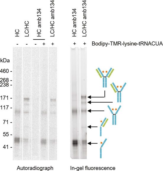
176   Marlitt Stech et al.

                         (CHO) cell lines, the drug is produced either by hemi-synthesis or
                         by chemical synthesis. One strategy to conjugate the latter to the
                         former is the amber suppression technology [3–6]. This technology
                         is based on the introduction of an amber stop codon into the gene
                         sequence at a desired position. By adding an engineered tRNA and
                         synthetase the ncaa is incorporated in the antibody sequence during
                         its synthesis exactly at the position of the amber stop codon. Fol-
                         lowing protein synthesis, the drug containing the corresponding
                         reactive group can be conjugated to the antibody by the reactive
                         group of the ncaa. The basis of the amber suppression technology is
                         an orthogonal tRNA/synthetase pair. The orthogonal synthetase is
                         engineered to amino-acylate specifically the ncaa at the 30 -end of
                         the orthogonal tRNA. The latter is engineered to recognize the
                         amber stop codon, thus allowing for the incorporation of the ncaa
                         in a site-specific manner. Most importantly, the orthogonal tRNA/
                         synthetase pair should not show cross-reactivities between endoge-
                         nous amino acids, tRNAs, and synthetases.
                              Orthogonal systems and the resulting products can be devel-
                         oped by using a CFPS system [5]. The CFPS system based on CHO
                         lysate contains microsomes derived from the endoplasmic reticu-
                         lum of CHO cells as previously described [7]. By translocating de
                         novo synthesized proteins into the lumen of these microsomes by
                         using a melittin signal peptide, post-translational modifications
                         such as disulfide bridge formation and glycosylation can be per-
                         formed [7]. Using this system, full-length post-translationally
                         modified antibodies can be produced within hours, making the
                         screening of orthogonal tRNAs, or synthetases, ncaa, antibody
                         candidates, and ADCs rapid and straightforward [5].
                              In this chapter, we describe the proof-of-concept for the cell-
                         free synthesis of antibodies containing ncaa. To allow for the site-
                         specific introduction of ncaa, the chosen model antibody contained
                         an amber stop codon at amino acid position 134 in the CH1
                         domain of antibody heavy chain (HC). In the first part, we
                         expressed      antibodies    containing     a   Bodipy-TMR-lysine,
                         pre-charged on a tRNA. Autoradiography and in-gel fluorescence
                         analysis showed the expression of the fluorescently labeled antibody
                         of interest. In the second part, we expressed antibodies containing
                         p-azido-L-phenylalanine (AzF) by using an orthogonal system,
                         composed of an engineered E. coli tRNA [8] amino-acylated by
                         an engineered E. coli tyrosine synthetase [9]. Autoradiography
                         confirmed the synthesis of suppression and full-length product.
                         Subsequently, the fluorophore DyLight 650-phosphine was conju-
                         gated to the antibody by Staudinger ligation. The successful conju-
                         gation was shown by in-gel fluorescence.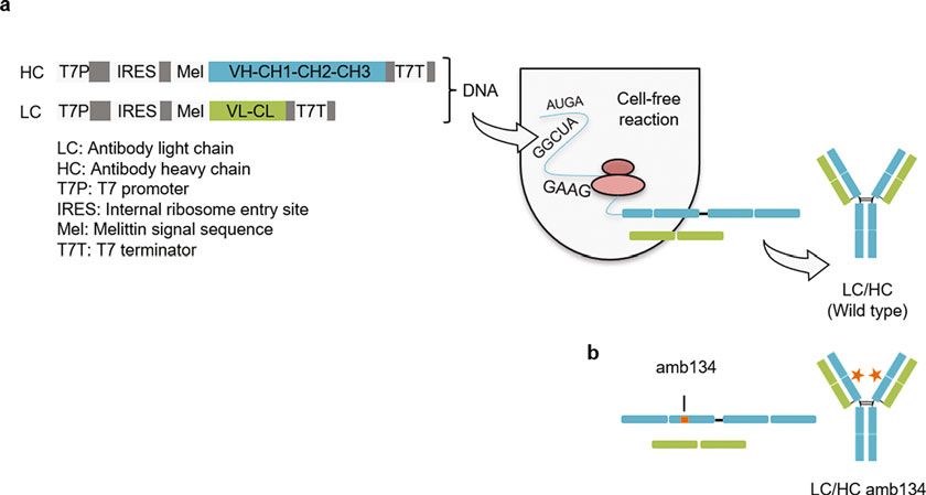
Eukaryotic Cell-Free Systems   177

2   Materials

                     Prepare all buffers and solutions using ultrapure water.

2.1 CFPS Using CHO    1. Ice pan.
Lysate                2. 1.5 mL reaction tubes.
                      3. 10 translation mix: 300 mM HEPES-KOH (pH 7.6),
                         2250 mM KOAc, 2.5 mM spermidine, 1 mM of each canonical
                         amino acid, and 39 mM Mg(OAc)2. Store at –80  C.
                      4. CHO lysate prepared as described previously [10, 11] (see Note
                         1). Shock-freeze in liquid nitrogen after every usage and store
                         at 80  C.
                      5. T7 RNA polymerase.
                      6. 5 energy mix: 100 mM creatine phosphate, 1.5 mM GTP,
                         1.5 mM CTP, 1.5 mM UTP, 8.75 mM ATP. and 0.5 mM m7G
                         (ppp)G cap analog. Store at – 80  C.
                           14
                      7.    C-leucine (200 dpm/pmol, 100 dpm/pmol). Store at
                           20  C.
                      8. Plasmid encoding antibody HC, plasmid encoding antibody
                         light chain (LC) (Fig. 1) (see Note 2). Store at 20  C.
                      9. Ultrapure water.
                     10. Thermomixer.
                     11. Phosphate-buffered saline (PBS) containing 0.2% n-Dodecyl
                         β-D-maltoside (DDM). Store at 4  C.

2.2 Preparation       1. Ice pan.
of Orthogonal         2. 1.5 mL reaction tubes.
Synthetase
                      3. DNA template encoding modified tyrosyl-tRNA-synthetase
                         (eAzFRS, including the mutations Thr37, Ser182, Ala183,
                         and Arg265 [9] and a C-terminal Strep-Tag) from E. coli,
                         cloned into a vector containing a T7 promotor. We used the
                         vector pQE2 vector (pQE2-eAzFRS-SII) (see Note 3).
                      4. E. coli expression system (RTS 500 E. coli HY Kit,
                         Biotechrabbit).
                      5. Thermomixer with RTS 500 thermomixer adapter.
                      6. 100 mM Isopropyl β-D-1-thiogalactopyranoside (IPTG).
                      7. Gravity flow Strep-Tactin® superflow mini-column (0.2 mL).
                      8. Strep-Tactin® Purification Buffer Set: 10 Washing Buffer
                         (1 M Tris-Cl, pH 8.0, 1.5 M NaCl, 10 mM EDTA), 10
                         Elution Buffer (1 M Tris-Cl, pH 8.0, 1.5 M NaCl, 10 mM
                         EDTA, 25 mM Desthiobiotin) and 10 Regeneration Buffer
178 Marlitt Stech et al.
Fig. 1 Schematic presentation of the template design of antibody heavy (HC) and light chain (LC) and their
rapid cell-free synthesis and assembly to functional IgG. (a) Template design without amber (amb) stop codon.
(b) Template design with amber stop codon to allow for the site-specific incorporation of a ncaa into cell-free
synthesized antibody HC. The amb stop codon TAG was positioned in the CH1 domain (replacing S134, EU
numbering) of HC. Orange asterisks indicate fluorescent dye conjugated to the incorporated ncaa.
(1 M Tris-Cl, 1.5 M NaCl, 10 mM EDTA, 10 mM HABA
(hydroxyl-azophenyl-benzoic acid)).
9. Zeba™ Spin Desalting Columns (7 K MWCO, 0.5 mL).
10. Amicon® Ultra Centrifugal Filters (10 K device, 0.5 mL).
11. Synthetase storage buffer: 50 mM HEPES pH 7.6, 10 mM
KOAc, 1 mM MgCl2, 4 mM DTT.
12. NanoDrop 2000c.
2.3 Preparation 1. Vector containing the nucleotide sequence of tRNATyrCUA
of Orthogonal tRNA (SupF Gene).
2.3.1 PCR Amplification 2. tRNATyrCUA-specific forward primer (5´ CgA gCT CgC
of the tRNA Gene CCA CCg gAA TTC 30 ) and 2´-OMe reverse primer (50 Tgg
Tgg Tgg ggg AAg gAT TCg 30 ).
3. 0.2 mL PCR tubes.
4. PCR cycler.
5. Taq DNA polymerase.
6. Taq buffer.
7. dNTPs.
8. 25 mM MgCl2.
9. Agarose gel electrophoresis chamber.Eukaryotic Cell-Free Systems 179
10. Agarose.
11. Rotiphorese 10 TBE buffer.
12. DNA stain.
13. DNA ladder.
14. PCR Purification Kit.
2.3.2 Transcription, 1. 5 transcription buffer: 400 mM HEPES-KOH, 0.5 mM sper-
Isolation, and Folding midine, 50 mM DTE and 75 mM MgCl2.
of tRNA 2. 5 NTP mix containing 18.75 mM ATP, 18.75 mM CTP,
18.75 mM UTP, and 7.5 mM GTP.
3. T7 RNA Polymerase.
4. DNaseI (1 U per μg plasmid DNA).
5. 10 MOPS buffer: 200 mM MOPS, 50 mM NaOAc, 10 mM
EDTA, pH 8.0.
6. MOPS sample buffer: 8% (v/v) formaldehyde, 12 mL formam-
ide, 2.4 mL 10 MOPS buffer, 0.05% (v/v) bromophenol
blue to a total volume of 24 mL.
7. TRIzol reagent.
8. High Performance Liquid Chromatography (HPLC) grade
Chloroform.
9. HPLC grade isopropanol.
10. 75% ethanol.
11. Cooled centrifuge.
12. NanoDrop 2000c.
13. PCR cycler.
2.4 Site-Specific 1. 100 μM Bodipy-TMR-lysine-tRNACUA (Biotechrabbit).
Incorporation Store at – 80 C.
of Non-canonical 2. 100 μM eAzFRS. Store at 80 C.
Amino Acids 3. 100 μM tRNATyrCUA. Store at 80 C.
4. 100 mM AzF (Bachem AG; Bubendorf, Schweiz). Store at
80 C.
5. Phosphate-buffered saline (PBS). Store at 4 C.
6. PBS containing 0.2% DDM. Store at 4 C.
2.5 Qualitative 1. SDS-PAGE sample buffer: 1 LDS buffer containing 106 mM
Protein Analysis Tris HCl, 141 mM Tris base, 2% LDS, 10% glycerol, 0.51 mM
EDTA, 0.22 mM SERVA Blue G, 0.175 mM Phenol Red,
pH 8.5.
2. 3–8% Tris acetate gels.180 Marlitt Stech et al.
3. Fluorescently labeled protein ladder for SDS-PAGE.
4. SDS-PAGE running buffer: 50 mM MES, 50 mM Tris Base,
0.1% SDS, 1 mM EDTA, pH 7.3.
5. SDS-PAGE gel tank system.
6. Radioactive ink.
7. Acetone.
8. Water bath.
9. Fluorescence/phosphorimager.
10. Gel dryer.
11. Phosphor screens.
3 Methods
3.1 Cell-free Batch-based cell-free reactions are carried out as coupled
Synthesis transcription-translation reaction, in which transcription and trans-
and Fluorescence lation take place simultaneously in the same reaction compartment
Labeling of IgG Using (“one-pot”).
Pre-Charged tRNA 1. Thaw all components of the cell-free reaction on ice. Mix all
3.1.1 Batch-Based CFPS components thoroughly before usage. Protect pre-charged
tRNA Bodipy-TMR-lysine-tRNACUA from light (see Note 4).
2. Pipet the following components on ice using a 1.5 mL reaction
vessel: 5 μL 10 translation mix (f.c. 1), 20 μL CHO lysate
(f.c. 40%), 1 μL T7 RNA polymerase (f.c. 1 U/μL), and 10 μL
5 energy mix (f.c. 1). Mix thoroughly after addition of each
component (see Note 5).
3. Add 2.5 μL of 200 dpm/pmol 14C-leucine (specific radioactiv-
ity of 66.67 dpm/pmol f.c.) for subsequent qualitative analysis
by autoradiography.
4. For fluorescence labeling, supplement the cell-free reaction
with 2 μM Bodipy-TMR-lysine-tRNACUA. Protect the trans-
lation mixture from light during pipetting and incubation.
5. Add HC and LC encoding plasmid at a final concentration of
60 nM each (see Note 6).
6. Adjust the final volume of the reaction mix with ultrapure water
to 50 μL (see Note 7).
7. Mix all components thoroughly and incubate the reaction at
27 C and 500 rpm for 3 h in a thermomixer (see Note 8). After
completing the cell-free reaction place reaction vessels on ice
and proceed with the procedure described in Subheadings 3.2
and 3.4 (Fig. 2).Eukaryotic Cell-Free Systems 181
Fig. 2 Batch-based cell-free synthesis and site-specific fluorescence labeling of
antibodies with Bodipy-TMR-lysine-tRNACUA by amber (amb) suppression.
Qualitative analysis of cell-free synthesized antibody heavy chain (HC) and
light chain (LC) was performed by SDS-Page followed by autoradiography (left
side) and in-gel fluorescence (right side) (analysis of MF). Cell-free synthesis
was performed in the presence of 14C-leucine and in the presence (+) or
absence () of Bodipy-TMR-lysine-tRNACUA. The amb stop codon TAG was
positioned in the CH1 domain (replacing S134, EU numbering) of HC. Orange
asterisks indicate fluorescent dye conjugated to the incorporated ncaa. Unas-
sembled LC (25.4 kDa) and termination product of HC amb134 (16.4 kDa) cannot
be visualized in the autoradiograph because of its low molecular weight
3.2 Fractionation 1. Centrifuge translation mixtures at 16,000g for 10 min at
of Translation 4 C. Take off the supernatant (SN1) and discard.
Mixtures 2. Carefully resuspend the microsomal pellet in PBS supplemen-
ted with 0.2% DDM by pipetting up and down for several
times. Incubate the solution for 45 min at room temperature
(RT) under intense agitation (see Note 9).
3. Centrifuge the solution at 16,000g for 10 min at 4 C to
separate soluble antibodies, located in the supernatant (super-
natant 2, SN2), from the microsomes. Take off the supernatant
(containing soluble antibodies) and place it on ice. Analyze
cell-free synthesized antibodies as described in Subheading 3.4.182 Marlitt Stech et al.
3.3 Cell-free 1. Enhanced tRNA synthetase is produced using the E. coli-based
Synthesis CECF-system “RTS500 ProteoMaster E. coli HY Kit.”
and Fluorescence 2. First, reconstitute the buffers and the E. coli lysate in reconsti-
Labeling of IgG Using tution buffer according to the manufacturer’s instructions.
Orthogonal System Pipet everything on ice and mix the buffers and solutions
thoroughly.
3.3.1 Preparation
of Orthogonal Synthetase 3. Prepare the reaction mixture as follows: Pipet 525 μL E. coli
lysate, 225 μL reaction mix, 270 μL amino acid mix without
methionine, 30 μL methionine, 11 μL IPTG and 39 μL tem-
plate pQE2-eAzFRS-SII containing 110 μg plasmid DNA, and
mix the solution thoroughly.
4. For the feeding mixture, pipet 7990 μL feeding mix, 110 μL
IPTG, 2650 μL amino acid mix without methionine, and
300 μL methionine. Mix the solution thoroughly.
5. Fill the reaction chamber (red lid) with the complete volume of
reaction mix and the feeding chamber (colorless lid) with the
complete volume of feeding mix. Insert the chamber into the
RTS 500 adapter in a thermomixer. Incubate the reaction at
30 C for 24 h at 1000 rpm. Harvest the reaction mix from the
reaction chamber and place the reaction mixture on ice.
6. In order to separate soluble from insoluble protein, centrifuge
the translation mix at 16,000 g for 10 min at 4 C. Harvest
the supernatant containing soluble eAzFRS by pipetting.
7. Subsequently, eAzFRS is purified via its C-terminal Strep-Tag.
Purification is performed using Strep-Tactin Gravity Flow Col-
umns (200 μL).
8. Equilibrate each column with 2 800 μL washing buffer and
add 500 μL of the supernatant from this step to each column.
9. Once the supernatant has completely entered the column, wash
each column 5 with 200 μL washing buffer (see Note 10).
10. To elute the synthetase, add 6 100 μL elution buffer to each
column. Collect each flow-through and analyze separately.
Afterwards, pool all fractions containing the target protein.
11. To regenerate the column, add 3 1 mL regeneration buffer
to each column, followed by addition of 2 800 μL of 1
washing buffer. For storage add 2 mL washing buffer and place
the columns at 4 C.
12. To exchange the elution buffer to synthetase storage buffer,
apply pooled elution fractions to Zeba™ Spin Desalting Col-
umns. First, remove the storage solution of the Zeba™ Spin
Desalting Column by centrifugation of the column at
1500 g for 1 min. Subsequently, add 300 μL synthetase
storage buffer to the resin bed and centrifuge the column at
1500 g for 1 min. After repeating this step 2 place theEukaryotic Cell-Free Systems 183
column into a new collection tube and apply 100 μL of the
pooled synthetase solution to each column. Collect target
proteins by a final centrifugation step at 2000 g for 2 min.
13. Concentrate target protein by using Amicon Centrifugal Filter
Devices 0.5 mL. Adjust the volume of the synthetase from step
12 to 500 μL with storage buffer and add this solution to the
concentrator. Centrifuge at 14,000 g for 10 min and 4 C.
Collect the flow-through. The concentration of the flow-
though can be determined by photometric measurement
using NanoDrop based on the calculated molecular mass of
the synthetase (48.6 kDa) and the extinction coefficient (54.3)
(see Note 11). Store synthetase at 80 C after shock freezing
in liquid nitrogen.
3.3.2 Preparation 1. First, suitable DNA templates of the tRNA gene need to be
of Orthogonal tRNA generated. For this, a reverse primer containing a 2’-OMe
group has to be used in order to prevent unspecific addition
of nucleotides to the 30 end by the T7 RNA polymerase.
2. The PCR reaction is composed of the following components:
1 Taq Buffer, 0.2 mM dNTP mix, 0.5 μM forward primer,
0.5 μM reverse primer, 2.5 mM MgCl2, 0.01 ng/μL plasmid,
and 0.04 U/μL Taq DNA polymerase.
3. Fill the reaction with ultrapure water to a final volume of
250 μL.
4. Use the following PCR program: (1) 5 min 95 C, (2) 30 s
95 C, (3) 30 s 52 C, (4) 10 s 72 C, (5) 10 min 72 C,
(6) cooling to 4 C. Repeat steps 2–4 30. Analyze generated
PCR products by agarose gel electrophoresis on a 2%
agarose gel.
5. Purify amplified tRNA PCR products by using a PCR Purifica-
tion Kit (see Note 12).
6. Apply 50 μL PCR product per column.
7. Elute in 20 μL ultrapure water.
8. Analyze DNA concentration using NanoDrop.
9. Thaw in vitro transcription components on ice. Mix all com-
ponents before using.
10. Pipet the reactions at RT. The transcription reaction is com-
posed of the following components: 100 μL 5 transcription
buffer (f.c. 1), 100 μL 5 NTP mix (f.c. 1), 25 μL 20
enzyme mix (f.c. 1), and 8 ng/μL (f.c.) template DNA. Fill
the reaction with water to the final volume of 500 μL (see Note
13). Incubate the reaction for 3–6 h at 37 C and 500 rpm.
11. After completing the reaction, centrifuge tRNA transcripts at
12,000 g for 1 min and collect the supernatant.184 Marlitt Stech et al.
12. Analyze tRNA transcripts by agarose gel electrophoresis (2%)
using 2 μL of the tRNA transcript (see Note 14).
13. Treat the transcription reaction with 1 U DNaseI per 1 μg
plasmid DNA for 10 min at 37 C and 500 rpm.
14. Add a three-fold volume of TRIzol to the transcription reac-
tion and mix carefully. TRIzol and chloroform shall be handled
with care and under the fume hood. Incubate for 5 min at RT.
15. Add chloroform (200 μL per 1 mL TRIzol) and mix for 15 s by
carefully inverting the tube. Incubate 2–3 min at RT. Centri-
fuge at 12,000 g, 4 C and 15 min.
16. Remove the aqueous phase and transfer to a fresh reaction tube
(see Note 15).
17. Add isopropanol (HPLC grade, 500 μL per 1 mL TRIzol) and
mix carefully, followed by incubation over night at 4 C.
18. Centrifuge at 15,000 g, 4 C for a minimum of 1 h. Remove
the supernatant and discard it.
19. Overlay the RNA pellet with ethanol (1 mL 75% per 1 mL
TRIzol) and incubate at 20 C for 30 min.
20. Centrifuge at 7.500g, 4 C for 10 min. Remove the superna-
tant quantitatively and air dry the pellet.
21. Resuspend the pellet in water (80 μL per 0.5 mL transcription
reaction).
22. Measure RNA concentration using NanoDrop and dilute to
100 μM.
23. To fold the RNA, use the following PCR program: 120 s
80 C, 30 s 75 C, 30 s 70 C, 30 s 65 C, 30 s 60 C, 30 s
55 C, 30 s 50 C, 30 s 45 C, 30 s 40 C, 30 s 35 C, 300 s
25 C, 4 C.
24. Shock-freeze tRNA in liquid nitrogen and store at 80 C.
3.3.3 Cell-free Synthesis 1. For the site-specific incorporation of ncaa into de novo synthe-
of IgG and Site-Specific sized antibodies cell-free reactions as described in Chap. 3,
Incorporation Subheading 1.1 need to be additionally supplemented with
of Non-canonical Amino an orthogonal tRNA/synthetase pair and the non-canonical
Acids (Ncaa) amino acids (ncaa).
2. Pipet the components in the following order and mix after
addition of each component: 5 μL 10 translation mix
(f.c. 1), 1.5 μL AzF (f.c. 3 mM), 20 μL CHO lysate
(f.c. 40%), 2.5 μL tRNATyrCUA (f.c. 5 μM), 1.5 μL eAzFRS
(f.c. 3 μM), 2.5 μL of 200 dpm/pmol 14C-leucine (specific
radioactivity of 66.67 dpm/pmol f.c.), 1 μL T7 RNA polymer-
ase (f.c. 1 U/μL), and 10 μL 5 energy mix (f.c. 1) (see Note
16).Eukaryotic Cell-Free Systems 185
3. Add HC and LC encoding plasmid at a final concentration of
60 nM each.
4. Adjust the final volume of the reaction mix with ultrapure water
to 50 μL. Incubate the reaction for 3 h at 27 C and 500 rpm
and protect the reaction from light during incubation.
5. After completing the reaction, centrifuge the translation mix-
ture at 16,000 g for 10 min at 4 C. Remove and discard the
resulting supernatant (SN1).
6. Wash the microsomal fraction (MF) with 200 μL PBS, centri-
fuge for 3 min at 16,000 g at 4 C, remove the supernatant
and resuspend the pellet in PBS including 0.2% DDM. Incu-
bate this solution for 45 min at RT under agitation.
7. Centrifuge for 10 min, 16,000 g and 4 C. Remove the
resulting supernatant (SN2) which contains solubilized anti-
bodies and place it on ice.
3.3.4 Labeling of IgG 1. Pipet the following components: 5 μL SN2 fraction, 1 μL
with Fluorescent Dye DyLight 650-phosphine (f.c. 10 μM), 4 μL ultrapure water,
(Staudinger Ligation) resulting in 10 μL final reaction volume. Incubate reactions for
1 h at 25 C und 600 rpm.
2. Analyze labeling reaction by autoradiography and in-gel fluo-
rescence as described in Chap. 3, Subheading 4. (Fig. 3).
3.4 Qualitative 1. Take a 6 μL aliquot of the sample (e.g., TM, MF, SN2) and mix
Protein Analysis with 6 μL 2 non-reducing LDS sample buffer. Incubate on a
shaker for 15 min at RT (see Note 17).
3.4.1 SDS-PAGE
and Autoradiography 2. Use 3–8% Tris acetate gels for gel electrophoresis. Pipet 12 μL
per gel pocket and run electrophoresis at 150 V for 1 h.
3. After electrophoresis, remove the gel from the plastic cassette
and put in 100 mL water for in-gel fluorescence analysis
(3.4.2).
3.4.2 In-Gel 1. Analyze the gel using Amersham Typhoon RGB Biomolecular
Fluorescence Analysis Imager. Place the gel onto the scanning surface and scan using
and Autoradiography the following parameters: 633 nm extinction and 670 nm
emission for DyLight-phosphine and 532 nm extinction and
580 nm emission for Bodipy-TMR-lysine.
2. Rinse the gel three times with deionized water to remove SDS
and buffer salts.
3. Dry gels for 90 min at 70 C using a vacuum filtration system.
4. Label the marker bands of the dried gel with radioactive ink.
5. Place dried gel into a phosphorimager cassette and incubate for
at least 3 days.186 Marlitt Stech et al.
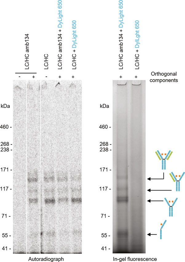
Fig. 3 Batch-based cell-free synthesis, site-specific introduction of ncaa by
amber (amb) suppression and subsequent fluorescence labeling of antibodies
with DyLight 650-phosphine. Qualitative analysis of cell-free synthesized anti-
body heavy chain (HC) and light chain (LC) was performed by SDS-Page followed
by autoradiography (left side) and in-gel fluorescence (right side) (analysis of
SN2). Cell-free synthesis was performed in the presence of 14C-leucine and with
(+) or without () supplementation of orthogonal components (synthetase
eAzPheRS-SII, tRNATyrCUA and ncaa p-azido-L-phenyalanine (AzF)). The amb
stop codon TAG was positioned in the CH1 domain (replacing S134, EU number-
ing) of HC. Orange asterisks indicate fluorescent dye conjugated to the
incorporated ncaa. Unassembled LC (25.4 kDa) and termination product of HC
amb134 (16.4 kDa) cannot be visualized in the autoradiograph because of its low
molecular weightEukaryotic Cell-Free Systems 187
6. Scan the screens using the Typhoon Trio + Variable Mode
Imager or Amersham Typhoon RGB Biomolecular Imager.
4 Notes
1. CHO lysates were prepared as described previously [10]. In
brief, CHO cells were grown in a Biostat B-DCU II bioreactor
(Sartorius Stedium Biotech GmbH) at 37 C using a chemically
defined and serum-free cell medium. Cells were grown to a
density of 3.5–5 106 cells/mL and harvested by centrifuga-
tion at 200 g for 5 min. Cells were washed twice and
resuspended in a HEPES-based homogenization buffer
(40 mM HEPES-KOH (pH 7.5), 100 mM NaOAc and
4 mM DTT). Resuspended CHO cells were lysed mechanically
by applying a 20-gauge needle and a syringe. By using the
syringe, cells were manually passed through the needle. After
cell disruption, the homogenate was centrifuged at 6500 g
for 10 min to remove cell nuclei and debris. The resulting
supernatant was applied to a Sephadex G-25 column which
was equilibrated with homogenization buffer. Elution fractions
with the highest RNA/protein ratios were pooled. In order to
remove the endogenous mRNA, cell lysates were mixed with
S7 micrococcal nuclease (f.c. 10 U/mL) and CaCl2 (f.c. 1 mM)
and incubated for 2 min at RT. Inactivation of micrococcal
nuclease was achieved by the addition of EGTA
(f.c. 6.7 mM). Afterwards, creatine kinase (f.c. 100 μg/mL)
was added to the lysate in order to ensure the regeneration of
ATP out of creatine phosphate. Aliquots of the CHO lysate
were immediately shock frozen in liquid nitrogen and subse-
quently stored at - 80 C until further usage.
2. Coding sequences of HC and LC should be N-terminally fused
to the melittin signal sequence to allow for the translocation of
de novo synthesized polypeptide chains into the lumen of the
microsomal vesicles [7]. Furthermore, HC and LC sequences
should be fused to regulatory sequences necessary for CFPS
(Fig. 1). The 50 untranslated region (50 UTR) of HC/LC tem-
plates contained a T7 promotor sequence and an internal ribo-
somal entry site (IRES from the intergenic region (IGR) of the
Cricket paralysis virus (CrPV), (Genbank accession
no. AF218039, nucleotides 6025–6216)) as regulatory
sequences to allow for efficient transcription based on T7
RNA polymerase and factor-independent translation initiation,
respectively. The 3’UTR contained a T7 terminator sequence
and a multiple cloning site for subsequent cloning. HC/LC
sequences were synthesized de novo by Geneart (Life technol-
ogies, Thermo Fisher) and cloned into pMA vector backbone.188 Marlitt Stech et al.
The sequence of the variable domains was kindly provided by
the lab of Michael Hust et al. (Technische Universit€at
Braunschweig) [12]. The position of the amber stop codon in
the CH1 domain at Serin 134 was chosen according to
Zimmerman et al. (2014) [4].
3. The template used for protein synthesis should contain a T7
promotor, ribosomal binding site, and T7 terminator such as
pIX3.0, pIVEX2.3d, and pIVEX2.4d vectors. Alternatively, a
T5 promotor as contained in pQE2 vectors can be used.
4. The fluorescent dye Bodipy is susceptible to light. Protect it
from light by using colored tubes or wrap the tubes with
aluminum foil.
5. It is very important to work in an RNase-free environment and
with RNase-free equipment. Use RNase-free filter tips for
pipetting and RNase-free reaction vessels. Pipette the compo-
nents in the listed order. Furthermore, it is recommended to
avoid repeated freeze-thaw cycles of all components. After
usage, shock-freeze the lysate in liquid nitrogen and store it at
80 C.
6. The DNA template used for CFPS should contain a T7 pro-
motor. We found that the applied DNA template concentration
is a potential parameter for optimization because template
concentration influences protein synthesis efficiency. Different
DNA templates may have different optimum concentrations
within the cell-free reaction. For the synthesis of the chosen
model antibody, we found that a 1:1 plasmid ratio of HC/LC,
each added at 60 nM, worked best.
7. Cell-free reactions are scalable. You can adjust the final volume
of the reaction according to the requirements of your
experiment.
8. In general, the optimal temperature of the CHO cell-free
system is 30 C [11] but the optimal incubation temperature
may be different for different proteins. For antibody synthesis,
27 C was found to result in the highest yields of active
antibodies.
9. Cell-free synthesized antibodies which have been translocated
and trapped inside the lumen of the microsomes can be released
by re-solubilization of the microsomal vesicles using PBS sup-
plemented with 0.2% DDM. It is important to thoroughly
resuspend the microsomes within the buffer in order to release
translocated antibodies quantitatively.
10. We find that it is beneficial to collect all fractions throughout
the purification, buffer exchange, and concentration procedure
(e.g., flow-through, washing fractions, elution fractions). Ali-
quots of these solutions should be diluted in SDS-PAGEEukaryotic Cell-Free Systems 189
sample buffer and analyzed by SDS-PAGE in order to monitor
the purity of the aminoacyl-tRNA-synthetase during the prep-
aration procedure.
11. We recommend to concentrate the synthetase up to a concen-
tration of 5 g/L to ensure a minimal final concentration of
100 μM. If necessary, repeat the concentration step.
12. We purify the PCR product using QIAquick PCR Purification
Kit and determine the concentration of the PCR product by
using a NanoDrop 2000c. For further analysis, prepare a 1%
(w/v) agarose gel and load 1 μL of the PCR product. The
expected band size is 123 bps.
13. It is important to work in an RNase-free environment. Use
RNase-free filter tips and reaction vessels.
14. Prepare a 2% (w/v) agarose gel. For sample preparation, mix
2 μL of the RNA with 6 μL MOPS sample buffer and load the
sample to the agarose gel. Use an RNA ladder. The expected
band size is around 200 bps.
15. After centrifugation, three phases will be visible: the aqueous
phase on top with approximately 50% of the total volume
containing the RNA; a middle interphase which is nearly invisi-
ble and below the red phenol/chloroform phase. Try to isolate
only the aqueous phase.
16. Reactive groups of ncaa are often sensitive to light and might
become instable upon light exposure. Thus, protect solutions
from light by using colored tubes or wrap the tubes with
aluminum foil.
17. The use of non-reducing sample buffer is important to main-
tain the disulfide bonds which connect the polypeptide chains
of the antibodies. Heating of samples before gel electrophore-
sis is not necessary.
Acknowledgments
The authors would like to thank Prof. Dr. Michael Hust for
providing the sequence of the antibody variable domains. Further-
more, we would like to thank Doreen Wüstenhagen and Dana
Wenzel (Fraunhofer IZI-BB, Potsdam-Golm) for their excellent
support regarding the preparation of the CHO lysates used in this
study. This work is supported by the European Regional Develop-
ment Fund (EFRE), the German Ministry of Education and
Research (BMBF, No. 031B0078A), and the German Research
Foundation (DFG Priority Programme 1623).190 Marlitt Stech et al.
References
1. KEGG DRUG Database. [Online] using a coupled in vitro transcription-
15.02.2021. https://www.genome.jp/kegg/ translation system based on CHO cell lysates.
drug/br08328.html Sci Rep 7:S12030
2. Davies SL, James DC (2009) Engineering 8. Edwards H, Schimmel P (1990) A bacterial
mammalian cells for recombinant monoclonal amber suppressor in Saccharomyces cerevisiae
antibody production. [Buchverf.] Mohamed is selectively recognized by a bacterial
Al-Rubeai. Cell line development. Springer aminoacyl-tRNA synthetase. Mol Cell Biol
Netherlands, Dordrecht. Bd. 6, S. 153–173 10:1633–1641
3. Axup JY (2012) Synthesis of site-specific anti- 9. Chin JW, Cropp TAS, Anderson JC et al
body-drug conjugates using unnatural amino (2003) An expanded eukaryotic genetic code.
acids. Proc Natl Acad Sci USA Science 301:964–967
109:16101–16106 10. Brödel AK, Wüstenhagen DA, Kubick S (2015)
4. Feng T, Lu Y, Manibusan A et al (2014) A Cell-free protein synthesis systems derived
general approach to site-specific antibody from cultured mammalian cells. Methods Mol
drug conjugates. Proc Natl Acad Sci U S A Biol (Clifton, NJ) 1261:129–140
111:1766–1771 11. Thoring L, Wüstenhagen DA, Borowiak M
5. Zimmerman ES, Heibeck TH, Gill A et al et al (2016) Cell-free systems based on CHO
(2014) Production of site-specific antibody- cell lysates: optimization strategies, synthesis of
drug conjugates using optimized non-natural "difficult-to-express" proteins and future per-
amino acids in a cell-free expression system. spectives. PLoS One 11:e0163670
Bioconjug Chem 25:351–361 12. Thie H, Toleikis L, Li J et al (2011) Rise and
6. VanBrunt MP, Shanebeck K, Caldwell Z et al fall of an anti-MUC1 specific antibody. PLoS
(2015) Genetically encoded Azide containing One 6:e15921
amino acid in mammalian cells enables site- 13. Thoring L, Dondapati SK, Stech M et al
specific antibody-drug conjugates using click (2017) High-yield production of "difficult-to-
cycloaddition chemistry. Bioconjug Chem express" proteins in a continuous exchange
26:2249–2260 cell-free system based on CHO cell lysates. Sci
7. Stech M, Nikolaeva O, Thoring L et al (2017) Rep 7:11710
Cell-free synthesis of functional antibodies
Open Access This chapter is licensed under the terms of the Creative Commons Attribution 4.0 International
License (http://creativecommons.org/licenses/by/4.0/), which permits use, sharing, adaptation, distribution
and reproduction in any medium or format, as long as you give appropriate credit to the original author(s) and the
source, provide a link to the Creative Commons license and indicate if changes were made.
The images or other third party material in this chapter are included in the chapter’s Creative Commons license,
unless indicated otherwise in a credit line to the material. If material is not included in the chapter’s Creative
Commons license and your intended use is not permitted by statutory regulation or exceeds the permitted use,
you will need to obtain permission directly from the copyright holder.You can also read