Polyphenol content and antioxidant activities of Prunus padus L. and Prunus serotina L. leaves: Electrochemical and spectrophotometric approach ...
←
→
Page content transcription
If your browser does not render page correctly, please read the page content below
Open Chemistry 2020; 18: 1125–1135
Research Article
Aleksandra Telichowska*, Joanna Kobus-Cisowska, Marta Ligaj, Kinga Stuper-Szablewska,
Daria Szymanowska, Mariusz Tichoniuk, Piotr Szulc
Polyphenol content and antioxidant activities of Prunus padus L.
and Prunus serotina L. leaves: Electrochemical and
spectrophotometric approach and their antimicrobial
properties
https://doi.org/10.1515/chem-2020-0121 2,2′-Azino-bis(3-ethylbenzothiazoline-6-sulfonic acid) dia-
received May 19, 2020; accepted July 28, 2020 mmonium salt (ABTS) cation radical (37.39b ± 3.81 mg TE/
g dw). The higher antioxidant potential of P. padus was
Abstract: The aim of the study was to compare the content
confirmed based on the oxidizing potentials of electroactive
of selected phytochemicals as well as the antioxidant and
compounds present in them. Stronger inhibition against
antimicrobial potential of the leaves of Prunus padus L. and
Enterococcus faecium and Klebsiella pneumoniae was found
Prunus serotina L., as there is very little research on this
for P. padus, whereas P. serotina extract was more potent
subject in the literature. Therefore, it is used to deepen against Enterococcus faecium bacterium. It has been shown
knowledge on this subject. In addition, an electrochemical that P. padus can be an attractive raw material with
test was also carried out, which was not yet available for the antioxidant and antimicrobial properties that can be used
above plants. Antibacterial studies have also been deepened on a much wider scale in food technology than its current
to include the analysis of new strains of bacteria and fungi, application.
which has not been studied earlier. The water extracts of
P. padus using the utra-performance liquid chromatography Keywords: Prunus, polyphenols, antioxidant, antibac-
(UPLC) system showed a higher content of both phenolic terial, electrochemical
acids and flavonols (651.77b ± 18.12 mg/100 g dw for acids
and 3.85b ± 0.08 mg/100 g dw for flavonols, respectively).
Ferulic and p-coumaric acids were the dominant polyphenols
in leaves. Extracts from P. padus showed higher activity 1 Introduction
against DPPH radical, which was 6.62b ± 0.06 mg TE/1 g dw,
Natural products of plant origin are gaining interest worldwide
as well as higher antioxidant capacity, measured using
due to component contents that may affect health. Bird cherry
(P. padus) and black cherry (P. serotina) are popular plants
found in many regions in the world. They have fruits with a
* Corresponding author: Aleksandra Telichowska, Foundation for bitter aftertaste, which are most often used as raw material for
the Education of Innovation and Implementation of Modern making tinctures. For infusions, not only bird cherry bark is
Technologies, 62-069, Dabrowka, Poland, e-mail: aleksandra. used, but also shoots, leaves, and leaf buds are used. In folk
telichowska@mail.up.poznan.pl
medicine, bird cherry was considered as a medicinal plant.
Joanna Kobus-Cisowska: Department of Gastronomy Sciences and
Bird cherry is a source of polyphenols. P. padus fruits
Functional Foods, Poznan University of Life Sciences, 60-637, Poznan,
Poland
contain mainly caffeic acid, chlorogenic acid, coumaric acid,
Marta Ligaj, Mariusz Tichoniuk: Department of Industrial Products ellagic acid, gallic acid, vanillic acid, and ferulic acid [1,2]. It
Quality and Packaging, Poznań University of Economics and Business, has also been shown that the fruits of P. padus additionally
61-875, Poznań, Poland contain organic acids, minerals, and vitamins, mainly
Kinga Stuper-Szablewska: Department of Chemistry, Poznan vitamin C. The most important flavonols found in the fruits
University of Life Sciences, Wojska Polskiego 28, 60-637, Poznan, of Prunus padus L. were quercitrin and quercetin, epicatechin
Poland
and catechin also dominated. In addition, quercetin
Daria Szymanowska: Department of Pharmacognosy, Poznan
derivatives, such as hyperoside, kaempferol, or isorham-
University of Medical Science, 60-781, Poznan, Poland; Department
of Biotechnology and Food Microbiology, Poznan University of Life netin, were also determined. Kaempferol glycosides ac-
Sciences, 60-627, Poznan, Poland counted for only 2%, and isoramnetine glycosides were
Piotr Szulc: Department of Agronomy, Poznan University of Life detected in less than 1 mg/kg fw [1]. On the other hand,
Sciences, 60-637, Poznan, Poland Prunus is considered as an invasive species. It contains
Open Access. © 2020 Aleksandra Telichowska et al., published by De Gruyter. This work is licensed under the Creative Commons Attribution
4.0 International License.1126 Aleksandra Telichowska et al.
cyanogenic glycosides such as prunazine and amygdalin. Podlasie, Poland (53° 13′ 14.865″ N 23° 51′ 9.327″ E). The
Cyanogenic glycosides are plant components and may be soil in the orchard was characterized by an average
toxic when consumed in large quantities due to the abundance of macronutrients. The approximate value of
hydrolytic release of hydrocyanic acid [3,4]. pH for soil, marked in 1 M KCl, was 6.13, and the content
It has been shown that the consumption of plant of humus was 1.14%. The average amount of precipita-
materials rich in polyphenols can have a health-promoting tion in the growing season was 317 mm per square meter,
effect as well as a positive effect on biochemical processes in with an average daily temperature of 14.4°C. The leaves
the body. Prunus padus L. has been demonstrated to be a were stored in frozen conditions (temperature = −28°C)
raw material containing polyphenols with antioxidant and until lyophilization and the extracts were prepared.
antimicrobial activities. The beneficial effects of Prunus Lyophilization was performed in a CHRIST 1–4 LSC
padus L. seed extracts were confirmed against pathogenic freeze dryer (Martin Christ Gefriertrocknungsanlagen
bacteria such as Staphylococcus aureus, Staphylococcus GmbH, Osterode am Harz, Germany) under constant
hominis, and Proteus mirabilis [5]. The leaves and branches conditions. The condensation temperature in the freeze
of bird cherry contain components that have a beneficial dryer was maintained at −28°C, the temperature on the
antidiabetic effect inhibiting alpha-glucosidase activity [6]. freeze dryer shelf at −20°C, and the product temperature
The positive effect of bird cherry on hypertension was also at −4°C. The entire process was carried out under
confirmed. This effectiveness is due to the presence of reduced pressure for 24 h. The leaves were extracted
hyperoside and chlorogenic acid as compounds that relax after grinding in Grindomix GM 200 (Retsch, Haan,
the smooth muscles of blood vessels [7]. Extracts of Germany) for 180 s at 1,700 × g at 21°C.
P. serotina fruits contain polar and nonpolar metabolites
with a vasodilating effect [7]. P. padus also contains
anthocyanins, cyanogenic glycosides, flavonoids, and
chlorogenic acid, which are important in the treatment of 2.2 Extraction
inflammation [8]. It has antibacterial and antifungal proper-
ties [5]. The beneficial antimicrobial effect was confirmed in Extraction with solvents such as water or mixture of water
extracts obtained from P. padus stems, indicating at the and alcohol is widely used to assess the content of
same time anti-inflammatory and analgesic effects. Strong biologically active compounds in plant raw materials.
anti-inflammatory properties result not only from the Polyphenols, vitamins, and minerals are easily extracted
inhibition of inflammatory mediators but also from the with polar solvents, enabling extracts with high antioxidant
properties that reduce inflammation edema [9]. Current activity. The water extract from P. padus (PPL) and
scientific research results have confirmed selectively bene- P. serotina (PSL) was obtained using water at 85°C, and
ficial properties of extracts from various anatomical parts of 1,000 mL of water was mixed with 50 g of raw material and
both P. serotina and P. padus; however, there are no reports extracted for 15 min. The extracts were filtered and
in the literature that would indicate to what extent these two centrifuged (800 × g, 15 min) each time. The fractions were
types of Prunus differ in terms of polyphenol content and decanted and filtered (Whatman 1:11 µm). The prepared
antioxidant activity measured spectroscopically and electro- extracts were stored in dark tubes until examination at 4°C.
chemically. Therefore, the main aim of the work was to
assess the antioxidant and antimicrobial properties of P.
padus and P. serotina leaves and thus to present the
possibility of creating values resulting from the application of 2.3 Color and osmolality of extract
bird cherry plant raw material as the source of polyphenols. measurement
Color of leaves extract was measured. Color measure-
ment was run in L × a × b × CEN unit system using
2 Materials and methods spectrometer CM-5 (Konica Minolta, Japan) according to
the methodology described by the device producer. As a
source of light, D 65 was applied, and color temperature
2.1 Materials
equaled 6,504 K. The observation angle of the standard
colorimetric observer was 10°. Measurements for each
The leaves of P. padus and P. serotina were harvested in sample was repeated fivefold. The instrument calibration
September 2019 in the orchard farm in Ozierany Małe in was performed with the use of a black pattern.Polyphenol content and antioxidant activities of P. padus L. and P. serotina L. leaves 1127
2.4 UPLC determination of phenolic acids equivalents (GAE) per 1 g (mg/1 g) of dry mass using
and flavonols the calibration curves of gallic acid.
The DPPH procedure was based on the reduction of
Phenolic compounds in water extract were analyzed after DPPH solution absorbance (2,2-diphenyl-1-picrylhy-
alkaline and acidic hydrolysis. The analysis was performed drazyl) at wavelength 517 nm in the presence of free
using an Acquity H class UPLC system equipped with a radicals [12]. Measurements were performed using the
Waters Acquity PDA detector (Waters, USA). Chromato- SP-830 Plus apparatus (Metertech, Taiwan). The percen-
graphic separation was performed on an Acquity UPLC® tage of DPPH radical scavenging was evaluated based on
BEH C18 column (100 mm × 2.1 mm, particle size 1.7 µm) the standard curve for y = 321.54x + 21.54 (R2 = 0.986)
(Waters, Ireland). The elution was carried out gradient and presented as mg TE/1 g dw of extract.
using following mobile phase composition: A, acetonitrile The ABTS cation radical scavenging activity was
with 0.1% formic acid; B, 1% aqueous formic acid mixture measured according to the Trolox Equivalent Antioxidant
(pH = 2). The eluent uptake rate was as follows: 0.4 mL/ Capacity test according to the methodology described by
min. Concentrations of phenolic compounds were deter- Kobus-Cisowska et al. (2020) [12]. Spectrophotometric
mined using an internal standard at wavelengths λ = measurement of the ability to scavenge ABTS+ formed
320 nm and 280 nm and the results were expressed as mg/ from ABTS (2,20-azinobis-(3-ethylbenzothiazoline-6-sul-
100 g d.m of Prunus leaves. Compounds were identified phonic acid) by oxidation with potassium persulfate was
based on a comparison of retention time of the analyzed carried out at a wavelength of 414 nm using SP-830 Plus
peak with the retention amount of standard to the analyzed apparatus (Metertech, Taiwan). The percentage rate of
samples and a repeated analysis. Detection level is 1 µg/g. ABTS+ scavenging was calculated from the standard curve
Retention times for phenolic acids were as follows: for y = 121.63x + 26.33 (R2 = 0.96) and expressed as mg TE/
protocatechuic acid 1.56 min, gallic acid 4.85 min, g dw of extract.
p-coumaric acid 8.06 min, 2,5-dihydroxybenzoic acid
9.55 min, 4-hydroxybenzoic acid 9.89 min, chlorogenic
acid 12.00 min, caffeic acid 15.20 min, syringic acid
15.60 min, sinapic acid 17.10 min, ferulic acid 19.00 min, 2.6 Ferric reducing
salicylic 17.85 min, t-cinnamic acid 20.00 min, and vanillic
acid 21.05 min. The retention time for flavonoids was as The antioxidant properties of the extracts were
follows: apigenin 1.10 min, vitexin 8.00 min, kaempferol determined using a ferric reducing/antioxidant power
11.00 min, luteolin 16.90 min, quercetin 17.00 min, narin- assay (FRAP method) according to the procedure
genin 17.50 min, rutin 19.00 min, and catechin described by O’Sullivan et al. [13]. FRAP reagent (2 mL;
19.50 min [10]. 0.01 mol TPTZ [2,4,6-tripyridyl-s-triazine] in 0.04 mol
HCl, 0.02 mol FeCl3·6H2O and 0.3 mol acetate buffer) was
added to 1 µL of each sample diluted in 999 µL distilled
H2O. A calibration curve was constructed using
2.5 Antioxidative potential analysis by FeSO4·7H2O. Samples were incubated for 30 min, and
spectrophotometric method the absorbance was measured at 593 nm (Metertech
SP880, Taiwan). Data were expressed as µM FeSO4/
The total phenolic content (TPC) of the obtained extracts 1 g dw of extract.
was determined using the method described by
Kulczyński et al. (2016) with minor modifications [11].
Aliquots of 100 µL diluted in 900 µL of 40% ethanol
(Sigma-Aldrich, Germany) were mixed with 1 mL of 2.7 Antioxidative potential analysis by
Folin–Ciocalteu reagent (Sigma-Aldrich, Germany), fol- electrochemical assay
lowed by the addition of 1 mL of 35% sodium carbonate
(POCH, Poland). Samples were vortexed for 5 s, and after The content of redox compounds in Prunus leaves
incubation in darkness at room temperature for 90 min, extracts was determined using square wave voltammetry
the absorbance of the reaction mixture was measured at (SWV). Voltammetric measurements were performed
765 nm against a blank. The TPC was expressed as using potentiostat PGSTAT12 with the GPES 4.9 control
milligram of gallic acid (Sigma-Aldrich, Germany) software (EcoChemie, The Netherlands). A three-1128 Aleksandra Telichowska et al.
electrode measuring system consisting of a reference (ATCC 13076), Listeria monocytogenes (ATCC 19115), and
electrode Ag/AgCl (3 M KCl) (Mineral, Poland), platinum Bacillus coagulans (GBI-30, 6086) as well as fungi of the
as an auxiliary electrode (Mineral, Poland), and carbon species Candida utillis (ATCC 9950), Aspergillus sp. and
paste as a working electrode (CPE) was used for the Fusarium sp. were propagated in Muller–Hinton medium
measurements. The CPE was developed according to a (Oxoid, UK) at 30°C (yeast) or 37°C (bacteria) for 24 h.
described procedure [14]. Carbon paste was made by Subsequently, to obtain a clear bacterial layer, the liquid
mixing graphite powder (Sigma) with mineral oil Mueller–Hinton agar medium was inoculated with a 10%
(Sigma) in the ratio of 70:30 (w/w). The surface of the 24 h indicator culture with an optical density of 0.5 on
CPE was renewed before use by removing the outer layer McFarland scale and poured into Petri dishes. A well was
of carbon paste on filter paper, application of fresh drilled in the surface of the solid medium inoculated with
paste, and polishing it to a smooth finish on a frosted indicator microorganisms, to which 50 microliters of the
glass microscope slide. Before electrochemical measure- extract was added. Plates were incubated under conditions
ment, the surface of CPE was treated with 0.05 M suitable for a given group of microorganisms for 24–48 h.
phosphate buffer mixed with 0.01 M KCl (pH 7.0) at a Then, the growth inhibition zone of indicator microorgan-
potential of +1.7 V for 60 s. After that, the electrodes were isms was measured (clearing around the application site of
immersed for 120 s in the solution containing extract the sample).
dissolved in phosphate buffer in the ratio 1:1 (v/v),
whether the SWV measurement in the range from −0.3 V
to +1.4 V was made. Applied SWV parameters were as
follows: step potential of 5 mV, frequency of 50 Hz, and 2.9 Statistical analysis
amplitude of 40 mV. Three repetitions of SWV measure-
ment for each extract were performed. SWV voltammo- Statistical analysis of all results was performed using
grams were smoothed using Sa the vitzky–Golay’s Microsoft Excel 2013 software (USA) and Statistica 13
method [15]. From SWV voltammograms, the baselines software (StatSoft, Poland). The electrochemical results
determined with moving average procedure were sub- were treated as an additional factor to the model based
tracted and finally were determined the data including on standard analytical techniques. The p values for
peak potential, peak height (current), peak area for each Levene’s test of independent variables were calculated.
signal, and the total peak areas. Based on our results
for Cornus mas extracts [16] an electrochemical index Ethical approval: The conducted research is not related
(EI) describing the electrochemical activity of tested to either human or animal use.
extracts, expressed as the total area of all redox
signals, in relation to 1 g dry matter of examined plant
material was also determined. With respect to the tested
samples, 1 mL of the extract was prepared from 0.063 g 3 Results
of leaves, which after dilution in the buffer gave a final
0.03125 g dry matter content of plant material in the
3.1 Characteristics of P. padus and P.
tested sample.
serotina extracts
Prunus extracts were physically and chemically char-
2.8 Antimicrobial activity testing using the acterized (Table 1). It was shown that the color of the
well-diffusion method tested extracts differed in terms of assessed parameters.
Parameter L* determining the brightness was 26.06 ±
Indicator microorganisms such as Gram-negative bacteria: 0.29 in the PPL sample and 34.59 ± 1.96 in the PSL
Klebsiella pneumoniae (ATCC 31488), Salmonella enteritidis sample. Parameter a*, responsible for the color change
(ATCC 860), Pseudomonas aeruginosa (ATCC 27853), and in the range from green to red, was 9.95 ± 0.04 for PPL
Acinetobacter baumannii (ATCC 19606) and Gram-positive and 14.69 ± 0.19 for PSL, whereas parameter b*
bacteria: Enterococcus faecium (ATCC 27270), Enterococcus responsible for the color change in the range from blue
faecium (ATCC 27270), Staphylococcus aureus (ATCC 25923), to yellow had lower values for PPL (1.80 ± 0.12) and
Lactobacillus fermentum (ATCC 14932), Clostridium butyricum higher for PSL (11.22 ± 0.22).Polyphenol content and antioxidant activities of P. padus L. and P. serotina L. leaves 1129
Table 1: Characteristics of the tested P. padus and P. serotina Table 2: Content of polyphenolic compounds in P. serotina and
leaves, given in CIE L*a*b* units and osmolality P. padus leaves
Sample PSL PPL PSL (mg/100 g PPL (mg/100 g
dw leaves) dw leaves)
Osmolality 0.171 ± 0.01
b
0.156 ± 0.01
a
(mOsm/kg H2O) Phenolic acids
Freezing −0.289a ± 0.01 −0.289a ± 0.01 Gallic acid 19.56a ± 0.64 22.3b ± 0.64
temperature (°C) 2,5- 14.52a ± 0.44 16.52b ± 0.24
L* 34.59b ± 1.96 26.06a ± 0.29 Dihydroxobenzoic acid
a* 14.69b ± 0.19 9.95a ± 0.04 4-Dihydroxobenzoic acid 23.6a ± 0.12 29.45b ± 0.13
b* 11.22b ± 0.22 1.80a ± 0.12 Caffeic acid 11.45a ± 0.61 13.65b ± 0.09
Syringic acid 5.62a ± 0.12 8.95b ± 0.04
Color p-Coumaric acid 103.6a ± 0.21 157.6b ± 8.33
Ferulic acid 185.3a ± 6.72 195.6b ± 5.64
Chlorogenic acid 29.5a ± 0.09 36.8b ± 0.24
Abbreviation: PPL, water extract from Prunus padus L. leaves, PSL, Sinapic acid 97.68a ± 0.39 147.5b ± 2.21
water extract from Prunus serotina L. leaves, results are mean t-Cinnamic acid 13.4a ± 0.08 20.8b ± 0.05
values of three determinations ± standard deviation. Values Vanillic acid 1.24a ± 0.06 2.6b ± 0.11
sharing the same letter in a line are not significantly different Salicylic acid ND ND
(P ≤ 0.05). Total phenolic acids 505.47a ± 9.48 651.77b ± 18.12
Flavonoids
Naringenina 0.13a ± 0.00 0.62b ± 0.02
The osmolality of the extracts indicates the freezing Vitexin ND ND
point of the extract and its differences relative to the Rutin ND 1.03 ± 0.02
freezing of water, which is a measure of the osmotic Quercetin 0.17b ± 0.01 0.13a ± 0.02
Apigenin ND ND
pressure of the tested extract. Extracts’ osmolality was
Kaempferol ND ND
0.156 mOsm/kg H2O for the PPL extract and 0.171 mOsm/
Luteolin ND ND
kg H2O for the PSL extract. Catechin 1.01a ± 0.01 2.07b ± 0.02
Total flavonoids 1.31a ± 0.02 3.85 b ± 0.08
Abbreviation: as in Table 1, ND, not detected, results are mean
values of three determinations ± standard deviation. Values
3.2 Phenolic acid and flavonoid contents sharing the same letter in a line are not significantly different
(P ≤ 0.05).
The content of phenolic acids and flavonols was
determined in the obtained extracts (Table 2). Qualitative
and quantitative characteristics of individual polyphe- leaves contained the highest content of catechins (1.01 ±
nols in the extracts differed between the samples. Higher 0.01 mg/100 g dw) among the flavonols tested.
phenolic acid contents were found in the PPL extract,
which was 651.77 ± 18.12 mg/100 g dw. The dominant
acids were p-coumaric acid 157.6 ± 8.33 mg/100 g dw, 3.3 Antioxidant potential analysis by
ferulic acid 195.6 ± 5.64 mg/100 g dw, and sinapic acid spectrophotometric method
147.5 ± 2.21 mg/100 g dw. The lowest amounts among the
tested acids were detected for vanillic acid 2.6 ± 0.11 mg/ The analyzed extracts were evaluated for their antioxidant
100 g dw and syringic acid 8.95 ± 0.04 mg/100 g dw. The potential by spectroscopic methods (Table 3). It was found
water extract of PSL leaves contained the highest content that extracts prepared from leaves of black cherry PSL and
of ferulic acid (185.3 ± 6.72 mg/100 g dw) and p-coumaric bird cherry PPL had different properties. The higher content
acid (103.6 ± 0.21 mg/100 g dw). The lowest levels were of these compounds was found in the extract of PPL leaves
determined for vanillic acid (1.24 ± 0.06 mg/100 g dw) (37.39 ± 3.81 mg GAE/g dw). In the FRAP test, PPL leaf
and syringic acid (5.62 ± 0.12 mg/100 g dw). PPL leaf extract also showed a 30% higher activity when compared
extract contained a higher concentration of flavonols with PSL. Test results were also complemented by deter-
(3.85 ± 0.08 mg/100 g dw) than PSL extract. PPL leaves mining the effect of the extracts using the DPPH radical test.
contained naringenin, rutin, quercetin, and dominant PPL extract was shown to scavenge radicals at 6.62 ±
catechin (2.07 ± 0.02 mg/100 g dw). The extract from PSL 0.06 mg TE/1 g dw, while DPPH anti-radical activity for PSL1130 Aleksandra Telichowska et al.
Table 3: Chemical structure of compounds analyzed
Phenolic acids R1 R2 R3 R4 R5
4-Hydroxobenzoic acid –H –H –H –OH –H
2,5-Dihydroxobenzoic acid –H –OH –H –H –OH
Gallic acid –H –OH –OH –OH –H
t-Cinnamic acid –H –H –H –H –H
Salicylic acid –OH –H –H –H –H
Syringic acid –H –OCH3 –OH –OCH3 –H
Vanillic acid –H –OCH3 –OH –H –H
Ferulic acid –H –H –OH –OCH3 –H
Caffeic acid –H –H –OH –OH –H
p-Coumaric acid –H –H –OH –OH –H
Sinapic acid –H -OCH3 –OH –OCH3 –H
Chlorogenic acid
Flavonoids
Naringenin
Vitexin
R1 R2
Apigenin –H –H
Kaempferol –OH –H
Quercetin –OH –OH
Luteolin –H –OH
Catechin –OH –H
Rutin o-gluk-ramn –OH
extract was slightly lower and amounted to 5.43 ± 0.07 mg 3.4 Antioxidant potential analysis by an
TE/1 g dw. These analyses were also confirmed by the tests electrochemical assay
carried out using the ABTS radical method, which also
showed higher activity of the solution from PPL leaves, The electrochemical activity of the extracts was deter-
where the value of aqueous extracts PPL was 9.65 ± mined using SWV. Electrochemical measurements
0.09 mg TE/g dw and was higher than for PSL extracts, showing the content of redox compounds in the extracts
where it was 8.55 ± 0.08 mg TE/g dw. demonstrated the presence of two signals onPolyphenol content and antioxidant activities of P. padus L. and P. serotina L. leaves 1131
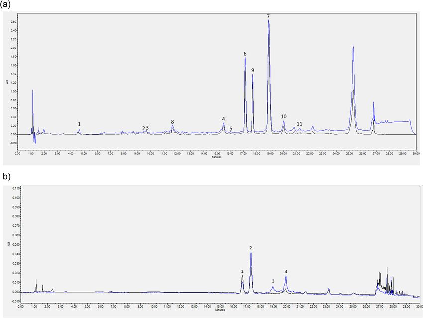
Figure 1: Comparison of chromatograms of phenolic acids for P. serotina and P. padus (a), comparision of chromatograms of flavonoids for
P. serotina and P. padus (b). (a) Comparison of chromatograms of phenolic acids for P. serotina (black line) and P. padus (blue line) leaves:
1 – gallic acid, 2 – 2,5-dihydroxobenzoic acid, 3 – 4-dihydroxobenzoic acid, 4 – caffeic acid, 5 – syringic acid, 6 – p-coumaric acid,
7 – ferulic acid, 8 – chlorogenic acid, 9 – sinapic acid, 10 – t-cinnamic acid, 11 – vanillic acid. (b) Comparison of chromatograms of
flavonoids for P. serotina (black line) and P. padus (blue line) leaves: 1 – quercetin, 2 – naringenin, 3 – rutin, 4 – catechin.
50 50
45
40 40
35
Current [ A]
Current [ A]
30 30
25
20 20
15
10 10
5
0 0
-0.4 -0.2 0.0 0.2 0.4 0.6 0.8 1.0 1.2 1.4 1.6 -0.4 -0.2 0.0 0.2 0.4 0.6 0.8 1.0 1.2 1.4 1.6
a) Potential [V]
b) Potential [V]
Figure 2: SWV signals for P. padus (a) and P. serotina (b) leaf extracts.1132 Aleksandra Telichowska et al.
Table 4: P. padus and P. serotina leaf extracts with FRAP, DPPH, Table 5: The electrochemical parameters determined for extracts
ABTS radicals and total polyphenol content from P. padus and P. serotina leaves
Sample/activity PSL PPL SWV parameters
TPC (mg GAE/g dw) 21.54 ± 3.34
a
37.39 ± 3.81
b
Sample PSL PPL
DPPH (mg TE/1 g dw) 5.43a ± 0.07 6.62b ± 0.06
Peak potential Ep [V] 0.325 ± 0.000 0.285 ± 0.000
ABTS (mg TE/g dw) 8.55a ± 0.08 9.65b ± 0.09
0.967 ± 0.010 0.950 ± 0.000
FRAP (µM FeSO4/1 g dw) 179.24a ± 3.66 233.20b ± 5.97
Peak current I [µA] 27.102 ± 0.871 29.065 ± 1.081
Abbreviation: as in Table 1. 1.267 ± 0.087 1.419 ± 0.225
Peak area EP × I [V × µA] 3.469 ± 0.425 4.280 ± 0.089
0.134 ± 0.014 0.171 ± 0.027
voltamperograms (Figure 1). In the PSL leaf extract, they Total peak current I [µA] 28.379a ± 0.957 30.484b ± 1.306
were located at +0.325 ± 0.000 and +0.967 ± 0.010 V, Total peak area [V × µA] 3.602a ± 0.439 4.451b ± 0.117
EI [V × µA/1 g dw] 115.267a ± 14.043 142.417b ± 3.737
while for PPL, these signals were slightly shifted toward
the negative potential, which was +0.285 ± 0.000 and Abbreviation: as in Table 1.
+0.955 ± 0.000 V, respectively. The dominant signal was
the first one registered in both cases, located in the literature, which, like in the current work, have
+0.3 V region (Figure 2). indicated the antioxidant potential of P. padus and
Table 4 summarizes electrochemical parameters P. serotina leaves, but also of fruits, bark, and flowers
determined for the analyzed extracts. The extract of [1–7]. As in the present study, other authors have
PPL leaves exhibited higher electrochemical activity, pointed out polyphenols as compounds that affect
expressed as the total area of redox signals, and the EI antioxidant potential. Current studies, however, do not
determined on its basis. The total signal area was 4.451 ± indicate differences in the content of specific biologically
0.117 V × µA, while EI reached the value of 142.417 ± active compounds in P. padus and P. serotina leaves, and
3.737. These values for PSL leaf extract were 3.602 ± most importantly, the work to date did not include both
0.439 V × µA and 115.267 ± 14.043, respectively. spectroscopic and electrochemical studies, allowing the
assessment of antioxidant activity. The use of electro-
chemical methods to assess the antioxidant potential is
3.5 Antimicrobial activity new in the field of bird cherry research. The aim of this
study was also to compare selected phytochemicals as
The influence of ground PSL and PPL leaves against well as to investigate the antioxidant and antimicrobial
indicator microorganisms of both Gram-positive and activity of bird cherry leaves, because there is very little
Gram-negative bacteria, as well as molds and fungi, was research on the subject in the literature. Antimicrobial
analyzed (Table 5). activity studies have been deepened to include pre-
The highest antimicrobial activity was demonstrated viously unknown strains of gram-positive and gram-
for PSL leaves against Enterococcus faecium (ATCC negative bacteria as well as fungi. The main phenolic
27270) (24 mm) and PPL leaves against Listeria mono- acid is chlorogenic acid, whose content in PPL was
cytogenes (ATCC 19115) (24 mm). The lowest activity of determined in earlier studies at the level of 1.39–1.94%
PSL leaf extract was demonstrated for bacteria from the dw [8]. Chlorogenic acid was also present in P. padus
Gram-negative group Acinetobacter baumannii (ATCC fruits at the level of 10.48 ± 0.28 mg/100 g fw [1]. Its
19606), and it was 8 mm. The extract also showed low presence in the fruits of P. serotina was also confirmed
activity against fungi, similarly as PPL leaf extract. by ref. [7]. Other authors determined the content of
individual polyphenols in one Prunus variety. These
leaves were not compared. Quercetin was determined in
P. serotina [7] but also in P. padus fruits (11.86 ± 2.36 mg/
4 Discussion 100 g fw) [1], indicating its importance in total antiox-
idant potential. Other studies demonstrated that rutin
Less known plants or under-utilized species have was primarily responsible for the properties of P. serotina
recently been the subject of great research interest due leaves [17] and fruits of P. padus (2.67 ± 1.02 mg/
to the presence of compounds exhibiting beneficial 100 g fw) [1]. It has been repeatedly suggested in the
health properties. There are studies available in the literature that polyphenol content determines thePolyphenol content and antioxidant activities of P. padus L. and P. serotina L. leaves 1133
Table 6: Micorbiological activity of P. padus and P. serotina leaves radical is one of the most reactive forms of ROS, which
extract can be generated by the Fenton reaction from H2O2 in the
presence of transition metal cations such as Fe2+. Natural
Indicator microorganisms PSL PPL plant sources are also sought for in the context of the
Growth
bactericidal and bacteriostatic activities since the
inhibition
zone (mm) overuse of antibiotics can have adverse effects. It has
been demonstrated that many substances present in
Gram-negative bacteria
rhizomes, fruits, leaves, or bark can exert a biocidal
Klebsiella pneumoniae (ATCC 18 ± 30 19 ± 30
31488)
effect [20]. The current study assessed the influence of
Salmonella enteritidis (ATCC 13076) 16 ± 30 14 ± 20 leaf compounds of P. serotina against indicator micro-
Pseudomonas aeruginosa (ATCC 15 ± 20 17 ± 30 organisms of both Gram-positive and Gram-negative
27853) bacteria, as well as molds and fungi (Table 6). The
Acinetobacter baumannii (ATCC 8 ± 10 15 ± 30 highest antimicrobial activity was demonstrated for
19606)
P. serotina (PSL) leaves against Enterococcus faecium
Gram-positive bacteria
Enterococcus faecium (ATCC 27270) 24 ± 40 22 ± 30 (ATCC 27270) (24 mm). Kumarasamy et al. (2004) [5] also
Staphylococcus aureus (ATCC 20 ± 30 15 ± 30 evaluated the activity of P. padus seed dichloromethane
25923) extracts against Enterococcus faecalis and showed weak
Lactobacillus fermentum (ATCC 18 ± 30 17 ± 30 growth inhibition of these bacterial strains as well as
14932)
Staphylococcus hominis. In the same study, the highest
Clostridium butyricum (ATCC 860) 19 ± 30 16 ± 30
Listeria monocytogenes (ATCC 23 ± 40 24 ± 40
antibacterial activity was demonstrated for methanol
19115) extract from P. padus seeds against Staphylococcus
Bacillus coagulans (GBI-30, 6086) 17 ± 30 19 ± 30 aureus (ATCC 25923) (1.0 × 10⁻⁴ mg/mL). In addition,
Fungi the extracts also showed the activity against methicillin-
Candida utillis (ATCC 9950) 8 ± 10 7 ± 10 resistant bacterial strains Staphylococcus aureus (ATCC
Aspergillus sp. 5 ± 10 3 ± 10
25923), Staphylococcus hominis, and Proteus mirabilis [5].
Fusarium sp. 3 ± 00 4 ± 10
The tested water extracts from PPL showed the highest
Abbreviation: PPL, water extract from Prunus padus L. leaves, PSL, activity against Listeria monocytogenes (ATCC 19115)
water extract from Prunus serotina L. leaves. (24 mm) and the lowest against fungi. Another study
evaluated the antimicrobial effect using methanol
extracts from the leaves and branches of P. padus.
antioxidant properties, whose mechanism of action can Both leaf and branch extract showed antimicrobial
be multidirectional. Polyphenols can inhibit the forma- activity against most Gram-positive bacteria tested;
tion of free radicals; they can scavenge them and however, only the branch extract exhibited any activity
increase the catalytic activity of endogenous enzymes against Gram-negative bacteria. The extract from
involved in free radical neutralization. Studies on bird P. padus branches was most active against Kocuria
cherry and the compounds it contains can be found in rhizophila (MIC = 125 µg/mL) [6]. The lowest activity of
the literature. However, there is no information on the P. serotina leaf extract (PSL) was demonstrated for
effect of bird cherry on reducing the degree of oxidative bacteria from the Acinetobacter baumannii (ATCC 19606)
damage caused by the OH˙ radical using an electro- Gram-negative group, and it was 8 mm. Low bird cherry
chemical DNA biosensor. The electrochemical method activity has been shown against fungi. The antioxidant
shows a significant advantage over other methods properties of bird cherry leaves depend on the content of
enabling the determination of 8-oxoguanine level, biologically active compounds, and their measurement
because it allows direct testing of DNA sample, without may be different depending on the methods used
the need for its hydrolysis, which is necessary for other (spectroscopic, electrochemical). The variation in the
highly sensitive methods [18]. The performed voltam- result of activity is most likely due to the affinity of the
metric measurement allowed determining the degree of extracted compounds for the reagents in the given
damage based on the observed changes in the signals of methods and specificity of action. Not only the composi-
DNA bases. Such changes in the guanine signal level are tion and the total content of individual compounds, but
commonly considered in this type of analysis, or less above all the mutual proportions, where, as the
frequently the appearance of nitrogen base-derived literature indicates, individual compounds may exhibit
signals, including 8-oxodG [19]. The OH˙ hydroxyl synergistic or antagonistic effects, which is important for1134 Aleksandra Telichowska et al.
both antioxidant and antimicrobial activities. Therefore, metabolite profiles and preliminary evaluation of anti-
it is necessary to test each raw material, because oxidant activity. Molecules. 2018;23(4):725. doi: 10.3390/
predicting activity for most raw materials is difficult or molecules23040725.
[2] Mikulic-Petkovsek M, Stampar F, Veberic R, Sircelj H. Wild
impossible. It has been shown that P. padus can be an
Prunus fruit species as a rich source of bioactive compounds.
attractive raw material with antioxidant and antimicro- J Food Sci. 2016;81:C1928–37.
bial properties that can be used on a much wider scale in [3] Drochioiu G, Arsene C, Murariu M, Oniscu C. Analysis of
food technology than its current application. cyanogens with resorcinol and picrate. Food Chem Toxicol.
2008;46:3540–5.
[4] Santos Pimenta LP, Schilthuizen M, Verpoorte R, Choi YH.
Quantitative analysis of amygdalin and prunasin in Prunus
serotina Ehrh. using 1H-NMR spectroscopy. Phytochem Anal.
5 Conclusions 2014;25:122–6.
[5] Kumarasamy Y, Cox PJ, Jaspars M, Nahar L, Sarker SD.
In recent years, there has been a very high interest in plant- Comparative studies on biological activities of Prunus padus
and P. spinosa. Fitoterapia. 2004;75:77–80.
derived products and their health-promoting properties. Bird
[6] Hyun TK, Kim HC, Kim JS. In vitro screening for antioxidant,
cherry (P. padus and P. serotina) is a new raw material that
antimicrobial, and antidiabetic properties of some Korean
can gain significance not only as an innovation in the native plants on Mt. Halla, Jeju Island. Indian J Pharm Sci.
development of functional food. It is common in many parts 2015;77:668–74.
of the world and does not require special cultivation [7] Luna-Vázquez FJ, Ibarra-Alvarado C, Rojas-Molina A, Rojas-
conditions. It can be used to design innovative dietary Molina JI, Yahia EM, Rivera-Pastrana DM, et al. Nutraceutical
value of black cherry prunus serotina ehrh. Fruits: antioxidant
supplements or functional foods. Food should not only
and antihypertensive properties. Molecules.
provide basic nutrients but should also be considered in 2013;18:14597–612.
terms of health benefits that can be obtained from it. Bird [8] Olszewska MA, Kwapisz A. Metabolite profiling and antiox-
cherry leaves are a good source of polyphenolic compounds idant activity of Prunus padus L. flowers and leaves. Nat Prod
with high antioxidant activity. Polyphenols showed a Res. 2011;25:1115–31.
protecting effect on cell structures against damage caused [9] Choi JH, Cha DS, Jeon H. Anti-inflammatory and anti-
nociceptive properties of Prunus padus. J Ethnopharmacol.
by free radicals, which contribute to faster aging of the body.
2012;144:379–86.
Current scientific research results confirm the beneficial [10] Stuper-Szablewska K, Kurasiak-Popowska D, Nawracała J,
properties of both P. serotina and P. padus extracts. It has Perkowski J. Response of non-enzymatic antioxidative me-
been demonstrated that bird cherry leaf extracts containing chanisms to stress caused by infection with Fusarium fungi
polyphenolic compounds have antioxidant and antibacterial and chemical protection in different wheat genotypes. Chem
Ecol. 2017;33:949–62.
properties. Thus, P. padus and P. serotina preparations can
[11] Kulczyński B, Kobus-Cisowska J, Kmiecik D, Gramza-
be a valuable raw material used in the food, pharmaceutical, Michałowska A, Golczak D, Korczak J. Antiradical capacity and
and cosmetics industries as a source of bioactive compounds polyphenol composition of asparagus spears varieties culti-
with multidirectional antioxidant activity. vated under different sunlight conditions. Acta Sci Pol Technol
Aliment. 2016;15:267–79.
Acknowledgments: The publication was co-financed [12] Kobus-Cisowska J, Szulc P, Szczepaniak O, Dziedziński M,
Szymanowska D, Szymandera-Buszka K, et al. Variability of
within the framework of the Ministry of Science and
Hordeum vulgare L. cultivars in yield, antioxidant potential,
Higher Education program as “Regional Initiative and cholinesterase inhibitory activity. Sustainability.
Excellence in years 2019–2020,” project number 005/ 2020;12:1938.
RID/2018/19. [13] O’Sullivan AM, O’Callaghan YC, O’Connor TP, O’Brien NM.
Comparison of the antioxidant activity of commercial honeys,
before and after in vitro digestion. Pol J Food Nutr Sci.
Conflict of interest: Authors declare no conflict of
2013;63:167–71.
interest. [14] Ligaj M, Tichoniuk M, Filipiak M. Detection of bar gene
encoding phosphinothricin herbicide resistance in plants by
electrochemical biosensor. Bioelectrochemistry.
2008;74:32–37.
[15]
References Press WH, Flannery BP, Teukolsky SA, Vetterling WT.
Savitzky–Golay smoothing filters. In: Numerical recipes in
Fortran 77: The art of scientific computing. Cambrigde, UK:
[1] Donno D, Mellano MG, De Biaggi M, Riondato I, Cambridge University Press; 1992.
Rakotoniaina EN, Beccaro GL. New findings in Prunus padus L. [16] Szczepaniak OM, Ligaj M, Kobus-Cisowska J, Maciejewska P,
Fruits as a source of natural compounds: Characterization of Tichoniuk M, Szulc P. Application for novel electrochemicalPolyphenol content and antioxidant activities of P. padus L. and P. serotina L. leaves 1135
screening of antioxidant potential and phytochemicals in nine, a biomarker of damage to DNA. Nucleic Acids Res.
Cornus mas extracts. CYTA J Food. 2019;17:781–9. 2002;30:1354–63.
[17] Luna-Vázquez FJ, Ibarra-Alvarado C, Rojas-Molina A, Romo- [19] Diculescu VC, Paquim AMC, Brett AMO. Electrochemical DNA
Mancillas A, López-Vallejo FH, Solís-Gutiérrez M, et al. Role of nitric sensors for detection of DNA damage. Sensors. 2005;5:377–93.
oxide and hydrogen sulfide in the vasodilator effect of ursolic acid [20] Kobus-Cisowska J, Szymanowska-Powałowska D,
and uvaol from black cherry prunus serotina fruits. Molecules. Szczepaniak O, Kmiecik D, Przeor M, Gramza-Michałowska A,
2016;21(1):78. doi: 10.3390/molecules21010078. et al. Composition and in vitro effects of cultivars of Humulus
[18] Bruskov VI, Malakhova LV, Masalimov ZK, Chernikov AV. Heat- lupulus L. Hops on cholinesterase activity and microbial
induced formation of reactive oxygen species and 8-oxogua- growth. Nutrients. 2019;11:1377.You can also read