Antioxidant and Antibacterial Activities of Magnesium Oxide Nanoparticles Prepared using Aqueous Extract of Moringa Oleifera Bark as Green Agents
←
→
Page content transcription
If your browser does not render page correctly, please read the page content below
OPEN ACCESS
Journal of Multidisciplinary Applied Natural Science Vol. 1 No. 1 (2021)                      https://doi.org/10.47352/jmans.v1i1.9
                                                                                                                  Research Article

Antioxidant and Antibacterial Activities of Magnesium Oxide
Nanoparticles Prepared using Aqueous Extract of Moringa
Oleifera Bark as Green Agents
Hanif Amrulloh , Awalul Fatiqin, Wasinton Simanjuntak, Hapin Afriyani, Annissa Annissa
Received : October 13, 2020    Revised : January 11, 2021       Accepted : January 25, 2021         Online : January 30, 2021

Abstract
In this research, Magnesium oxide nanoparticles (MgONPs) was prepared from MgCl2 solution using aqueous extract of M. oleifera
bark as green agent. Preparation procedure involved mixing of MgCl2.6H2O solution and the aqueous extract of M. oleifera bark,
followed by drop wise addition of NaOH solution. The formation of MgO nanoparticles in this synthesis was confirmed using UV-
Vis absorption. The spherical crystal structure of MgO nanoparticle confirmed XRD analysis. The average particle size of the
synthesized MgO nanoparticles measured between 60 - 100 nm using SEM and TEM images and PSA results. MgONPs
synthesized showed good antioxidant activity and antibacterial activity against S. aureus, E. faecalis, E. coli, and S. dysenteriae
bacteria.

Keywords antibacterial, antioxidant, bark extract, green synthesis, magnesium oxide nanoparticles

1. INTRODUCTION                                                     [11]. In biomedical applications, there are evidence
                                                                    reports showing MgONPs have antioxidant [12],
   Green method is a method that uses extracts                      antibacterial [13]–[15], antifungal [12][16], and
from plants as agents to form nanoparticles. This                   anticancer [16] activities.
method provides a variety of resources in the                           Moringa Oleifera (M. oleifera) is a multipurpose
process of nanoparticle synthesis [1]–[3]. In                       tropical plant from the Moriceae family which is
addition, there are many other advantages in the use                spread throughout India, Asia, and sub-Saharan
of natural materials in the synthesis of                            Africa [17]. The use of Moringa extract has been
nanoparticles. The fact that the use of biodegradable               widely used in synthesizing metal nanoparticles and
raw materials as a synthesis of nanoparticles has no                metal oxides. Moringa flower extract reportedly can
harmful impact on the environment, leads to a green                 be used to prepare palladium [18], silver [19], and
technology policy, is first and foremost                            hydroxyapatite [20] nanoparticles. Moringa leaf
environmentally friendly. Second, the efficiency of                 extract to prepare silver [21], zinc oxide [22],
synthesis, which promotes further production and                    titanium oxide [23] and nickel oxide [24]
further production through the availability of                      nanoparticles. Phytochemical analysis of water
abundant natural raw materials. Thirdly, the                        extracts of M. oleifera bark shows that Moringa
nanoparticles produced are biocompatible in                         wood bark contains compounds such as alkaloids,
comparison        with     conventionally      seized               phenolic acids, terpenoids, and flavonoids [25].
nanoparticles [4].                                                  These compounds play a role in the chelation
   Magnesium oxide nanoparticles (MgONPs) are                       process and are able to reduce metal ions to
multipurpose metal oxide nanoparticles which have                   nanoparticles [26].
many applications in various fields. MgONPs have                        The effect of M. oleifera bark water extract for
been widely used as catalysts [2][5][6] and catalyst                preparation of MgO nanoparticles was examined in
supports for various organic reactions [7],                         this presence research. In this presence study. This
adsorbents [8]–[10] and electrochemical biosensors                  research has been confirmed, since the special use
Copyright Holder:
                                                                    of these plant components for nanoparticle
© Amrulloh, H., Fatiqin, A., Simanjuntak, W., Afriyani, H.,         preparation is still limited. The MgONPs
  Annissa, A. (2021)                                                characteristics were evaluated through UV-Vis
                                                                    absorption spectroscopy, X-Ray (XRD), Electron
First Publication Right:
Journal of Multidisciplinary Applied Natural Science                scanning (SEM). Electron transmission microscopy
                                                                    (TEM) and particle size analyzer (PSA). The results
This Article is Licensed Under:                                     of this analysis were evaluated. The bioactivity of
                                                                    MgONPs was subsequently examined as

                                                               44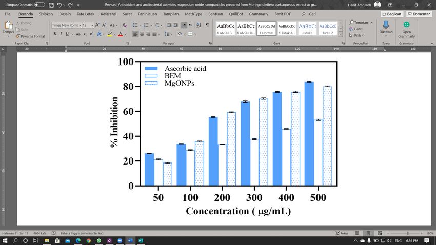
J. Multidiscip. Appl. Nat. Sci.

Table 1. Phytochemicals analysis of M. oleifera bark aqueous extract
     Chemical constituents             Testing Methods                   M. oleifera bark aqueous extract
             Alkaloids                 Dragendroff's test                                +
             Flavonoids                   Shinoda test                                   +
              Saponins                     Foam test                                     +
           Carbohydrate                  Anthrone test                                   +
            polyphenols                    Puncal-D                                      -
              Proteins                   Ninhydrin test                                  -
           Asam Amino                     Millon's test                                  -
              phenolics                Ferric chloride test                              +
             Triterpens                  Salkowski test                                  -
          Anthraquinones                Borntragges test                                 -
+ = present, - = absence

antioxidant and antibacterial.                               and alkaloids. Total phenolic was estimated using
                                                             the Follin-Ciocalteu test [27], and the result was
2. MATERIALS AND METHOD                                      expressed as microgram per milligram (µg/mg)
                                                             gallic acid equivalent (GAE). Total flavonoid
2.1. Materials                                               content was determined by the colorimetric AlCl3
   Fresh M. oleifera bark were collected from the            method, using catechin as standard and expressed as
plants that grow naturally around the City of Metro,         microgram per milligram (µg/mg) equivalent of
Lampung, Indonesia during September 2019.                    catechin (CE) [28].
Laboratory grade magnesium chloride hexahydrate
(MgCl2.6H2O), Folin-Ciocalteu reagents, sodium               2.2.3. Preparation of MgO nanoparticles
carbonate (Na2CO3), gallic acid, catechin,                   (MgONPs)
aluminum chloride (AlCl3), sodium nitrite                       To synthesize nanoparticles, an aqueous extract
(NaNO2), and sodium hydroxide (NaOH) were                    of M. oleifera barks 50 mL was mixed with
purchased from Merck Sigma-Aldrich Reagent Pte,              MgCl2.6H2O solution 1 mM 50 mL in a beaker with
Singapore.                                                   heating to 90 °C and stirred at 600 rpm. The 1 M
                                                             NaOH solution was added drop wise until the color
2.2. Methods                                                 of the mixture faded out and precipitate was
2.2.1. Plant extract                                         formed. The mixture was left for 3 hours to
    A fresh, M. oleifera sample washed by floating           maximize the synthesis process. MgONPs
water, dried and then poured into powder and stored
at room temperature under direct sunlight. In 100
mL of distilled water, four grams of M. oleifera
bark powder was soaked and heated for 20 minutes
at 60 ° C. The mixture was then filtered 1 hour with
Whatman Filter Paper 1 to isolate the extract from
the residue. The mixture was left.

2.2.2 Phytochemicals analysis
   The aqueous extract was subjected to
phytochemical analysis to detect the presence of
carbohydrates,    amino       acids,     glycosides,          Figure 1. Total phenolic and flavonoid content in
polyphenols, saponins, steroids, flavonoids, tannins,         M. oleifera bark aqueous extract, Notes: value is
                                                                                 mean ± SD

                                                        45
J. Multidiscip. Appl. Nat. Sci.
synthesized was centrifuged at 7500 rpm at room             reference. DPPH 0.1 mM solution is was prepared
temperature and re-dispersed in deionized water and         by dissolving in ethanol. 1 mg of ascorbic acid is
methanol (99%) to remove biological residues. The           was dissolved in 1 mL of methanol. Dilution was
process was repeated twice, and the solid was dried         carried out to make a standard solution of ascorbic
at 100 °C. The solid was subjected to calcination at        acid with different concentrations (50-500 μg). For
600 °C for 5 hours was used to optimize the                 each tube containing a standard solution of ascorbic
formation of oxides.                                        acid (200 μl), 1 mL of 0.1 mM DPPH solution was
                                                            added and continued with the addition of 800 μl 50
2.2.4. Characterizations of MgO nanoparticles               mM Tris-HCl buffer (pH7.4). The final volume is
(MgONPs)                                                    adjusted to 4 mL using ethanol. Stock solutions for
   Several techniques were used to describe MgO             BEM and MgONPs were synthesized prepared by
nanoparticles (MgONPs). The confirmation of                 dissolving 1 mg of each sample in 1 mL of a
MgONPs synthesis was based on the change in the             suitable solvent (BEM using methanol and
color of the mixture during the reaction and                MgONPs using DMSO).
recorded with UV-Visible spectroscopy (Analytic                Different aliquots of stock solution (50-500 μg)
Jena Specord 200 Plus) by scanning the spectrum in          was added to separate tube, and the final volume is
the range of 200 - 800 nm. The morphology of                adjusted to 2 mL using ethanol. A total of 1 mL of
nanoparticles was studied with scanning electron            0.1 mM DPPH solution and 800 μl 50 mM Tris-
microscopy (SEM; FEI Inspect-S50). The size and             HCl buffer (pH7.4) was added to each tube. The
morphology of the MgONPs were investigated by               control was made by mixing 1 mL DPPH 0.1 mM,
transmission electron microscopy (TEM; Jeol Jem             800 μL 50 mM Tris-HCl buffer (pH7.4), and 2 mL
1400) and the average particle size by the particle         ethanol. Absorbance was recorded after incubation
size analyzer (PSA; Horiba SZ 100z). The crystal            for 30 minutes at room temperature, measured by
structure of the synthesized MgONPs nanoparticles           UV-Vis spectrophotometer at 517 nm. The
was confirmed by x-ray diffraction (XRD;                    percentage of antioxidant activity (% Inhibition)
PANAnalytical Expert Pro).                                  was calculated using the following equation:

2.2.5. Antioxidant activity                                                                                 (1)
   Antioxidant activity of Moringa oleifera bark
aqueous extract (BEM) and MgONPs was evaluated                The mean and standard deviation (SD) were
through (2,2-diphenyl-1-picrylhydrazyl) DPPH                calculated based on triplicate measurements by
radical testing in accordance with the procedure            repeating three times.
described by Das et al. [29], using ascorbic acid as
                                                            2.2.6. Antibacterial activity
                                                            2.2.6.1. Microorganism and inoculum preparation
                                                               The antibacterial activity of BEM and MgONPs
                                                            nanoparticles was evaluated against both gram-
                                                            positive (S. aureus and E. faecalis) and gram-
                                                            negative (E. coli and S. dysenteriae) obtained from
                                                            the microbiology laboratory of Airlangga
                                                            University. Bacterial cultures for testing were
                                                            cultivated on nutrient agar (NA) tilted by selecting
                                                            a colony from the Mueller-Hinton agar plate
                                                            (MHA) after 24 hours.
                                                               A single bacterial or fungal colony is selected
                                                            and transferred to the Mueller-Hinton (MHB) broth
                                                            using a sterilized loop, followed by shaking at 100
                                                            rpm at a temperature of 37 °C overnight for a
Figure 2. UV-Vis spectrum of M. oleifera aqueous            normalized population. The optical density of
          extract and MgO nanoparticles

                                                       46
J. Multidiscip. Appl. Nat. Sci.
Figure 3. (a) diffraction pattern, (b) SEM image, (c) TEM image and (d) particles size distribution of syn-
thesized MgONPs
bacterial or fungal suspense was maintained at the severely. Each well was subsequently supplemented
standard 0.5 for MacFarland by adding sterilized with 10 μL resazurin solution. The plate was
MHB for the test of antibacterial and antifungal covered with film to avoid dehydration and
activity. The inoculum is therefore composed in incubated for 24 hours at 37 ° C. The change in
about 106-107 CFU / mL of several fungi or color was noticed visually. A change in blue to rose
bacteria. color signaling growth of the cells was considered.
In case of a color change, the MIC was registered at
2.2.6.2. Minimum inhibition concentration (MIC) the lowest concentration. The mixtures between the
determination sterile distilled water and the DMSO solvent and
The resazurin microtiter test was used to assess the nutrient broth were used as adverse controls by
the minimum concentration of inhibition. This Streptomycin (antibacterial) (10 μg / 500 μL).
approach has been chosen as the easiest and most
economical way to simultaneously scan for multiple 3. RESULT AND DISCUSSION
microorganism isolates and produces satisfactory
performance. A 270 mg tablet of resazurin in 40 mL 3.1. Phytochemical screening of M. oleifera bark
of sterile distilled water was prepared to dissolve aqueous extract
the resazurin solution. The test was conducted in Different methods are used to qualitatively test
aseptic conditions in 96-well plates. The samples phytochemical compounds contained in moringa
were transferred into the plate well with a volume oleifera (BEM) extracts. The test method used
of 100 μL containing 600 mg / mL. Subsequently, refers to the report Das et al. [29], Qualitative
the checked sample was applied to all other wells evaluations of several chemical contents are shown
by 50 μL of bacterial suspense and diluted in Table 1.
47J. Multidiscip. Appl. Nat. Sci.
Table 1 shows BEM containing phytochemical structure of the synthesized MgO nanoparticulate
compounds such as alkaloids, flavonoids, saponins, were studied using X-ray diffraction technologies.
and phenolics. These compounds play a role in the The XRD patterns of MgO nanoparticles
chelation process and are able to reduce metal ions manufacturing are shown in Figure 3(a). MgONPs
to nanoparticles [26]. showed a high intensity peak with two peaks =
Total phenolic and flavonoid levels in BEM 42.915 and 62.304 and a low intensity of 31.636,
21.65 ± 4.25 (µg / mg GAE) and 55.31 ± 3.82 (µg / 74.729 and 78.629. The obtained results have been
mg CE) respectively in dry powder, as shown in verified using XRD data (No: 78-0430) from
Figure 1. Extracts of some parts of the plant contain JCPDS. A high purity of synthesized MgO
different constituents with distinct functional nanoparticles does not appear as major peaks from
groups, which can act as reducing or chelating Mg or other impurities observed on the
agents in the formation of nanoparticles. Total diffractogram. The average crystalline (D) diameter
phenolic and flavonoid levels in bark water extracts was calculated with the formula of Scherrer
are lower than total phenolic and flavonoid levels in (equation 2) for (200) planes of 20 – 30 nm.
water extracts of other M. oleifera plants that have
been previously reported. Siddhuraju and Becker (2)
[30] reported total phenolic content in leaves is
74.30 ± 9.00 (µg / mg GAE), while Mohammed and Where K is a constant dimension depending on
Manan [31] reported total phenolic content in ore is the particular geometry of the target, λ is the
101.79 ± 2.89 (µg / mg GAE). The total flavonoid wavelength of X-ray radiation, β is the full width at
content in leaf water extract was reported by half maximum (FWHM) of the significant peaks in
Okumu et al. [32] amounted to 79.13 ± 13.04 (µg / radians, and θ is the Bragg's angle. SEM has been
mg CE). used to carry out morphological tests of MgO
nanoparticles synthesized with extract. Figure 3(b)
3.2. Characterization shows an image of the MgO nanoparticles scanning
3.2.1. UV-Vis spectroscopy of BEM and MgONPs electric microscopy (SEM) showing that the
synthesized resulting MgO nanos are in the form of spherical
Synthesis of MgO nanoparticles using Moringa particle of 20 to 80 nm.
aqueous extract followed by a change in color Figure 3 (c) and (d) shows TEM and PSA of
during the synthesis process. The color of the MgO nanoparticles prepared. The results of particle
solution changes from clear (MgCl2 solution) to size analysis (PSA) show that the MgO with
dark brown when added with Moringa extract. After particle size in the range of nanometer with
adding NaOH, the color of the solution changes to relatively narrow distribution was produced. The
brighter, indicating the formation of MgO and Mg samples obtained have the particle size in the range
(OH)2 complexes in the solution. Spectrum of 60-100 nm. The existence of spheres particles
adsorption of MgO nanoparticles measured in the
range of 200-800 nm. Figure 2 shows the UV
spectrum with a sharp peak at around 290 nm,
which confirms the formation of MgO nanoparticles
[28]. Besides, the precursor ion Mg2+, MgCl2 salt
does not show a spectrum at the specified
wavelength. The existence of a peak of about 280 -
290 nm can be attributed to the formation of metal
oxide nanoparticles after the addition of plant
extracts and NaOH solution [28].
3.2.2. Size and morphology of MgONPs synthesized
In order to correctly assess the atomic position
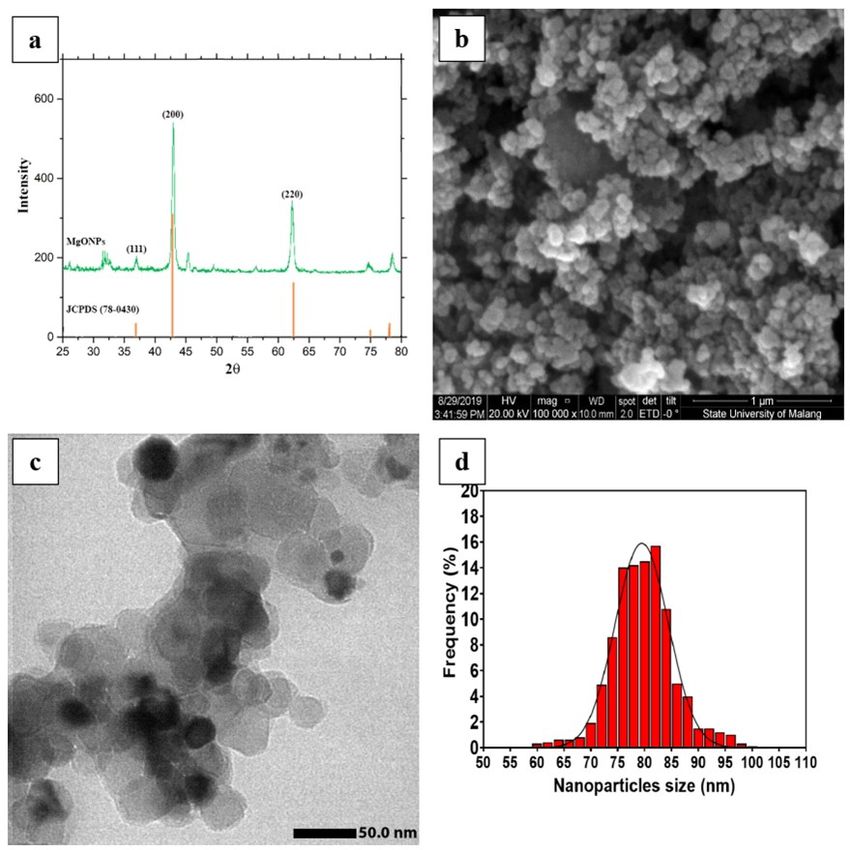
Figure 4. Antioxidant activity of BEM and
in the lattice structure, a crystalline stage and
MgONPs
48J. Multidiscip. Appl. Nat. Sci.
compound and accepts hydrogen or electrons from
BEM or MgO nanoparticles. This test is often used
for the antioxidant activity of compounds present in
medicinal plant extracts [37]. The antioxidant
activity of MgONPs tends to be higher than that of
BEM (Figure 5). Several workers have reported
similar observations of antioxidant activity by ZnO
[38], CuO [39], and MgO [28] nanoparticles.
3.4. Antibacterial
The potential antibacterial activity of BEM and
MgONPs was evaluated against gram-positive
Figure 5. Antibacterial activity of BEM and
bacteria (S. aureus and E. faecalis) and gram-
MgONPs
negative (E. coli and S. dysenteriae) clinically
with a size ranging from 60-100 nm was supported isolated in vitro. MIC values of the presence of
by the results of the transmission electron BEM against S. aureus; E. faecalis; E. coli, and S.
microscope (TEM). Based on these results, MgO dysenteriae are in the range 300 - 550 g/mL (Figure
nanoparticles have been successfully produced 5). The antibacterial activity of MgONPs
using aqueous extracts of bark of M. oleifera plant, nanoparticles was observed with MIC values (300-
providing an alternative method for synthesizing 550 μg/mL). If we compare the susceptibility
MgO nanoparticles. Amrulloh et al [33] reported between the bacteria tested, S. dysenteriae is very
the use aqueous extract of Moringa leaves as a susceptible to BEM and MgONPs compared to
green agent in synthesizing MgO nanoparticles. The other test bacteria. Medicinal plants such as M.
water extract sample of Moringa leaves contains oleifera, because of their antibacterial activity, are
phytochemical compounds such as alkaloids, traditionally used to treat various diseases [40].
flavonoids, saponins, carbohydrates, polyphenols, The antibacterial activity of MgONPs
proteins, amino acids, and phenolics with phenolics nanoparticles against gram-positive and gram-
and flavonoids contents of 34.75 ± 4.03 (μg / mg negative test bacteria showed different results. The
GAE) and 74.28 ± 4.82 (μg / mg CE), respectively. difference between gram-positive and gram-
The structure of the synthesized MgO nanoparticles negative bacteria is mainly in the structure of their
was confirmed by the spherical structure. The cell walls. Gram-positive bacteria have a thick layer
average particle size of the synthesized MgO of peptidoglycan without an outer membrane and
nanoparticles measured between 40 - 70 nm. MgO contain teichoic acid. In contrast, gram-negative
nanoparticles synthesized from leaf aqueous extract bacteria have a thin layer of peptidoglycan with an
have narrower particle size distribution than that of outer membrane that contains lipopolysaccharides.
MgO produced using bark aqueous extract, which Because of this difference, each type of bacteria
was attributable to the presence of antioxidants-rich shows a different sensitivity [41].
compounds (e.g. flavonoids, phenolic acids,
reducing sugars, etc.) [34][35]. They are 4. CONCLUSION
biomolecules that play a vital role as outstanding
bio-reducing and/or bio-capping agents towards Green synthesis of MgO nanoparticles that were
generation of the nanoparticles [36]. prepared using M. oleifera bark aqueous extract was
successful. The formation of MgO nanoparticles in
3.3. Antioxidant this synthesis was confirmed using UV-Vis
The antioxidant activity of BEM and MgONPs absorption. The spherical crystal structure of MgO
was assessed by DPPH testing using ascorbic acid nanoparticles confirmed XRD analysis. The average
as a positive control. The free radical capture particle size of the synthesized MgO nanoparticles
activity of DPPH BEM and MgONPs is directly measured between 60 - 100 nm using SEM and
related to their concentration. DPPH is a stable TEM images and PSA results. Our study shows that
49J. Multidiscip. Appl. Nat. Sci.
MgO nanoparticles synthesized show a good REFERENCES
antioxidant activity, ascorbic acid, as a comparison.
We also demonstrated that water extracts of M. [1] L. Katata-Seru, T. Moremedi, O. S. Aremu,
oleifera bark and MgO nanoparticles have good and I. Bahadur. (2018). “Green synthesis of
antibacterial activity against S. aureus, E. faecalis, iron nanoparticles using Moringa oleifera
E. coli, and S. dysenteriae with MIC values (300 - extracts and their applications: Removal of
550 g/mL). nitrate from water and antibacterial activity
against Escherichia coli”. Journal of
AUTHOR INFORMATION Molecular Liquids. 256 : 296–304. 10.1016/
j.molliq.2017.11.093.
Corresponding Author [2] Y. Abdallah, S. O. Ogunyemi, A. Abdelazez,
Hanif Amrulloh — Department of Mathematic M. Zhang, X. Hong, E. Ibrahim, A. Hossain,
Education, Institute for Islamic Studies Ma’arif H. Fouad, B. Li, and J. Chen. (2019). “The
NU (IAIMNU), Metro-34111 (Indonesia) ; Green Synthesis of MgO Nano-Flowers Using
orcid.org/0000-0001-7458-9258 Rosmarinus officinalis L. (Rosemary) and the
Email: Antibacterial Activities against Xanthomonas
amrulloh.hanif@iaimnumetrolampung.ac.id oryzae pv. oryzae”. BioMed Research
International. 2019 : 5620989.
Authors 10.1155/2019/5620989.
Awalul Fatiqin — Department of Biology, [3] G. Sharma, R. Soni, and N. D. Jasuja. (2017).
Islamic State University Raden Fatah, Palembang “Phytoassisted synthesis of magnesium oxide
-30126 (Indonesia); nanoparticles with Swertia chirayaita”.
orcid.org/0000-0001-7799-2835 Journal of Taibah University for Science. 11
Wasinton Simanjuntak — Department of (3): 471–477. 10.1016/j.jtusci.2016.09.004.
Chemistry, Lampung University, Bandar [4] G. Pal, P. Rai, and A. Pandey. (2019). in “A.
Lampung- 3514 (Indonesia); K. Shukla and I. Siavash (ed) Green
orcid.org/0000-0001-8152-5084 Synthesis, Characterization and Applications
Hapin Afriyani — Department of Chemistry, of Nanoparticles”. Elsevier Inc, Amsterdam.
Lampung University, Bandar Lampung- 3514 10.1016/C2017-0-02526-0.
(Indonesia); [5] B. Wang, X. Xiong, H. Ren, and Z. Huang.
orcid.org/0000-0002-6092-127X (2017). “Preparation of MgO nanocrystals and
Annissa Annissa — Department of Public catalytic mechanism on phenol ozonation”.
Health, Faletehan University, Serang-42161 RSC Advances. 7 (69): 43464–43473.
(Indonesia); 10.1039/c7ra07553g.
[6] M. B. Gawande, P. S. Branco, K. Parghi, J. J.
ACKNOWLEDGEMENT Shrikhande, R. K. Pandey, C. A. A.
Ghumman, N. Bundaleski, O. M. N. D.
This research supported by the Ministry of Teodorod, and R. V. Jayaram. (2011).
Religious Affairs Republic Indonesia through “Synthesis and characterization of versatile
collaboration research BOPTN UIN Raden Fatah MgO-ZrO2 mixed metal oxide nanoparticles
Palembang No: B-383/Un.09/PP.06/05/2019. and their applications,” Catalysis Science &
Furthermore, acknowledgment also expressed for Technology. 1 (9): 1653–1664. 10.1039/
the full support from Laboratorium Sentral Mineral c1cy00259g.
& Material Maju Universitas Negeri Malang, [7] J. Chen, M. Zhang, C. Pang, F. Xiang, M.
Direktorat Riset & Pengabdian Masyarakat Zhu, X. Ma, G. Chang, and W. Yin. (2020).
Universitas Indonesia, and Laboratorium TEM “Hydrophilic Pd/MgO Nanosystem for the
Jurusan Kimia Universitas Gajah Mada for Highly Efficient Aqueous-Phase Catalysis of
technical contributions on the research projects. Suzuki-Miyaura Reactions”. Industrial &
Engineering Chemistry Research. 59 (1): 81–
50J. Multidiscip. Appl. Nat. Sci.
87. 10.1021/acs.iecr.9b05248. 10.1063/5.0002154.
[8] V. Srivastava, Y. C. Sharma, and M. [15] S. C. De La Rosa-García, P. Martínez-Torres,
Sillanpää. (2015). “Green synthesis of S. Gómez-Cornelio, M. A. Corral-Aguado, P.
magnesium oxide nanoflower and its Quintana, and N. M. Gómez-Ortíz. (2018).
application for the removal of divalent “Antifungal activity of ZnO and MgO
metallic species from synthetic wastewater”. nanomaterials and their mixtures against
Ceramics International. 41 (5): 6702–6709. colletotrichum gloeosporioides strains from
10.1016/j.ceramint.2015.01.112. tropical fruit”. Journal of Nanomaterials.
[9] Z. Camtakan, S. Erenturk, and S. Yusan. 2018. 10.1155/2018/3498527.
(2012). “Magnesium oxide nanoparticles: [16] B. Mangalampalli, N. Dumala, and P. Grover.
Preparation, characterization, and uranium (2019). “Toxicity assessment of magnesium
sorption properties,” Environmental Progress oxide nano and microparticles on cancer and
& Sustainable Energy. 31 (4): 536–543. non-cancer cell lines”. The Nucleus. 62 (3):
10.1002/ep.10575. 227–241. 10.1007/s13237-019-00298-9.
[10] A. A. Pilarska, Ł. Klapiszewski, and T. [17] A. F. A. Razis, M. D. Ibrahim, and S. B.
Jesionowski. (2017). “Recent development in Kntayya. (2014). “Health benefits of Moringa
the synthesis, modification and application of oleifera”. Asian Pacific Journal of Cancer
Mg(OH)2 and MgO: A review”. Powder Prevention. 15 (20): 8571–8576. 10.7314/
Technology. 319 : 373–407. 10.1016/ APJCP.2014.15.20.8571.
j.powtec.2017.07.009. [18] K. Anand, C. Tiloke, A. Phulukdaree, B.
[11] H. R. Raveesha, S.Nayana, D. R. Vasudha, J. Ranjan, A. Chuturgoon, S. Singh, R. M.
P. S. Begum, S. Pratibha, C. R. Ravikumara, Gengan. (2016). “Biosynthesis of palladium
N. Dhananjaya. (2019). “The electrochemical nanoparticles by using Moringa oleifera
behavior, antifungal and cytotoxic activities flower extract and their catalytic and
of phytofabricated MgO nanoparticles using biological properties”. Journal of
Withania somnifera leaf extract”. Journal of Photochemistry and Photobiology B: Biology.
Science: Advanced Materials and Devices. 4 165 : 87–95. 10.1016/
(1): 57–65. 10.1016/j.jsamd.2019.01.003. j.jphotobiol.2016.09.039.
[12] K. Kandiah, T. Jeevanantham, and B. [19] M. R. Bindhu, M. Umadevi, G. A. Esmail, N.
Ramasamy. (2019). “Reliability of antioxidant A. Al-Dhabi, and M. V. Arasu. (2020).
potential and in vivo compatibility with “Green synthesis and characterization of
extremophilic actinobacterial-mediated silver nanoparticles from Moringa oleifera
magnesium oxide nanoparticle synthesis”. flower and assessment of antimicrobial and
Artificial Cells, Nanomedicine, and sensing properties”. Journal of
Biotechnology. 47 (1): 862–872. Photochemistry and Photobiology B: Biology.
10.1080/21691401.2019.1580287. 205. 10.1016/j.jphotobiol.2020.111836.
[13] A. M. Azzam, M. A. Shenashen, B. B. [20] M. Sundrarajan, S. Jegatheeswaran, S.
Mostafa, W. A. Kandeel, and S. A. El-Safty. Selvam, N. Sanjeevi, and M. Balaji. (2015).
(2019). “Antibacterial Activity of Magnesium “The ionic liquid assisted green synthesis of
Oxide Nano-hexagonal Sheets for Wastewater hydroxyapatite nanoplates by Moringa
Remediation”. Environmental Progress & oleifera flower extract: A biomimetic
Sustainable Energy. 38 (s1): S260–S266. approach”. Materials & Design. 88 : 1183–
10.1002/ep.12999. 1190. 10.1016/j.matdes.2015.09.051.
[14] M. Suma, G. N. Sushma, M. Zikriya, and Y. [21] J. S. Moodley, S. B. N. Krishna, K. Pillay,
F. Nadaf. (2020). “Photocatalytic and Sershen, and P. Govender. (2018). “Green
antibacterial approach of green synthesised synthesis of silver nanoparticles from
MgO nanoparticles”. 3rd International Moringa oleifera leaf extracts and its
Conference on Condensed Matter and antimicrobial potential”. Advances in Natural
Applied Physics. 2220 : 020072. Sciences: Nanoscience and Nanotechnology.
51J. Multidiscip. Appl. Nat. Sci.
9 (1): 015011. 10.1088/2043-6254/aaabb2. [29] P. E. Das, A. F. Majdalawieh, I. A. Abu-
[22] K. Elumalai, S. Velmurugan, S. Ravi, V. Yousef, S. Narasimhan, and P. Poltronieri.
Kathiravan, and S. Ashokkumar. (2015). (2020). “Use of a hydroalcoholic extract of
“Green synthesis of zinc oxide nanoparticles moringa oleifera leaves for the green
using Moringa oleifera leaf extract and synthesis of bismuth nanoparticles and
evaluation of its antimicrobial activity”. evaluation of their anti-microbial and
Spectrochimica Acta Part A: Molecular and antioxidant activities,” Materials. 13 (4).
Biomolecular Spectroscopy. 143 : 158–164. 10.3390/ma13040876.
10.1016/j.saa.2015.02.011. [30] P. Siddhuraju and K. Becker. (2003).
[23] V. Sivaranjani and P. Philominathan. (2016). “Antioxidant properties of various solvent
“Synthesize of Titanium dioxide nanoparticles extracts of total phenolic constituents from
using Moringa oleifera leaves and evaluation three different agroclimatic origins of
of wound healing activity”. Wound Medicine. drumstick tree (Moringa oleifera Lam.)
12 : 1–5. 10.1016/j.wndm.2015.11.002. leaves”. Journal of Agricultural and Food
[24] A. A. Ezhilarasi, J. J. Vijaya, K. Kaviyarasu, Chemistry. 51 (8): 2144–2155. 10.1021/
M. Maaza, A. Ayeshamariam, and L. J. jf020444+.
Kennedy. (2016). “Green synthesis of NiO [31] S. Mohammed and F. A. Manan. (2015).
nanoparticles using Moringa oleifera extract “Analysis of total phenolics , tannins and
and their biomedical applications: flavonoids from Moringa oleifera seed
Cytotoxicity effect of nanoparticles against extract”. Journal of Chemical and
HT-29 cancer cells”. Journal of Pharmaceutical Research. 7 (1): 132–135.
Photochemistry and Photobiology B: Biology. [32] M. O. Okumu, J. M. Mbaria, L. W. Kanja, D.
164 : 352–360. 10.1016/ W. Gakuya, S. G. Kiama, and F. O. Ochola.
j.jphotobiol.2016.10.003. (2016). “Phytochemical profile and
[25] H. P. N. Sholapur and B. M. Patil. (2013). antioxidant capacity of leaves of Moringa
“Pharmacognostic and phytochemical oleifera ( Lam ) extracted using different
investigations on the bark of Moringa oleifera solvent systems”. Journal of Pharmacognosy
Lam.”. Indian Journal of Natural Products and Phytochemistry. 5 (4): 302–308.
and Resources. 4 (1): 96–101. [33] H. Amrulloh, A. Fatiqin, W. Simanjuntak, H.
[26] C. Tiloke and A. A. Chuturgoon. In “V. C. Afriyani, and A. Annissa. (2021).
Kalia and A. K. Saini (eds) Engineering for “Bioactivities of Nano-scale Magnesium
Bioactive Compounds: Strategies and Oxide Prepared Using Aqueous Extract of
Processes”. Springer Singapore, Singapore. Moringa Oleifera Leaves as Green Agent”.
[27] R. Magesh, R. M. Poorani, V. Karthikeyan, Advances in Natural Sciences: Nanoscience
K. Sivakumar, and C. Mohanapriya. (2015). and Nanotechnology.
“Proportionate phytochemical screening and [34] M. Hariram, S. Vivekanandhan, V. Ganesan,
assessment of antioxidant potency on selected S. Muthuramkumar, A. Rodriguez-uribe, A.
species of lamiaceae family”. International K. Mohanty, M. Misra. (2019). “Tecoma
Journal of Pharmacognosy and stans flower extract assisted biogenic
Phytochemical Research. 7 (5): 1066–1072. synthesis of functional Ag-Talc
[28] B. Das, S. Moumita, S. Ghosh, M. I. Khan, D. nanostructures for antimicrobial applications”.
Indira, R. Jayabalan, S. K.Tripathy, A. Bioresource Technology Reports. 7. 10.1016/
Mishra, and P. Balasubramanian. (2018). j.biteb.2019.100298.
“Biosynthesis of magnesium oxide (MgO) [35] D. T. C. Nguyen, H. H. Dang, D. V. N. Vo, L.
nanoflakes by using leaf extract of Bauhinia G. Bach, T. D. Nguyen, and T. Van Tran.
purpurea and evaluation of its antibacterial (2021). “Biogenic synthesis of MgO
property against Staphylococcus aureus”. nanoparticles from different extracts (flower,
Materials Science and Engineering: C. 91 : bark, leaf) of Tecoma stans (L.) and their
436–444. 10.1016/j.msec.2018.05.059. utilization in selected organic dyes treatment”.
52J. Multidiscip. Appl. Nat. Sci.
Journal of Hazardous Materials. 404.
10.1016/j.jhazmat.2020.124146.
[36] E. R. Essien, V. N. Atasie, E. U. Udobang,
and G. Umanu. (2019). “Preparation of
monodispersed and cytotoxic silver
nanoparticles using Launaea taraxacifolia leaf
extract”. Journal of Nanostructure in
Chemistry. 9 (4): 259–268. 10.1007/s40097-
019-00316-x.
[37] S. Dewanjee, M. Gangopadhyay, N.
Bhattacharya, R. Khanra, and T. K. Dua.
(2015). “Bioautography and its scope in the
field of natural product chemistry”. Journal of
Pharmaceutical Analysis. 5 (2): 75–84.
10.1016/j.jpha.2014.06.002.
[38] T. Safawo, B. V. Sandeep, S. Pola, and A.
Tadesse. (2018). “Synthesis and
characterization of zinc oxide nanoparticles
using tuber extract of anchote (Coccinia
abyssinica (Lam.) Cong.) for antimicrobial
and antioxidant activity assessment”.
OpenNano. 3 : 56–63. 10.1016/
j.onano.2018.08.001.
[39] R. Dobrucka. (2018). “Antioxidant and
Catalytic Activity of Biosynthesized CuO
Nanoparticles Using Extract of Galeopsidis
herba”. Jounal of Inorganic and
Organometallic Polymers and Materials. 28
(3): 812–819. 10.1007/s10904-017-0750-2.
[40] K. Z. Khor, V. Lim, E. J. Moses, and N.
Abdul Samad. (2018). “The in Vitro and in
Vivo Anticancer Properties of Moringa
oleifera”. Evidence-based Complementary
and Alternative Medicine. 2018.
10.1155/2018/1071243.
[41] N. Y. T. Nguyen, N. Grelling, C. L.
Wetteland, R. Rosario, and H. Liu. (2018).
“Antimicrobial Activities and Mechanisms of
Magnesium Oxide Nanoparticles (nMgO)
against Pathogenic Bacteria, Yeasts, and
Biofilms”. Scientific Reports. 8 (1). 10.1038/
s41598-018-34567-5.
53You can also read