Low CETP activity and unique composition of large VLDL and small HDL in women giving birth to small for gestational age infants - Nature
←
→
Page content transcription
If your browser does not render page correctly, please read the page content below
www.nature.com/scientificreports
OPEN Low CETP activity and unique
composition of large VLDL
and small HDL in women giving
birth to small‑for‑gestational age
infants
Marie Cecilie Paasche Roland1,2, Kristin Godang3, Pål Aukrust4,5,6, Tore Henriksen1,2,5 &
Tove Lekva4*
Cholesteryl ester transfer protein (CETP) regulates high density lipoproteins (HDL)-cholesterol (C)
and HDL-C is essential for fetal development. We hypothesized that women giving birth to large-for-
gestational-age (LGA) and small-for-gestational age (SGA) infants differed in longitudinal changes in
lipoproteins, CETP activity and HDL-C and that placentas from women with higher or lower circulating
HDL-C displayed differential expression of mRNAs involved in cholesterol/nutrient transport, insulin
signaling, inflammation/ extracellular matrix (ECM) remodeling. Circulating lipids and CETP activity
was measured during pregnancy, NMR lipidomics in late pregnancy, and associations with LGA
and SGA infants investigated. RNA sequencing was performed in 28 placentas according to higher
and lower maternal HDL-C levels. Lipidomics revealed high triglycerides in large VLDL and lipids/
cholesterol/cholesteryl esters in small HDL in women giving birth to SGA infants. Placentas from
women with higher HDL-C had decreased levels of CETP expression which was associated with mRNAs
involved in cholesterol/nutrient transport, insulin signaling and inflammation/ECM remodeling. Both
placental and circulating CETP levels were associated with growth of the fetus. Low circulating CETP
activity at 36–38 weeks was associated with giving birth to SGA infants. Our findings suggest a link
between increased maternal HDL-C levels, low CETP levels both in circulation and placenta, and SGA
infants.
Abbreviations
AC Abdominal circumference
CETP Cholesteryl ester transfer protein
GDM Gestational diabetes mellitus
HDL High density lipoproteins
LDL Low density lipoproteins
LGA Large-for-gestational age
SGA Small-for-gestational age
TG Triglycerides
Being born too large (large-for-gestational age, LGA) or too small (small-for gestational-age, SGA) is associ-
ated with perinatal mortality and also with subsequent adult obesity, diabetes and cardiovascular disease for
the child1,2. Fetal growth is a result of multiple factors including maternal nutrition and metabolism, the fetal
genetic potential for growth and placental properties including its metabolism and endocrine function, blood
1
National Advisory Unit for Women’s Health, Oslo University Hospital, Rikshospitalet, Oslo, Norway. 2Department
of Obstetrics, Oslo University Hospital, Rikshospitalet, Oslo, Norway. 3Section of Specialized Endocrinology,
Department of Endocrinology, Oslo University Hospital, Rikshospitalet, Oslo, Norway. 4Research Institute of
Internal Medicine, Oslo University Hospital, Rikshospitalet, Oslo, Norway. 5Faculty of Medicine, University of Oslo,
Oslo, Norway. 6Section of Clinical Immunology and Infectious Diseases, Oslo University Hospital, Rikshospitalet,
Oslo, Norway. *email: tove.lekva@ous-research.no
Scientific Reports | (2021) 11:6213 | https://doi.org/10.1038/s41598-021-85777-3 1
Vol.:(0123456789)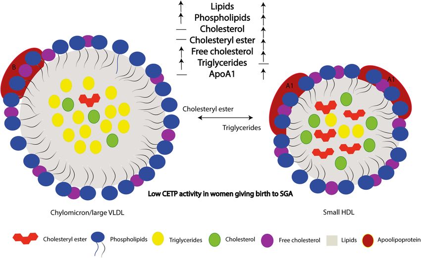
www.nature.com/scientificreports/
perfusion and structural features. Previous clinical studies have shown a direct association between maternal
plasma cholesterol levels and birthweight3–5.
Cholesterol and in particular high-density lipoprotein cholesterol (HDL-C) has essential roles in fetal devel-
opment and in early gestation maternal cholesterol is transferred to the fetus across the placenta whereas in late
pregnancy the fetus is able to synthesize its own c holesterol6–8. Importantly, more than 50% of fetal total choles-
DL9, which is different from the maternal circulation where low-density lipoprotein (LDL)
terol is carried by H
represents the major class of lipoproteins. The maternal plasma cholesterol is taken up by the syncytiotropho-
blast facing the maternal blood, followed by entrance into the subsyncytial endothelial cells of the fetoplacental
vasculature wherefrom cholesterol is effluxed or secreted by specific transport proteins into the fetal circulation.
The cholesterol transport may be mediated both by receptor-dependent and independent mechanisms besides
that placenta may make its own c holesterol10.
HDL has diverse functions, including reverse cholesterol and lipid transport, role in inflammation through
its anti-inflammatory properties, in hemostasis through its ability to modulate platelet function and seems also
to be linked to insulin resistance. All these properties may indeed influence pregnancy outcomes. HDL function
is related to the protein cargo carried by HDL (ApoA1, ApoA2, apoC, apoE, apoM, apoA4, lecithin–cholesterol
acyltransferase (LCAT), cholesteryl ester transfer protein (CETP) and paraoxonase 1 (PON1)11. However, there
is a difference in the protein cargo between fetal and maternal HDL, and proteins involved in lipid metabolism,
inflammatory pathways and innate immunity were differentially expressed between the mother and the fetus9.
Fetal and maternal HDL undergoes constant remodeling, that is dependent on the metabolic status of the mother
and/or the fetus12,13. Inflammation is accompanied by changes in HDL lipid composition resulting in reduction
of phospholipid and cholesteryl esters and an increase in triglycerides (TG)14.
Mice with apolipoprotein (apo) A1 deficiency, being the major protein in the HDL particles, displayed reduced
fetal growth in dams with lower maternal cholesterol levels15,16. It has been hypothesized that the ability of the
placenta to take up HDL-cholesterol and transport it to the fetus is compromised in women giving birth to
SGA, and a reason for the high maternal HDL-cholesterol levels in these women17. Notably, even if the infant is
synthesizing cholesterol, a lack of transfer of maternal cholesterol may impact g rowth10.
Cholesteryl ester transfer protein (CETP) is a glycoprotein that catalyzes the exchange of cholesteryl esters
for TG between HDL and apolipoprotein B (apoB) containing lipoproteins and decreases HDL-C18. Increased
TG content in the HDL particle may therefore be explained by increased CETP activity and CETP mediated
replacement of cholesteryl esters by TGs in the HDL core results in lower plasma HDL-C levels. The composi-
tion of HDL and thereby partly its function during fetal growth is highly associated with CETP activity. To our
knowledge there is no data in the literature of the function of CETP in the placenta. Interestingly data obtained
from public repositories shows that CETP expression in the placenta is among the highest of all tissues, increases
markedly during gestation and is increased in endothelial progenitor cells isolated from cord blood.
We hypothesized that placentas from women with higher and lower circulating maternal HDL-C displayed
differential expression of mRNAs involved in cholesterol/nutrient transport, insulin signaling and inflamma-
tion/extracellular matrix (ECM) remodeling. We also hypothesized that women giving birth to LGA and SGA
infants displayed differences in longitudinal changes in maternal lipids and CETP activity. To elucidate the role
of HDL-C and CETP and their potential interaction in fetal growth we investigated in a prospective cohort study
(STORK) (i) differences in longitudinal change in lipids (cholesterol including HDL-C and TG) in women giv-
ing birth to LGA and SGA infants, (ii) the RNA expression by RNA sequencing in 28 placentas from women
divided into higher and lower maternal HDL-C circulating levels at week 36–38, (iii) CETP mRNA expression
and its association with cholesterol/nutrient transport, insulin signaling and inflammation/ECM remodeling in
the placenta and (iv) CETP activity in plasma measured multiple times during pregnancy of 300 women and its
association with LGA and SGA babies, (v) lipoprotein composition in plasma of 160 women at week 36–38 and
its association with LGA and SGA babies with particular focus of HDL particles.
Methods
The STORK study is a prospective longitudinal cohort study in which 1031 low-risk women of Scandinavian
heritage were followed throughout their pregnancy and gave birth at Oslo University Hospital Rikshospitalet
between 2002 and 200819. The exclusion criteria were multiple pregnancies, known pre-gestational diabetes and
any severe chronic diseases (lung, cardiac, gastrointestinal or renal). Each pregnant woman had four study-
related antenatal visits at weeks 14–16, 22–24, 30–32, and 36–38. A 75 g OGTT was performed on all women
at 14–16 and 30–32 weeks of gestation. The follow-up study was performed 5-year after the index pregnancy20.
Three hundred women agreed to participate and subjects included in this study are participants included in
the follow-up study using samples from pregnancy (i.e. a sub-study of the STORK study). Blood samples were
drawn in the morning between 07:30 and 08:30 after an overnight fast, centrifuged and stored at − 80 °C. EDTA
plasma were stored on ice before centrifuged. Written informed consent was obtained from all study participants.
All clinical investigations were conducted in accordance with the principles enshrined in the Declaration of
Helsinki. The study was approved by the Regional Committee for Medical Research Ethics of Southern Norway
in Oslo, Norway.
AGA, LGA and SGA infants. Infants were divided according to birth weight: appropriate-for-gestational
age (AGA), between 10 and 90th percentile, LGA, > 90th percentile and SGA < 10th percentile, adjusted for ges-
tational age and fetal sex, according to Norwegian reference curves21.
Intrauterine measurements of fetal growth. Ultrasound measurement was performed and head cir-
cumference, femur length and abdominal circumference (AC) at 22–24, 30–32, and 36–38 weeks was recorded
Scientific Reports | (2021) 11:6213 | https://doi.org/10.1038/s41598-021-85777-3 2
Vol:.(1234567890)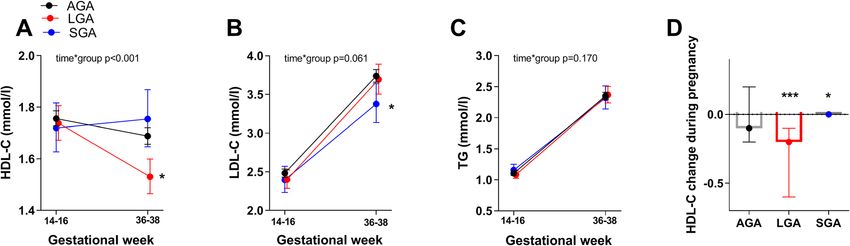
www.nature.com/scientificreports/
as previously described19. The measurements were done by three people. The measurements of each participant
were done by the same operator each time to minimize interobserver differences. All measurements were done
three times and the average value was used.
Lipoproteins and lipids. Lipoproteins and lipids were measured in the 1031 women at an accredited clini-
cal chemistry laboratory by immunoassay (IMMULITE 2000; Siemens Healthcare GmbH, Erlangen, Germany)
at Oslo University Hospital, Rikshospitalet22. Total cholesterol, HDL-C and triglycerides were measured at weeks
14–16 and 36–38 during pregnancy. LDL-C was determined by Friedewald’s formula23. A commercially high–
throughput proton NMR metabolomics platform (Nightingale Health Ltd., Helsinki, Fin) was used to quan-
tify lipoprotein subclass profile with lipid concentrations, abundant proteins and various low-molecular-weight
metabolites in fasting EDTA plasma at gestational weeks 36–38 in a subset of the women giving birth to 90
AGA, 48 LGA and 22 SGA infants. The subclass sizes were defined by their average diameter: extremely large
(XXL) very low density lipoprotein (VLDL) chylomicrons (> 75 nm), extra-large (XL), large (L), medium (M),
small and extra-small (XS) VLDL (64.0, 53.6, 44.5, 36.8, and 31.3 nm), intermediate lipoprotein IDL (28.6 nm),
L, M, and S LDL subclasses (25.5, 23.0 and 18.7 nm), and XL, L, M and S HDL subclasses (14.3, 12.1, 10.9 and
8.7 nm). The components phospholipids (PL), cholesterol, cholesteryl esters (CE), free cholesterol (FC) and TG
of the lipoprotein subclasses were quantified. The mean size for VLDL, LDL and HDL particles was calculated
by weighting the corresponding subclass diameters with their particle concentrations.
Measurement of CETP activity. Plasma CETP activity was measured in duplicate using commercially
available kit (MAK106) from Sigma-Aldrich (St. Louis, MO), as previous published24. The reaction mixture
contained a donor molecule that was a fluorescent self-quenching neutral lipid as well as an acceptor molecule.
Five µL of diluted plasma sample was added to the reaction mixture and incubated for 3 h at 37 °C in a black 384
well plate. CETP-mediated transfer from donor to acceptor resulted in an increase in fluorescence intensity with
an excitation wavelength of 465 nm and emission of 535 nm as read by the fluorescent plate reader. The CV for
the analysis was < 13%. All 4 samples from one person were analyzed on the same plate.
Collection, storage and RNA extraction of placental biopsies. Blocks of 2–4 cm were taken from
the placental parenchyma, briefly washed in phosphate buffer saline, snap frozen in liquid nitrogen, and stored
in − 80 °C until homogenization. Half of the biopsy was homogenized in TRIzol reagent (Invitrogen, Life Tech-
nologies) on ice with a tissue grinder (Sigma Aldrich). Total RNA was extracted using TRIzol reagent (Inv-
itrogen, Life Technologies) and purified with RNeasy microkit columns (Qiagen, Netherlands) as previously
published25. The electropherograms from the bioanalyzer and RIN values showed satisfying RNA quality, as
previously published25. Samples chosen for RNAseq were of satisfying quality with a small percentage of con-
tamination by maternal decidual and nucleated blood cells (~ 2%). Furthermore, the most abundantly expressed
genes identified in the term placenta were ones known to be involved in placental function, with good overlap
with transcripts identified in previous studies.
RNAseq. Samples were selected as previously described25. Sequencing libraries were prepared from 500 ng
of total RNA using the TruSeq RNA sample preparation reagents (Illumina, San Diego, CA) according to the
manufacturer’s instructions, with fragmentation for 4 min at 94 °C. The libraries were sequenced using 125 bp
paired-end sequencing on an Ilumina HiSeq 2000. We recorded an average 22.3 million (range, 20.2–24.6 mil-
lion) paired reads per sample. Fastq files were generated using bcl2fastq (v1.8.4). Sequence reads were mapped
to the reference genome (hg19/GRCh38) using TopHat2 (v2.0.13) and Bowtie2 (v.2.2.3.0). Library sizes and
standard deviations for input into TopHat were calculated empirically by aligning 1,000,000 reads to an index
built from human cDNA sequence. Sequence alignment was guided using only previously annotated gene mod-
els downloaded from Ensembl (http://www.ensembl.org; Homo_sapiens.GRCh38.79.gtf). On average, there was
73.6% concordant read pair mapping (range 69.6–76.6), with a mean unique mapping of 94.1%. Raw expres-
sion counts were calculated per gene using featureCounts (http://bioinf.wehi.edu.au/featureCounts/) and the
same gtf file which was used for the read alignment. Differential expressed genes in RNAseq data was tested
using DESeq226 package for R dividing the samples between median HDL-C levels from the 1031 samples at
36–38 weeks of gestation. Outlier detection (Cook distance cutoff) and filtering out low expressed genes was
performed using the default method in DESeq2, as previously published25.
Statistical analysis. Statistical analyses were conducted using SPSS for Windows, version 21.0 (Chicago,
IL, USA). In general, data are expressed as mean ± SD when normally distributed and median (25th, 75th per-
centile) when skewed. Differences in the temporal course of CETP, HDL-C, LDL-C and TG during pregnancy
between birth weight groups was evaluated with repeated measures ANOVA a priori, and with Bonferroni
adjusted t-tests between different groups a posteriori. Data from this analysis are expressed as estimated mar-
ginal means and 95% confidence intervals. The birth weight groups (SGA, LGA and AGA infants), birth weight
and abdominal circumference (AC) were adjusted for gestational age and fetal sex. Associations between CETP
expression/CETP activity and AC/delta AC and mRNA transcripts were evaluated by Spearman correlation. The
association between CETP activity and AC/delta AC were also adjusted in linear regression models by age, BMI,
fetal sex, gestational diabetes (GDM), and preeclampsia. Unadjusted and adjusted (age, BMI, fetal sex, GDM and
preeclampsia) logistic regression model were performed on the CETP activity data comparing SGA vs AGA.
Linear regression models were used on the lipidomics data comparing AGA with SGA/ LGA infants adjust-
ing for age, fetal sex, GDM, preeclampsia and BMI at weeks 14–16 with adjustment for multiple comparisons,
and data presented as standardized estimated marginal means and 95% confidence interval. Two-tailed p-val-
Scientific Reports | (2021) 11:6213 | https://doi.org/10.1038/s41598-021-85777-3 3
Vol.:(0123456789)www.nature.com/scientificreports/
ues < 0.05 were considered significant, except for interactions analysis where p < 0.01 was considered significant.
The RNAseq results were presented as log2fold and unadjusted p-values are presented.
Results
Table 1 shows the characteristic of the study population including a total of 1031 women with 810 AGA, 142
LGA and 76 SGA newborns. In the total cohort the women giving birth to LGA infants were taller, had a longer
duration of pregnancy, were less frequently primipara, gave birth to boys more frequent, had a higher BMI
at 14–16 weeks and had a lower HDL-C at 36–38 weeks, compared to AGA. The women giving birth to SGA
infants were shorter, had a shorter pregnancy duration, were more frequently primipara, gave birth to girls more
frequent, had a lower BMI at 14–16 weeks and had a lower LDL-C at 36–38 weeks, compared to AGA. In the
sub-study cohort of 300 women we found the same pattern except for the lack of differences in height, gestational
age and LDL-C for SGA compared to AGA infants.
HDL‑C circulating levels are changing differently in pregnancy in mothers giving birth to LGA
and SGA infants. Dividing our cohort according to birthweight into AGA, LGA and SGA infants, and
adjusting for age and BMI, we found an interaction between time and group and a significant lower HDL-C at
36–38 weeks in women giving birth to LGA infants (Fig. 1A). Also, mothers giving birth to SGA infants had
lower levels of LDL-C (Fig. 1B) at 36–38 weeks, while no difference in TG (Fig. 1C) was found between groups.
As shown in Fig. 1D the change of HDL-C during pregnancy was significantly different for both LGA and SGA
infants compared to AGA infants with a larger decrease in HDL-C in women giving birth to LGA infants, but
not in women giving birth to SGA infants, compared to AGA infants.
CETP mRNA levels are lower in placentas from women with the highest HDL‑C levels at
36–38 weeks. As a model to investigate the possible effects or consequence of the maternal circulating
HDL-C level on mRNAs in the placenta, we divided HDL-C levels from the 1031 women at week 36–38 weeks
by the median, and used the cut-off to investigate the differentially expressed mRNAs from the RNAseq of 28
placenta biopsies (Table 1, Supplementary File). Many interesting mRNAs were regulated in relation to HDL-C
levels including those involved in lipid metabolism and inflammation/ECM remodeling (Table 2). Importantly,
CETP was one of the top-regulated mRNAs on the list and was decreased in the placentas from the group of
mothers with the highest HDL-C levels (Fig. 2A). We found associations between CETP mRNA in the placenta
and factors important for growth of the fetus; glucose transport [glucose transport protein 4 (GLUT4), insulin
receptor (INSR)], amino acid transport [L-amino acid transporter(LAT) 1 and 3, System A amino acid trans-
porter 3 (SNAT4)], fatty acid transport [fatty acid binding protein (FABP) 4 and 5, fatty acid transport protein 1
(FATP1), CD36, cholesterol transport (ATP binding cassette subfamily A member 1 (ABCA1), and G1 (ABCG1)]
and inflammation/ECM remodeling [desmin (DES), matrix Gla protein (MGP), matrix metallopeptidase 28
(MMP28), TNF receptor superfamily, member 4 (TNFRSF4), interleukin 22 receptor subunit alpha 2 (IL22RA2)].
We found strong associations with CETP in all these pathways (Fig. 2B). Comparing mRNAs from LGA vs AGA
infants (Table 1, Supplementary File) we found no group differences in the mRNAs regulated in the higher and
lower HDL-C groups. Nonetheless, the fact that down-regulation of CETP was strongly associated with high
HDL-C levels could suggest a role for HDL-C in the regulation of all these genes.
CETP mRNA and circulating levels are associated with abdominal circumference of the
fetus. Our findings from the RNAseq analyses of placenta showed a down-regulation of CETP in the pla-
centa of women with the highest HDL-C levels, suggesting an interaction between HDL-C and CETP, of which
expression of the latter was associated with pathways involved in glucose and lipid metabolism as well as inflam-
mation. To further elucidate these issues, we next investigated how the intrauterine fetal growth was associated
with CETP expression in placenta and plasma. Abdominal circumference (AC) of the fetus at 36–38 week and
the difference in AC from week 30–32 to 36–38, both adjusted for gestational age and sex, were associated with
CETP mRNA expression in the placentas (r = 0.30, p = 0.127 and r = 0.47, p = 0.014), respectively; n = 28. Adjust-
ing the associations in a linear regression model with age and BMI did not change the results, p = 0.084 and
p = 0.036, respectively. Circulating CETP activity at 36–38 weeks in the women was not associated with AC at
week 36–38, but associated with the difference in AC from week 30–32 to 36–38, (r = 0.16, p = 0.009, n = 278)
(Fig. 3). Adjusting the associations in a linear regression model with age and BMI did not change the results,
neither did adjusting with gestational diabetes, preeclampsia or fetal sex (data not shown). No associations were
found between CETP and head circumference and fetal length.
CETP circulating levels are lower in pregnant women giving birth to SGA infants. Analyzing
the circulating CETP activity in women during pregnancy in 300 women divided by their birth of AGA, LGA
and SGA infants, we found a lower CETP activity at 36–38 weeks in the women giving birth to SGA infants
(Fig. 4A). Performing a logistic regression investigating CETP activity at 36–38 weeks comparing SGA vs. AGA
infants, and adjusting for age, fetal sex, GDM, preeclampsia and BMI, we found almost a two times higher risk
of giving birth to SGA infants when the CETP activity at 36–38 weeks decreased 1 log per standard deviation
(Fig. 4B). However, CETP activity at week 36–38 did not predict SGA infants compared to LGA infants or pre-
dict LGA infants compared to AGA infants (data not shown). Investigating the two HDL-C groups separately
we found a significant association between CETP activity and birthweight in the group with the highest HDL-C;
r = 0.19, p = 0.022, but not in the lowest HDL-C group; r = -0.03, p = 0.688.
Scientific Reports | (2021) 11:6213 | https://doi.org/10.1038/s41598-021-85777-3 4
Vol:.(1234567890)www.nature.com/scientificreports/
Total AGA infants LGA infants SGA infants
N 1031 810 142 76
Age (years) 31.2 ± 3.9 31.2 ± 3.9 31.8 ± 3.6 31.4 ± 3.6
Height (cm) 169 ± 6 169 ± 6 170 ± 6** 167 ± 6**
Gestational age (week) 39.9 ± 1.8 39.9 ± 1.7 40.5 ± 1.2*** 39.1 ± 2.7**
Primipara, n (%) 545 (53) 441 (55.1) 45 (31.7)*** 59 (77.6)***
BMI 14–16 week (kg/m2) 23.8 (21.8, 26.3) 23.6 (21.6, 26.1) 25.4 (23.8, 27.4)*** 22.5 (20.7, 24.6)**
Birth weight (g) 3587 ± 575 3525 ± 422 4435 ± 267*** 2674 ± 469***
Preterm, n (%) < 34/34–37 weeks 18 (1.7)/38 (3.7) 13 (1.6)/30 (3.7) 0/0* 5 (6.6)/7 (9.2)**
Preeclampsia, n (%) 38 (3.7) 26 (3.2) 8 (5.6) 4 (5.3)
Gestational diabetesa, n (%) 244 (24.8) 181 (23.3) 54 (40.0)*** 9 (12.7)*
Male newborns, n (%) 548 (53.3) 417 (51.5) 107 (75.4)*** 24 (31.6)**
Total C 14–16 weeks (mmol/l) 4.9 (4.3–5.4) 4.9 (4.4–5.5) 4.8 (4.3–5.4) 4.9 (4.2–5.3)
LDL-C 14–16 weeks (mmol/l) 2.52 (2.07–3.02) 2.53 (2.08–3.04) 2.51 (2.14–3.02) 2.45 (1.81–2.83)
HDL-C 14–16 weeks (mmol/l) 1.8 (1.5–2.1) 1.8 (1.5–2.1) 1.7 (1.5–2.0) 1.8 (1.5–2.2)
TG 14–16 weeks (mmol/l) 1.09 (0.89–1.39) 1.09 (0.89–1.38) 1.10 (0.88–1.46) 1.06 (0.88–1.41)
Total Chol 36–38 weeks (mmol/l) 6.7 (5.9–7.5) 6.7 (5.9–7.5) 6.5 (5.5–7.6) 6.6 (5.5–7.4)
LDL-C 36–38 weeks (mmol/l) 3.79 (3.15–4.52) 3.81 (3.20–4.52) 3.68 (2.87–4.77) 3.67 (2.75–4.13)*
HDL-C 36–38 weeks (mmol/l) 1.7 (1.4–2.0) 1.7 (1.4–2.0) 1.5 (1.3–1.8)*** 1.8 (1–4-2.2)
TG 36–38 weeks (mmol/l) 2.37 (1.92–2.91) 2.35 (1.89–2.90) 2.45 (1.97–3.01) 2.29 (1.81–2.79)
CETP activity, N 300 227 49 23
Age (years) 32.2 ± 3.9 32.3 ± 4.0 32.4 ± 3.3 31.6 ± 3.0
Height (cm) 169 ± 6 169 ± 6 171 ± 6* 167 ± 6
Gestational age (wk) 40.0 ± 1.5 40.0 ± 1.5 40.5 ± 1.2* 40.0 ± 2.1
Primipara, n (%) 150 (50.0) 116 (51.8) 16 (32.7)* 18 (78.3)***
BMI 14-16w (kg/m2) 23.6 (21.6, 26.0) 23.6 (21.7, 25.8) 25.3 (22.6, 27.1)** 21.2 (20.3, 23.2)***
Birth weight (g) 3631 ± 539 3553 ± 375 4405 ± 258*** 2740 ± 365***
Preterm, n (%) < 34/34–37 weeks 1 (0.3) / 12 (4.0) 0 (0) / 12 (5.3) 0 (0)/0 (0) 1 (4.3) / 0 (0)**
Preeclampsia, n (%) 10 (3.3) 6 (2.6) 1 (2.0) 3 (13)**
Gestational diabetesa, n (%) 72 (24.5) 53 (23.7) 18 (38.3)* 1 (4.5)*
Male newborns, n (%) 155 (51.8) 115 (50.7) 33 (67.3)* 7 (30.7)
Total C 14–16 weeks (mmol/l) 4.8 (4.3–5.4) 4.8 (4.2–5.4) 4.8 (4.3–5.3) 5.0 (4.6–5.3)
LDL-C 14–16 weeks (mmol/l) 2.42 (2.00–3.96) 2.41 (1.99–2.95) 2.48 (2.15–3.10) 2.50 (2.00–2.93)
HDL-C 14–16 weeks (mmol/l) 1.8 (1.6–2.1) 1.8 (1.6–2.1) 1.7 (1.5–2.0) 1.8 (1.7–2.2)
TG 14–16 weeks (mmol/l) 1.04 (0.85–1.32) 1.04 (0.84–1.30) 1.04 (0.87–1.39) 1.06 (0.82–1.33)
Total Chol 36–38 weeks (mmol/l) 6.6 (5.9–7.5) 6.6 (5.9–7.5) 6.4 (5.6–7.5) 6.8 (6.1–7.8)
LDL-C 36–38 weeks (mmol/l) 3.81 (3.03–4.66) 3.84 (3.05–4.66) 3.76 (3.10–4.75) 3.54 (2.89–4.63)
HDL-C 36–38 weeks (mmol/l) 1.7 (1.4–2.0) 1.7 (1.4–2.0) 1.5 (1.3–1.9)* 2.0 (1.5–2.3)
TG 36–38 weeks (mmol/l) 2.28 (1.77–2.76) 2.20 (1.76–2.71) 2.37 (1.88–2.88) 2.33 (1.91–2.79)
Placenta RNAseq, N 28 20 8
Age (years) 31.7 ± 4.5 30.8 ± 4.5 33.9 ± 3.6
Height (cm) 169 ± 6 169 ± 6 170 ± 6
Gestational age (wk) 39.7 ± 0.8 39.8 ± 0.9 39.9 ± 0.7
Primipara, n (%) 13 (46.4) 11 (55.0) 2 (25.0)
BMI 14–16 weeks (kg/m2) 25.1 (22.0, 30.2) 25.1 (20.7, 30.2) 26.1 (22.6, 30.6)
Birth weight (g) 3737 ± 504 3475 ± 295 4391 ± 243***
Preterm, n (%) < 34/34–37 weeks 0 (0) 0 (0) 0 (0)
Preeclampsia, n (%) 10 (35.7) 7 (35.0) 3 (37.5)
Gestational diabetesa, n (%) 13 (46.4) 9 (45.0) 4 (50.0)
C-sections, n (%) 8 (28.6) 5 (25.0) 3 (37.5)
Male newborns, n (%) 15 (53.6) 9 (45.0) 6 (75.0)
Total C 14–16 weeks (mmol/l) 5.0 (4.6–5.8) 4.9 (4.2–6.3) 5.1 (4.8–5.5)
LDL-C 14–16 weeks (mmol/l) 2.53 (1.95–3.41) 2.55 (1.95–3.66) 2.46 (1.88–3.05)
HDL-C 14–16 weeks (mmol/l) 1.8 (1.6–2.4) 1.6 (1.5–2.4) 2.3 (1.8–2.5)
TG 14–16 weeks (mmol/l) 1.22 (0.79–1.62) 1.22 (0.78–1.69) 1.16 (0.90–1.38)
Total Chol 36–38 weeks (mmol/l) 6.5 (5.9–8.6) 6.4 (5.9–9.0) 6.6 (5.8–7.9)
LDL-C 36–38 weeks (mmol/l) 3.82 (2.75–5.02) 3.91 (2.81–5.83) 3.59 (2.48–4.79)
Continued
Scientific Reports | (2021) 11:6213 | https://doi.org/10.1038/s41598-021-85777-3 5
Vol.:(0123456789)www.nature.com/scientificreports/
Total AGA infants LGA infants SGA infants
HDL-C 36–38 weeks (mmol/l) 1.6 (1.4–2.1) 1.5 (1.4–2.3) 2.0 (1.7–2.1)
TG 36–38 weeks (mmol/l) 2.52 (2.09–3.41) 2.54 (2.09–3.44) 2.41 (1.35–3.32)
Table 1. Characteristics of the study population. AGAappropriate-for-gestational age, LGA large-for-
gestational age, SGA small-for-gestational age, C cholesterol, TG triglycerides. aWHO2013, with representative
OGTT. Data given as mean ± SD when normal distributed and median (25th, 75th) when skewed distributed.
*p < 0.05, **p < 0.01, ***p < 0.001.
Figure 1. (A–C) Cholesterol and triglyceride levels at week 14–16 and week 36–38 during pregnancy between
AGA (appropriate for gestational age) n = 731, LGA (large for gestational age) n = 135 and SGA (small for
gestational age) n = 64 infants, adjusted for age and BMI at week 14–16. Data is given as estimated marginal
means and 95% CI. (D) HDL change during pregnancy between AGA, LGA and SGA infants. Data is given as
median (25th, 75th). *p < 0.05, ***p < 0.001 different from AGA infants.
mRNA Name p-value Log2fold
Lipid metabolism
AGPAT2 1-Acylglycerol-3-phosphate O-acyltransferase 2 0.002 − 0.61
PLA2G2A Phospholipase A2 group IIA 0.025 − 0.62
RBP4 Retinol binding protein 4 0.003 0.97
APOL4 Apolipoprotein L4 0.001 0.68
CETP Cholesteryl ester transfer protein 0.006 − 0.70
APOE Apolipoprotein E 0.059 − 0.37
Inflammation/ECM remodeling
DES Desmin < 0.001 − 0.72
MMP9 Matrix metallopeptidase 9 0.004 − 0.74
COMP Cartilage oligomeric matrix protein 0.034 − 0.68
PTX3 Pentraxin 3 0.037 − 0.67
MGP Matrix Gla protein 0.005 − 0.63
MMP28 Matrix metallopeptidase 28 0.013 − 0.61
CNN1 Calponin 1 0.010 − 0.70
IL22RA2 Interleukin 22 receptor subunit alpha 2 0.012 0.68
TNFRSF4 Tumor necrosis factor receptor superfamily, member 4 0.008 − 0.65
PRELP Proline and arginine rich end leucine rich repeat protein 0.015 − 0.52
Fetal HDL proteome
IGHG1 Immunoglobulin heavy constant gamma 1 < 0.001 1.07
IGLC2 Immunoglobulin lambda constant 2 < 0.001 1.26
Table 2. Some of the mRNAs involved in lipid metabolism and inflammation/ECM remodeling from the
RNAseq analysis showing downregulated and upregulated mRNAs in placenta of women with the highest
HDL-C levels.
Scientific Reports | (2021) 11:6213 | https://doi.org/10.1038/s41598-021-85777-3 6
Vol:.(1234567890)www.nature.com/scientificreports/
Figure 2. (A) CETP mRNA expression in the term placentas (n = 28) of mothers with circulating higher and
lower HDL-C (median (25th, 75th) at week 36–38, p-value from RNAseq analysis. (B) CETP mRNA expression
and associations with mRNAs involved in nutrient transport, insulin signaling and inflammation/ECM
remodeling in the total 28 placentas, and divided in placentas from mothers with circulating higher and lower
HDL-C at 36–38 weeks. Numbers represent correlation coefficient (r) and p-value (p).
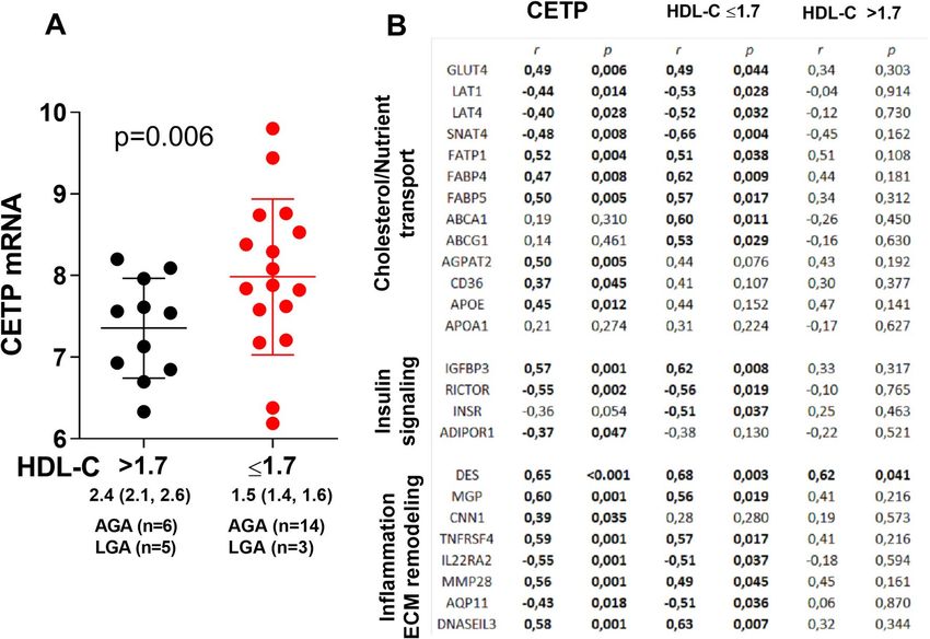
Unique lipoprotein composition in women giving birth to SGA infants. To get a better under-
standing of the composition of the lipoproteins in the study group we used a commercially available lipidomics
analysis from a subset of the women (n = 160) at weeks 36–38. We found major differences in many of the mark-
ers in women giving birth to AGA vs LGA and SGA infants, adjusted for age, fetal sex, GDM, preeclampsia and
BMI (Figs. 5, 6). First of all, HDL-C was found lower in the LGA infant group which corresponded to the first
analyses of the whole cohort. Briefly, the lipidomics data show that women giving birth to SGA infants have
unique composition of the lipoproteins, with high levels of TG in VLDL and high levels of lipids/cholesterol/
cholesteryl ester in the medium and small HDL, which corresponds to the low CETP activity (Fig. 7). These
analyses also show that women giving birth to SGA infants have higher ApoA1 and HDL particle concentration,
with no difference in the size of HDL, showing a unique composition of the medium and small HDL compared
to AGA. Moreover, we found that the ABCA1 and ABCG1 in the placenta were associated with CETP in the
women with the lower HDL-C but not in the women with the higher HDL-C. Finally, exploring the composition
of the lipoproteins in more details (Fig. 6) several interesting findings were revealed. First, we found higher levels
of total VLDL, lipids, PL, FC and TG in XXL-VLDL, TG in XL-VLDL and L-VLDL in the SGA infant group.
Second and most importantly, total HDL, lipid, PL, cholesterol, CE and FC were higher in M-HDL and S-HDL
for the SGA infant group, while cholesterol and CE were lower in M-HDL in the LGA infant group, indicating
a unique composition of S-HDL in the women giving birth to SGA infants that are of major importance for the
role HDL plays in cholesterol efflux. The main findings are illustrated in Fig. 7.
Discussion
In the present study we found decreased levels of CETP expression in the placentas of women with the highest
HDL-C at week 36–38. The CETP expression was associated with mRNAs involved in cholesterol/nutrient trans-
port, insulin signaling and inflammation/ECM remodeling. Moreover, both placental and circulating CETP levels
were positively associated with growth of the fetus, reflected as increase in AC during third trimester and notably,
the regulation of CETP in placenta was strongly related to maternal HDL-C levels. Also, the women giving birth
to SGA infants had a lipoprotein composition with high levels of TG in VLDL and lipids/cholesterol/cholesteryl
esters in medium and small HDL at week 36–38, corresponding to decreased CETP activity. The modulation of
the composition of small HDL particle which also include regulation of ApoA1 may be of particular importance.
Finally, low circulating CETP activity during pregnancy at 36–38 weeks was associated with giving birth to SGA
infants. Our findings suggest a link between increased maternal HDL-C levels, particularly high cholesteryl
Scientific Reports | (2021) 11:6213 | https://doi.org/10.1038/s41598-021-85777-3 7
Vol.:(0123456789)www.nature.com/scientificreports/
Figure 3. Correlation plot between (A) CETP mRNA in the placenta and abdominal circumference (AC) and
the difference in AC between week 30–32 to 36–38, (B) CETP activity in plasma and AC and the difference
between AC from week 30–32 to 36–38.
Figure 4. (A) CETP activity during pregnancy in women giving birth to AGA (appropriate for gestational age)
n = 208, LGA (large for gestational age) n = 45 and SGA (small for gestational age) n = 22 infants, adjusted for age
and BMI at week 14–16. *p < 0.05 between SGA and AGA infants. (B) Logistic regression showing univariate
and adjusted (age, BMI, fetal sex, gestational diabetes and preeclampsia) models giving birth to SGA infants
compared to AGA infants, odds ratio (OR), when the CETP activity is low at 36–38 weeks, using standardized
values.
Scientific Reports | (2021) 11:6213 | https://doi.org/10.1038/s41598-021-85777-3 8
Vol:.(1234567890)www.nature.com/scientificreports/
Figure 5. Concentration of total cholesterol, triglycerides, phospholipids, cholesteryl esters, free cholesterol,
lipids, particle concentration, particle size at week 36–38 between women giving birth to LGA and SGA vs. AGA
infants. Data is normalized and given as standardized estimated marginal means and 95 confidence interval
adjusted for age, fetal sex, gestational diabetes, preeclampsia and BMI at week 14–16. *p < 0.05, **p < 0.01,
***p < 0.001.
ester/cholesterol/lipid in medium and small HDL, low CETP levels both in circulation and placenta and SGA
infants, potentially involving altered regulation of metabolic and inflammatory pathways within the placenta.
In pregnancy HDL is present both in the maternal and the fetal circulation. Herein we show that maternal
HDL-C level may be a predictor of birth weight and represent a possible link to aspects of placental function. The
reduced fetal growth in fetuses from ApoA1−/− mice (low HDL-C) was not mediated by a change in gene expres-
sion of ApoA1 or cholesterol synthesis in the fetus itself, and suggested that factors in the maternal circulation
were responsible for the change in fetal growth by changing placental function or transport across the placenta
irectly15,16. In the present study, we show that a large decrease in HDL-C during pregnancy
affecting the fetus d
was associated with giving birth to LGA infants, while no decrease in HDL-C during pregnancy was associated
with giving birth to SGA infants, compared to a modest decrease in AGA, after adjusting for maternal age and
BMI. These data are also supported by previous studies showing increased HDL-C levels at 24–26 weeks gesta-
tion in women giving birth to SGA infants17. The authors suggest that the elevated HDL-C concentrations were
a consequence of placental dysfunction that could diminish the transport of HDL across the placenta to the fetus
and thereby raising maternal levels.
Our study shows that women giving birth to SGA infant have higher HDL particle concentration, but with no
difference in the size of HDL. However, we found a unique composition of the medium and small HDL compared
to AGA. Several studies have found lipid-free ApoA1 and the smaller forms of HDL to be the main acceptors
of cholesterol efflux via ABCA1 pathway in m acrophages27. The cholesteryl ester/lipid rich small HDL in the
women giving birth to SGA infants could suggest that the cholesterol efflux is decreased in these women and
that they have an unfavorable lipid profile which may in turn have effects on the placenta/fetal growth. Increase
in the lipid content of HDL is thought to decrease its capacity to remove cellular cholesterol and small, lipid-
poor HDL particles thus represent more efficient cholesterol acceptors28. The lipid/cholesterol rich HDL in the
maternal circulation may impact the placenta in that also the cholesterol efflux in the placenta is compromised.
The cholesterol efflux proteins ABCA1 and ABCG1 control the transport of cholesterol from the maternal
circulation to the fetus, and vice versa29. Indeed, we found that the ABCA1 and ABCG1 in the placenta were
associated with CETP in the women with the lower HDL-C but not in the women with the higher HDL-C. Of
relevance to our findings, a decrease of placental ABCA1 expression and cholesterol transport activity has been
observed in preeclampsia30.
Notably, we found large differences in the mRNA profile of the placenta between mothers with higher and
lower HDL-C levels. As a human model to investigate the possible relation of maternal HDL-C levels on mRNA
Scientific Reports | (2021) 11:6213 | https://doi.org/10.1038/s41598-021-85777-3 9
Vol.:(0123456789)www.nature.com/scientificreports/
Figure 6. Composition of total (P), lipid (L), phospholipid (PL) cholesterol (C), cholesteryl ester (CE), free
cholesterol (FC) and triglycerides (TG) in chylomicrons and very-low-density lipoprotein (VLDL), large density
lipoprotein (LDL), intermediate-density lipoprotein (IDL) and high-density lipoprotein (HDL) at week 36–38
between women giving birth to AGA and LGA and AGA and SGA infants. Data is normalized and given as
standardized estimated marginal means and 95% confidence interval adjusted for age, fetal sex, gestational
diabetes, preeclampsia and BMI at week 14–16. *p < 0.05, **p < 0.01, ***p < 0.001.
expression in the placenta, we used previous RNAseq data from 28 placentas divided according to maternal
HDL-C levels (divided by the median) and investigated differentially expressed genes. mRNAs involved in
lipid metabolism and related to inflammation/ECM remodeling were regulated, and we speculate that this
could influence placental environment and communication/transport between maternal and fetal interfaces.
The inflammation/ECM remodeling genes were mostly upregulated in the placentas of women with low HDL-C,
potentially contributing to these women being more metabolic unbalanced which may influence the inflamma-
tion/remodeling in the placenta. It is known that the fetal HDL differs from the maternal HDL, however the full
HDL proteome in each circulation and its associations is u nknown9. Interestingly, we found increased expression
of immunoglobulin heavy constant gamma 1 (IGHG1) and immunoglobulin lambda constant 2 (IGLC2) increased
in the placentas of women with the highest HDL-C. These mRNAs have been shown to be part of the fetal HDL
proteome, and low abundant in the maternal HDL proteome, and linked to immune response9. The impact
of the increased levels of these mRNAs is, however, at present unknown. However, investigating the RNAseq
data of differentially expressed genes between LGA vs AGA infants we found almost no overlapping mRNAs
comparing the list with placenta divided into HDL-C plasma levels, which may suggest that these mRNAs are
modulated by HDL-C.
CETP was one of the top regulated placental genes and was decreased in the placentas from women with the
highest HDL-C group further indicating an interaction between HDL and CETP. Placental CETP mRNA was also
associated with change of AC of the fetus. At the molecular levels, CETP expression was associated with regula-
tion of mRNAs involved in cholesterol/nutrient transport, insulin signaling and inflammation/ECM remodeling,
and interestingly the association was strongest in the placentas of women with the lowest HDL-C levels. We have
previously published adiponectin receptors including adiponectin receptor 1 (ADIPOR1) in the placenta and its
association with nutrient transport and fetal growth in LGA infants31. Herein we found CETP in the placenta
negatively associated with ADIPOR1. CETP was also associated with mRNAs involved in cholesterol transport
Scientific Reports | (2021) 11:6213 | https://doi.org/10.1038/s41598-021-85777-3 10
Vol:.(1234567890)www.nature.com/scientificreports/
Figure 7. Illustration of the main findings. Low CETP activity in the circulation of women giving birth to SGA
infants, followed by low cholesteryl ester and triglyceride exchange between chylomicron/large VLDL and small
HDL, resulting in high triglycerides, lipids, phospholipids and free cholesterol in the chylomicrons/large VLDL
and high phospholipids, lipids, cholesteryl ester, cholesterol, free cholesterol and ApoA1 in the small HDL
compared to the composition in women giving birth to AGA infants.
including APOE, which is a constituent of VLDL and essential ligand for the uptake an clearance of atherogenic
lipoproteins and have been found to be secreted to the maternal circulation from the placenta32. Further, CETP
was associated with mRNAs involved in nutrient t ransport33 including the glucose transporter, GLUT434, and
mRNAs involved in fatty acid t ransport35. In the liver, CETP mRNA levels are increased in response to high cel-
lular cholesterol c ontent36 and many binding sites in the CETP promoter have been identified for several tran-
scription factors that regulate its activity37. Although our findings could suggest that CETP could be involved in
placental-mediated fetal growth, these issues will have to be further clarified as the role of CETP in the placenta
is not known. It is a possibility that these changes are driven by HDL-C with CETP as one important mediator.
CETP activity has been shown to be elevated in normal pregnancy compared to non-pregnant individuals38,39.
Our study includes a larger population and in addition one extra time point closer to term. At week 36–38, we
found CETP activity decreased in the whole cohort, and most in the women giving birth to SGA infants. In
contrast, a small study found high CETP activity in both maternal and cord sera of SGA infants40, while another
study found lower CETP mass but a higher cholesteryl ester transfer in neonatal plasma of SGA infants41. Sur-
prisingly, in the women giving birth to LGA infants and with the largest decrease in HDL-C during pregnancy,
we did not find CETP activity increased during pregnancy, compared to AGA. Further, we did find associations
between CETP and change in AC of the fetus, but, these associations were not as strong as for adiponectin in
our previous study, where our focus was LGA infants and we found that maternal adiponectin levels are lowest
when the fetus has the highest AC. This may suggest that the CETP activity in the maternal circulation is not as
important for growth in LGA infants, compared to in the SGA infants. Indeed, we found that CETP activity at
36–38 weeks was associated with SGA infants, and not LGA infants.
Our study has some limitations. Overall, not all markers are measured in all samples and studies are per-
formed on sub-cohorts. The RNAseq was originally not designed for investigating mRNAs that differed in placen-
tas of women with higher and lower maternal HDL-C. These placentas were chosen to investigate differentially
expressed mRNAs between placentas from women with preeclampsia and gestational diabetes compared to
women without these states. In addition, we did not have any SGA infants in the RNAseq cohort and the small
sample size did not allow a proper correction for the several possible confounding factors. Only one biopsy was
taken from each placenta and the collection was performed by different individuals. While the technique for
placental biopsy was standardized, we cannot exclude the possibility that biopsies were taken at different sites
and that regional differences may have accounted for differences in differentially expressed genes. There were
too few overlapping samples between RNAseq and the lipidomics data to perform any analysis. Further, we only
have CETP activity data of 300 women. Also, we can not exclude some degree of sample degradation before this
activity assay, although all samples have been stored in − 80 °C and been treated the same under preanalytical,
storage and analytical phase. However, our cohort is a well characterized cohort with clinical data and markers
in the blood at several timepoints during pregnancy.
To conclude, our findings suggest a link between increased maternal HDL-C levels, low CETP levels both in
circulation and placenta and association with the risk of giving birth to a SGA infant.
Scientific Reports | (2021) 11:6213 | https://doi.org/10.1038/s41598-021-85777-3 11
Vol.:(0123456789)www.nature.com/scientificreports/
Data availability
All data generated or analysed during this study are included in this published article (and its Supplementary
Information file).
Received: 3 August 2020; Accepted: 4 March 2021
References
1. Visentin, S. et al. Early origins of adult disease: Low birth weight and vascular remodeling. Atherosclerosis 237, 391–399 (2014).
2. Law, C. M. et al. Fetal, infant, and childhood growth and adult blood pressure: A longitudinal study from birth to 22 years of age.
Circulation 105, 1088–1092 (2002).
3. Mudd, L. M., Holzman, C. B. & Evans, R. W. Maternal mid-pregnancy lipids and birthweight. Acta Obstet. Gynecol. Scand. 94,
852–860 (2015).
4. Kulkarni, S. R. et al. Maternal lipids are as important as glucose for fetal growth: Findings from the Pune Maternal Nutrition Study.
Diabetes Care 36, 2706–2713 (2013).
5. Sattar, N. et al. Lipid and lipoprotein concentrations in pregnancies complicated by intrauterine growth restriction. J. Clin. Endo-
crinol. Metab. 84, 128–130 (1999).
6. Gallo, L. A., Barrett, H. L. & Dekker Nitert, M. Review: Placental transport and metabolism of energy substrates in maternal obesity
and diabetes. Placenta 54, 59–67 (2017).
7. Lin, D. S., Pitkin, R. M. & Connor, W. E. Placental transfer of cholesterol into the human fetus. Am. J. Obstet. Gynecol. 128, 735–739
(1977).
8. Pitkin, R. M., Connor, W. E. & Lin, D. S. Cholesterol metabolism and placental transfer in the pregnant Rhesus monkey. J. Clin.
Investig. 51, 2584–2592 (1972).
9. Sreckovic, I. et al. Distinct composition of human fetal HDL attenuates its anti-oxidative capacity. Biochim. Biophys. Acta 1831,
737–746 (2013).
10. Woollett, L. A. Review: Transport of maternal cholesterol to the fetal circulation. Placenta 32(Suppl 2), S218–S221 (2011).
11. Shah, A. S., Tan, L., Long, J. L. & Davidson, W. S. Proteomic diversity of high density lipoproteins: Our emerging understanding
of its importance in lipid transport and beyond. J. Lipid Res. 54, 2575–2585 (2013).
12. Marsche, G., Saemann, M. D., Heinemann, A. & Holzer, M. Inflammation alters HDL composition and function: Implications for
HDL-raising therapies. Pharmacol. Ther. 137, 341–351 (2013).
13. Scholler, M. et al. Phospholipid transfer protein is differentially expressed in human arterial and venous placental endothelial cells
and enhances cholesterol efflux to fetal HDL. J. Clin. Endocrinol. Metab. 97, 2466–2474 (2012).
14. Kitchens, R. L., Thompson, P. A., Munford, R. S. & O’Keefe, G. E. Acute inflammation and infection maintain circulating phos-
pholipid levels and enhance lipopolysaccharide binding to plasma lipoproteins. J. Lipid Res. 44, 2339–2348 (2003).
15. McConihay, J. A., Honkomp, A. M., Granholm, N. A. & Woollett, L. A. Maternal high density lipoproteins affect fetal mass and
extra-embryonic fetal tissue sterol metabolism in the mouse. J. Lipid Res. 41, 424–432 (2000).
16. Rebholz, S. L. et al. Studies in genetically modified mice implicate maternal HDL as a mediator of fetal growth. FASEB J. 32, 717–727
(2018).
17. Kramer, M. S. et al. Maternal lipids and small for gestational age birth at term. J. Pediatr. 163, 983–988 (2013).
18. Filippatos, T. D., Kei, A. & Elisaf, M. S. Anacetrapib, a new CETP inhibitor: The new tool for the management of dyslipidemias?.
Diseases 5, 21 (2017).
19. Roland, M. C. et al. Fetal growth versus birthweight: The role of placenta versus other determinants. PLoS ONE 7, e39324 (2012).
20. Lekva, T. et al. Beta-cell dysfunction in women with previous gestational diabetes is associated with visceral adipose tissue distri-
bution. Eur. J. Endocrinol. 173, 63–70 (2015).
21. Skjaerven, R., Gjessing, H. K. & Bakketeig, L. S. Birthweight by gestational age in Norway. Acta Obstet. Gynecol. Scand. 79, 440–449
(2000).
22. Roland, M. C. P., Lekva, T., Godang, K., Bollerslev, J. & Henriksen, T. Changes in maternal blood glucose and lipid concentrations
during pregnancy differ by maternal body mass index and are related to birthweight: A prospective, longitudinal study of healthy
pregnancies. PLoS ONE 15, e0232749 (2020).
23. Friedewald, W. T., Levy, R. I. & Fredrickson, D. S. Estimation of the concentration of low-density lipoprotein cholesterol in plasma,
without use of the preparative ultracentrifuge. Clin. Chem. 18, 499–502 (1972).
24. Ueland, T. et al. Elevated cholesteryl ester transfer protein activity early in pregnancy predicts prediabetes 5 years later. J. Clin.
Endocrinol. Metab. 105, dgz119 (2019).
25. Lekva, T. et al. Gene expression in term placentas is regulated more by spinal or epidural anesthesia than by late-onset preeclampsia
or gestational diabetes mellitus. Sci. Rep. 6, 29715 (2016).
26. Love, M. I., Huber, W. & Anders, S. Moderated estimation of fold change and dispersion for RNA-seq data with DESeq2. Genome
Biol. 15, 550 (2014).
27. Heinecke, J. W. Small HDL promotes cholesterol efflux by the ABCA1 pathway in macrophages: Implications for therapies targeted
to HDL. Circ. Res. 116, 1101–1103 (2015).
28. Kontush, A. & Chapman, M. J. Antiatherogenic small, dense HDL—guardian angel of the arterial wall?. Nat. Clin. Pract. Cardiovasc.
Med. 3, 144–153 (2006).
29. Kallol, S. & Albrecht, C. Materno-fetal cholesterol transport during pregnancy. Biochem. Soc. Trans. 48, 775–786 (2020).
30. Baumann, M. et al. Placental ABCA1 and ABCG1 expression in gestational disease: Pre-eclampsia affects ABCA1 levels in syn-
cytiotrophoblasts. Placenta 34, 1079–1086 (2013).
31. Lekva, T. et al. Large reduction in adiponectin during pregnancy is associated with large-for-gestational-age newborns. J. Clin.
Endocrinol. Metab. 102, 2552–2559 (2017).
32. Melhem, H. et al. Placental secretion of apolipoprotein A1 and E: The anti-atherogenic impact of the placenta. Sci. Rep. 9, 6225
(2019).
33. Brett, K. E., Ferraro, Z. M., Yockell-Lelievre, J., Gruslin, A. & Adamo, K. B. Maternal-fetal nutrient transport in pregnancy patholo-
gies: The role of the placenta. Int. J. Mol. Sci. 15, 16153–16185 (2014).
34. James-Allan, L. B., Arbet, J., Teal, S. B., Powell, T. L. & Jansson, T. Insulin stimulates GLUT4 trafficking to the syncytiotrophoblast
basal plasma membrane in the human placenta. J. Clin. Endocrinol. Metab. 104, 4225–4238 (2019).
35. Winterhager, E. & Gellhaus, A. Transplacental nutrient transport mechanisms of intrauterine growth restriction in rodent models
and humans. Front. Physiol. 8, 951 (2017).
36. Jiang, X. C., Agellon, L. B., Walsh, A., Breslow, J. L. & Tall, A. Dietary cholesterol increases transcription of the human cholesteryl
ester transfer protein gene in transgenic mice. Dependence on natural flanking sequences. J. Clin. Investig. 90, 1290–1295 (1992).
37. Le Goff, W., Guerin, M., Petit, L., Chapman, M. J. & Thillet, J. Regulation of human CETP gene expression: Role of SP1 and SP3
transcription factors at promoter sites -690, -629, and -37. J. Lipid Res. 44, 1322–1331 (2003).
Scientific Reports | (2021) 11:6213 | https://doi.org/10.1038/s41598-021-85777-3 12
Vol:.(1234567890)www.nature.com/scientificreports/
38. Iglesias, A., Montelongo, A., Herrera, E. & Lasuncion, M. A. Changes in cholesteryl ester transfer protein activity during normal
gestation and postpartum. Clin. Biochem. 27, 63–68 (1994).
39. Winkler, K. et al. Low density lipoprotein (LDL) subfractions during pregnancy: Accumulation of buoyant LDL with advancing
gestation. J. Clin. Endocrinol. Metab. 85, 4543–4550 (2000).
40. Kim, S. M. et al. Cord and maternal sera from small neonates share dysfunctional lipoproteins with proatherogenic properties:
Evidence for Barker’s hypothesis. J. Clin. Lipidol. 11, 1318-1328 e1313 (2017).
41. Kaser, S. et al. Lipoprotein profile and cholesteryl ester transfer protein in neonates. Metabolism 50, 723–728 (2001).
Acknowledgements
The sequencing service was provided by the Norwegian Sequencing Centre (http://www.sequencing.uio.no), a
national technology platform hosted by the University of Oslo and supported by the Functional Genomics and
Infrastructure programs of the Research Council of Norway and the Southeastern Regional Health Authorities.
Author contributions
K.G., P.A., T.H. contributed to conception and design, and reviewed/edited manuscript. M.C.P.R., T.L., contrib-
uted to conception and design, researched data, and wrote the manuscript.
Funding
The project has been financially supported by grants from The National Advisory Unit for Women’s Health
(MCPR), Diabetesforbundet (TL), Freia (TL) and Blix (TL). The funders had no role in study design, data col-
lection and analysis, decision to publish, or preparation of the manuscript.
Competing interests
The authors declare no competing interests.
Additional information
Supplementary Information The online version contains supplementary material available at https://doi.
org/10.1038/s41598-021-85777-3.
Correspondence and requests for materials should be addressed to T.L.
Reprints and permissions information is available at www.nature.com/reprints.
Publisher’s note Springer Nature remains neutral with regard to jurisdictional claims in published maps and
institutional affiliations.
Open Access This article is licensed under a Creative Commons Attribution 4.0 International
License, which permits use, sharing, adaptation, distribution and reproduction in any medium or
format, as long as you give appropriate credit to the original author(s) and the source, provide a link to the
Creative Commons licence, and indicate if changes were made. The images or other third party material in this
article are included in the article’s Creative Commons licence, unless indicated otherwise in a credit line to the
material. If material is not included in the article’s Creative Commons licence and your intended use is not
permitted by statutory regulation or exceeds the permitted use, you will need to obtain permission directly from
the copyright holder. To view a copy of this licence, visit http://creativecommons.org/licenses/by/4.0/.
© The Author(s) 2021
Scientific Reports | (2021) 11:6213 | https://doi.org/10.1038/s41598-021-85777-3 13
Vol.:(0123456789)You can also read