Evaluation of the Stomatognathic System before and after Osteopathic Manipulative Treatment in 120 Healthy People by Using Surface ...
←
→
Page content transcription
If your browser does not render page correctly, please read the page content below
International Journal of
           Environmental Research
           and Public Health

Article
Evaluation of the Stomatognathic System before and
after Osteopathic Manipulative Treatment in 120
Healthy People by Using Surface Electromyography
Andrea Manzotti 1,† , Chiara Viganoni 1 , Dorina Lauritano 2, *,† , Silvia Bernasconi 1 ,
Alice Paparo 1 , Rachele Risso 1 and Alessandro Nanussi 1,2
 1    SOMA–osteopathic Institute of Milan, 20126 Milan, Italy; andreamanzotti@soma-osteopatia.it (A.M.);
      bernasconi.s@hotmail.it (S.B.); alice.paparo9@gmail.com (A.P.); super-ginger@hotmail.it (R.R.);
      nanussi@tiscalinet.it (A.N.)
 2    Department of Medicine and Surgery, Centre of Neuroscience of Milan, University of Milano-Bicocca,
      20126 Milan, Italy; c.viganoni@campus.unimib.it
 *    Correspondence: dorina.lauritano@unimib.it; Tel.: +39-335-679-0163
 †    These authors contributed equally to this work.

 Received: 13 March 2020; Accepted: 29 April 2020; Published: 7 May 2020

 Abstract: Objective: To investigate the action of osteopathic manipulative treatment on the muscular
 activity of the stomatognathic apparatus by using surface electromyography (sEMG). Material and
 Methods: Surface electromyography (sEMG) was performed on the masseter and anterior temporalis
 muscles of 120 subjects (73 F; 47 M), both at time T0 and T2. The sample was divided into
 three randomized groups of 40 subjects each: control, placebo, and osteopathic manipulative
 treatment (OMT). In the T1 interval between the two evaluations, the control group was not treated,
 the placebo group underwent a placebo treatment, and the OMT group underwent manipulative
 treatment. The mean value of each measurement and its coefficient of variation, between time
 T0 and T2, were calculated for both the intragroup (OMT, placebo, control) and the intergroup
 (OMT-placebo, OMT-control). Outcomes: In 40% of the subjects, statistically significant improvements
 were highlighted in the OMT. Whereas, the statistically significant results of the placebo and control
 groups were 7.5% and 17.5%, respectively, of which more than 75% moved away from the physiological
 range, showing a worsening of the muscular activity. This analysis showed statistically significant
 variations (p ≤ 0.05) in the OMT group compared to the placebo and the control groups. Conclusions:
 OMT determines variations of the activity of masticatory muscles.

 Keywords: osteopathic manipulative treatment; masticatory muscles; electromyography; occlusion;
 temporomandibular joint

1. Introduction
     The relationship between dental occlusion, body posture, and temporomandibular disorders
(TMD) is a controversial topic in dentistry. At present, literature data are mostly based on the effects of
dental occlusion on head and body posture, but limited information is available on the inverse effects
of posture on dental occlusion [1].
     What is known is that TMDs have a multifactorial etiology that includes a broad range of
phenotypic risk factors, as reported in the Orofacial Pain Prospective Evaluation and Risk Assessment
Study (OPPERA) [2], and that it is a complex disorder often characterized by the co-occurrence of
multiple conditions. Hence, it is no longer appropriate to consider it solely as a localized orofacial
pain condition as the majority of people with chronic TMDs present a multisystem disorder with

Int. J. Environ. Res. Public Health 2020, 17, 3250; doi:10.3390/ijerph17093250    www.mdpi.com/journal/ijerph
Int. J. Environ. Res. Public Health 2020, 17, 3250 2 of 10
overlapping co-morbidity [3]. For this reason, the authors consider an interdisciplinary approach
essential for the treatment of TMDs.
Furthermore, TMDs are the second most common musculoskeletal condition after chronic low
back pain, as reported by the National Institute of Dental and Craniofacial Research. Thus, the need
for first-line conservative treatment like osteopathic manipulative treatment (OMT) is recognized [4].
Osteopathic medicine is a noninvasive, drug-free manual medicine, classified as a complementary
and alternative medicine. Its goal is the care of the individual in its entirety and works through manual
manipulation techniques [5]. The OMT is a hands-on approach that has the objective of improving
structures that inhibit the body’s function or homeostasis [6].
Alterations in the masticatory muscles, neck muscles, and occlusion can be considered causal factors
of imbalance of the postural muscular chains [7]. Therefore, therapies that seek occlusal restoration,
such as muscle relaxation techniques, should lead to an improvement in the overall equilibrium of the
neuromuscular system and in the posture [8]. Furthermore, recent studies have shown that trigeminal
afferents and alterations in the stomatognathic system are related to proprioception, visual stabilization,
and postural stability [9,10].
The objective of this study was to investigate the action of osteopathic manipulative treatment on
the muscular activity of the stomatognathic apparatus and on mandibular posture compensations by
means of surface electromyography (sEMG).
Although the use of this device as a stand-alone diagnostic tool has raised strong negative criticism
because of the absence of normative values [11], its role in a controlled research setting is recognized to
have scientific merit [12].
Furthermore, the focus of this research was not to diagnose nor to treat TMDs, but to validate the
effectiveness of OMT in terms of the change in muscular patterns when testing healthy subjects, so the
choice of this instrument is related to its noninvasiveness.
2. Materials and Methods
The study was a randomized double-blind controlled trial (RCT) and the sample was made up
of 120 healthy adult subjects (47 males and 73 females) between 19 and 62 years old (the average
sample age was: 25.64 years), who all voluntarily participated in the study. As a result of the lack of
research in this field, we did not conduct a power analysis to determine the sample size needed for this
study. We decided to involve 120 patients considering this number sufficient for the representativeness
of the sample for the validation of OMT. The power of the study was calculated retrospectively on
the main outcome, which we discuss in the Data Analysis section. All the subjects were evaluated
at the Institute of Osteopathy of Milan ‘S.O.M.A.’ (viale Sarca No. 336-20126, Milan MI). This study
was approved by the Institutional Review Board of the Institute of Osteopathy of Milan ‘S.O.M.A’,
following the principles of Helsinki for human experimentation. The sample was selected on a series
of inclusion criteria, such as the absence of orthodontic therapy in progress or during the previous
year, temporomandibular disorders, pain of the oral cavity, mobile protheses, bite, surgery or accidents
which occurred in the previous six months, and finally, the presence of at least 24 healthy dental
elements. After examining the description of the methods and objectives, the subjects signed an
informed consent to proceed with the execution of the dental and osteopathic evaluations, with the
analysis through surface electromyography, and finally, with the osteopathic treatment. Furthermore,
a consent was signed by each subject for the processing of personal and sensitive data. The 120 subjects
suitable for the study were randomly divided into three groups of 40 subjects each, using a random
number generator. The sample was divided as follows:
1. Treatment group (12 males, 28 females);
2. Placebo group (17 males, 23 females);
3. Control group (18 males, 22 females).Int. J. Environ. Res. Public Health 2020, 17, 3250 3 of 10
An extraoral and intraoral clinical examination of the stomatognathic system was carried out
on each subject. An orthodontic evaluation was performed to the detect the presence of craniofacial
asymmetries, malocclusions following Angle’s classification, discrepancies between centric occlusion
(CO) and centric relation (CR), cross-bite, alterations of overjet, and overbite. Then, a functional
examination of mandibular movements and Temporomandibular Joint (TMJ) was carried out following
the criteria proposed by the Italian Association of Gnathology (AIG), illustrated in Table 1, based on
the research from the Diagnostic Criteria for Temporomandibular Disorders (RDC/TMD) Axis I [13].
Table 1. The criteria used to assess the absence of disfunctions.
1. Straight opening pattern without pain
2. Maximum Mouth Opening > 40 mm
MANDIBULAR MOVEMENTS 3. Lateral and Protrusive movements without pain and restrictions
4. Elastic Assisted Mouth Opening (End-Feel)
1. Absence of noises (clicking, popping, crepitus)
TMJ 2. Symmetric movements
3. Absence of pain with palpation
After the functional examination, at time T0, each patient underwent the first evaluation of the
masticatory muscles with sEMG.
The study protocol was divided into three different time groups:
1. T0: the first evaluation with sEMG;
2. T1 (30 min): OMT, placebo treatment, waiting;
3. T2: the second evaluation with sEMG.
2.1. Evaluation with Surface Electromyography
Surface electromyography (sEMG) is a noninvasive technique used to analyze the function of the
masticatory muscles which, being located in a superficial position, are easily accessible using surface
electrodes [14,15]. This exam is painless and innocuous [16].
The sEMG recordings were made using a Teethan wireless system; disposable, pre-gelled,
silver/silver chloride bipolar surface electrodes [17], (dimensions 41 mm × 21 mm, diameter 10 mm
and interelectrode distance 21 ± 1 mm), were placed on the muscular stomach parallel to the muscle
fibers [18], according to the recommendations of sEMG for the noninvasive assessment of muscles
(SENIAM) [19]. To standardize the EMG potentials of the analyzed muscles, two 10 mm thick rolls
were positioned on the mandibular premolars and molars of each participant, and the 5-s maximum
voluntary clenching (MVC) was recorded [20,21]. For the following measurements, the subject was
invited to clench as hard as possible for 5 s, maintaining the same level of contraction. The participants
sat on a rigid chair, positioned at a distance of 3 m from a smooth wall without visual interferences,
with the head unsupported and they were asked to maintain a natural standing position.
Four sets of tests were performed on all subjects at a time T0 and then at a time T2:
1. Sitting with eyes closed in MVC on cotton rolls (COT);
2. Sitting with eyes opened in MVC on cotton rolls;
3. Orthostatic open-eyed position in MVC on cotton rolls;
4. Sitting with eyes closed in MVC (the intercuspal position).
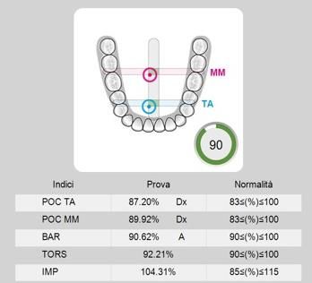
The sEMG data analysis was automatically performed by the software. For each participant,
the potentials of the analyzed muscles recorded during the MVC tests were expressed as a percentage
of the mean potential recorded during the standardization test (MVC on COT) (unit µV/µV × 100)
(see Figure 1).Int. J. Environ. Res. Public Health 2020, 17, 3250 4 of 10
Int. J. Environ. Res. Public Health 2020, 17, x 4 of 10
Int. J. Environ. Res. Public Health 2020, 17, x 4 of 10
Figure 1.Figure 1. Electromyographic
Electromyographic indexesand
indexes and range
range of
of reference.
reference.POCPOCTA: TA:
percentage overlapping
percentage overlapping
coefficient of right and left Temporalis; POC MM: percentage overlapping coefficient of right and left
coefficient of right and left Temporalis; POC MM: percentage overlapping coefficient of right and left
Masseter
Figure muscles; BAR: occlusal
1. Electromyographic barycenter; TORS:of
Torsion index;POC
IMP: impact index.
Masseter muscles; BAR: occlusal indexes and TORS:
barycenter; range reference.
Torsion TA:impact
index; IMP: percentage
index.overlapping
coefficient of right and left Temporalis; POC MM: percentage overlapping coefficient of right and left
To assess the muscle symmetry, the EMG waves of paired muscles (right and left masseter and
Masseter
themuscles;
To temporalis)
assess muscle BAR: occlusal barycenter;
symmetry, the EMG TORS: Torsion index; muscles
IMP: impact index.and left masseter and
were compared by computing thewaves
percentageof paired
overlapping (right
coefficient (POC). When the
temporalis) were
muscles compared
contract by computing
with perfect symmetry,the percentage
a POC overlapping
of 83%–100% coefficient
is obtained. The balance(POC). When the
of the
To assess the muscle symmetry, the EMG waves of paired muscles (right and left masseter and
musclescontractile
contract with
activity perfect symmetry,
of contralateral a POC and
temporalis of 83–100% is obtained.
masseter muscles The balance
(for example, of the and
left masseter contractile
temporalis) were compared by computing the percentage overlapping coefficient (POC). When the
activity right temporalis), expressed
of contralateral temporalisby theand Torsion index muscles
masseter (TORS), ranges between 0%
(for example, left(complete
masseter lateral
and right
muscles contract with perfect symmetry, a POC of 83%–100% is obtained. The balance of the
displacing
temporalis), force)
expressed and
by 100%
the (no lateral
Torsion displacing
index (TORS),force). The
ranges Barycenter
between index
0% (BAR)
(complete was considered
lateral displacing
contractile activity
to investigate of occlusal
if the contralateral temporalis
barycenter was in and masseter
an anterior muscles (for
or posterior example,
position. left masseter
It identifies the mostand
force)right
and 100% (no lateral
temporalis), displacing
expressed by theforce).
TorsionTheindex
Barycenter
(TORS), index
ranges(BAR) was considered
between 0% (complete to lateral
investigate
prevalent pair of masticatory muscles as the percentage ratio of the difference between temporalis
if thedisplacing
occlusal barycenter
force) standardized
and masseter and 100%was(noinpotentials
an anterior
lateral or posterior
displacing
(normal Theposition.
force).90%–100%).
range TheItindex
Barycenter identifies
(BAR)
intensity the
wasmost
of muscle prevalent
considered
work,
to masticatory
pair of investigate
expressed by if the
theimpact
occlusal
muscles as barycenter
the
index wasnormally
percentage
(IMPACT), in an anterior
ratio ofranges or posterior
the difference
from position.
between
100%–115%. High Itvalues
identifies
temporalis the most
and
are typicalmasseter
prevalent
standardized pair
in bruxist of masticatory
potentials
patients. (normal
Finally, muscles
the range
asymmetryas theindex
percentage
90–100%). ratio
The intensity
(ASIM) of the
assessed ofdifference
the muscle work,
symmetry between
between temporalis
expressed
right and by the
impactand masseter
left
index muscular standardized
(IMPACT), (−10 10) (see Figure
from range
2). The90%–100%).
100–115%. advantage ofThe
High values intensity
using these
are of in
indexes
typical muscle
lies in work,
bruxist thepatients.
expressed by of
possibility theevaluating
impact index (IMPACT),
the symmetry, ornormally
asymmetry, ranges from 100%–115%.
conditions High values
of muscular activity [22]. are typical
Finally, the asymmetry index (ASIM) assessed the symmetry between right and left muscular activity
in bruxist patients. Finally, the asymmetry index (ASIM) assessed the symmetry between right and
(−10 < (%) > 10) (see Figure 2). The advantage of using these indexes lies in the possibility of evaluating
left muscular activity (−10 < (%) > 10) (see Figure 2). The advantage of using these indexes lies in the
the symmetry, or asymmetry, conditions of muscular activity [22].
possibility of evaluating the symmetry, or asymmetry, conditions of muscular activity [22].
Figure 2. Electromyographic evaluation of muscles asymmetry expressed with the relative ASIM index
and through a visual representation of the progressive recruitment of Temporalis muscles in blue and
Masseter muscles in Pink.Int. J. Environ. Res. Public Health 2020, 17, 3250 5 of 10
2.2. Osteopathic Evaluation and OMT-Osteopathic Manipulative Treatment
Each subject underwent an osteopathic evaluation with the aim of observing the posture in static
position, performing a dynamic objective examination, and finding areas of tension, mobility restrictions,
and greater tissue density.
The subjects from the control group then waited 30 min for the second evaluation and the ones
from the placebo group underwent a placebo treatment during the T1 interval.
The subjects of the treatment group also underwent an osteopathic manipulative treatment lasting
30 min.
The treatment included three standardized techniques to be applied to all subjects and three
black-box techniques chosen by the operator for the individual subject, based on the previously
performed osteopathic assessment.
The standard techniques applied were the direct intraoral and extraoral inhibition of the masseter
muscles, direct inhibition of temporalis muscles, and direct inhibition of the external and internal
pterygoid muscles.
At time T2, all the subjects were again subjected to surface electromyography.
2.3. Data Analysis
An inferential analysis was carried out using the SPSS software version 25 (IBM, Milan,
Italy), and R studio version 1.1.463. The values obtained from the evaluations at times T0 and T2
were compared both with the intragroup (OMT, placebo, control) and intergroup (OMT-placebo,
OMT-control). The T-test was used in order to compare the averages obtained by studying the
three groups and to find out if the differences from T0 and T2 were significant (for a p-value ≤ 0.05).
Each index has its own reference range which corresponds to an optimal physiological range.
The power of the study was calculated retrospectively on the main outcome, the variable POC TA
starting with the sample size (total = 120, 40 in each group), the significance level (α = 0.05), and with
three different effect sizes between groups after the intervention (OMT vs. placebo = 0.19; placebo vs.
control = 0.42; OMT vs. control = 0.51). The effect size was calculated through Cohen’s D test. In the
comparison of OMT with the placebo group, the power was 0.18, for the placebo vs. control group,
the power was 0.62, and for the OMT vs. control group, the power was 0.80.
3. Results
OMT intragroup: in 40% of the subjects, statistically significant changes were found. In the sEMG
examinations, the variations detected involved POC TA, POC MM, TORS, and BAR. The average
POC TA recorded an improvement in all the measurements, whereas POC MM showed a significant
improvement just in the orthostatic position. The average TORS and BAR improved from the T0 to T2
evaluation, although without catching the physiological range identified from the software.
Placebo intragroup: the statistically significant changes highlighted in the sEMG evaluations were
POC TA and IMPACT, only in 7.5% of the subjects, while the IMPACT index remained within the
physiological range, the average POC TA moved away from it, so we can state that there was a
worsening in the symmetry of the temporalis muscles activity.
Control intragroup: in 17.5% of the measurements, ASIM, TORS, and BAR recorded statistically
significant variations remaining in the physiological ranges.
4. Discussion
The results show a statistical significance between the electromyographic indexes measured in T0
and T2.
The data obtained by analyzing each group showed the greatest number of statistically significant
results in the OMT group, as shown in Figure 3; indeed 40% of the subjects of this group improved their
muscular activity and no worsening was noted. Several studies show how tissue inhibition reducesInt. J. Environ. Res. Public Health 2020, 17, 3250 6 of 10
pain, increases joint mobility, eliminates adhesions between muscle fibers, improves local circulation,
andJ. induces
Int. overall
Environ. Res. Public relaxation [23].
Health 2020, 17, x 6 of 10
Significative Changes
Significance No Significance
N
40
Int. J. Environ. Res. Public Health 2020, 17, x 6 of 10
35
30
25 Significative Changes
20
Significance No Significance
N
15
10
40
5
35
0
30 OMT Control Placebo
25
20
Figure 3. Number
Number of
of subjects
subjects that
that presented
presented significative
significative variations of their muscular
muscular activity between
15
T0 and T2.
10
5
In the placebo and control0
groups, we found less statistically significant changes compared to the
OMT group. In the placebo group justOMT 7.5% of measurements
Control changed, 75% of which can be considered
Placebo
as a worsening of the sEMG indexes; whereas in the control group, 17.5% of the measurements
presented significant changes, 85% of which were considered worsening by the software, as shown in
Figure 3. Number of subjects that presented significative variations of their muscular activity between
Figure 4.
T0 and T2.
Figure 4. Quality of the significant changes inside each group, considering electromyographic
indexes: all the variations in OMT group can be considered improvements whereas the ones in
Control and Placebo groups seem more randomly distributed, with a worsening of the absolute value
of sEMG indexes.
4. Discussion
Figure
The 4.4.Quality
results Quality
show ofof
athethe significant
statistical changes
significance
significant changes inside
between
inside each
the group,
each group, considering
electromyographic
considering electromyographic
indexes measured
electromyographic indexes: in
T0 andindexes:
all all the variations
the variations
T2. in OMT in OMTcan
group group can be considered
be considered improvements
improvements whereas whereas
the onesthe ones in
in Control
Control
and and
Placebo Placebo
groups groups
seem seem
more more randomly
randomly distributed,
distributed, with with
a a worsening
worsening
The data obtained by analyzing each group showed the greatest number of statistically of of
the the absolute
absolute value
value of
of
sEMGsEMG indexes.
indexes.
significant results in the OMT group, as shown in Figure 3; indeed 40% of the subjects of this group
improved their muscular activity and no worsening was noted. Several studies show how tissue
Analyzing the results obtained by comparing the OMT and placebo groups, there were six
4. Discussion
inhibition reduces pain, increases joint mobility, eliminates adhesions between muscle fibers,
statistically significant changes.
improves local circulation,
The results and induces
show a statistical overall between
significance relaxation [23].
the electromyographic indexes measured in
Additionally, the comparison between the OMT and control groups showed seven statistically
In
T0 and T2.the placebo and control groups, we found less statistically significant changes compared to
significant changes.
the OMT
The datagroup. In the placebo
obtained group just
by analyzing each7.5%
group of measurements changed,
showed the greatest 75% ofof
number which can be
statistically
considered as a worsening
significant results in the OMTofgroup,the sEMG
as shownindexes; whereas
in Figure in the
3; indeed 40%control group, 17.5%
of the subjects of this of the
group
measurements
improved their presented significant
muscular activity andchanges, 85% ofwas
no worsening which were
noted. considered
Several studiesworsening
show howby the
tissue
software,
inhibitionasreducesshown in Figure
pain, 4.
increases joint mobility, eliminates adhesions between muscle fibers,
Analyzing
improves the results and
local circulation, obtained
inducesbyoverall
comparing the OMT
relaxation [23]. and placebo groups, there were six
statistically significant
In the placebo andchanges.
control groups, we found less statistically significant changes compared to
the OMT group. In the placebo group just 7.5% of measurements changed, 75% of which can beInt. J. Environ. Res. Public Health 2020, 17, 3250 7 of 10
From the intergroup analysis, we can observe that there were more significant changes in the
OMT group compared to the placebo and the control groups (Figure 3).
Initial studies have found muscular massage to be effective for persistent back pain [24],
and physiotherapy interventions have already been reported in literature as efficacious for patients
with TMD pain and restricted motion [25,26].
Given the results of this study, we can consider OMT as a valid aid to improve the muscular activity
of the stomatognathic system in preparation for a gnathological intervention. This is in light of the
influence that OMT can have on muscular tension and postural compensation. Some of the variations
highlighted in the control and placebo groups can be related either to the impossibility of avoiding
emotional interferences, and/or to the muscular fatigue that might occur after the first evaluation of
patients, and on them having to wait for a second one. The relevance of psychosocial factors indeed
has been widely underlined in literature, so much so that psychosocial factors are considered as
important for the treatment outcome of TMD, as are initial pain intensity and physical diagnoses [27].
Although this was not our objective, we have to keep in mind the influence of cognitive, behavioral,
and emotional aspects on strategies implemented by patients to maintain adequate functioning [28],
and that they may also play a role in this case.
What is interesting is the significant minority of variations in the muscular activity of the
placebo group. Indeed, despite the lack of a specific therapeutic action, a placebo treatment can
elicit real psychobiological responses [29]. Clinically relevant evidence demonstrates that placebo
effects can have meaningful therapeutic effects [30], therefore these outcomes should suggest that
only a real manipulative treatment can cause more constant, and apparently, positive variations in
the electromyographic indexes. Other studies have also shown that complementary and alternative
medicine (CAM) treatments, such as massage, spinal manipulation, and mobilization, are significantly
more efficacious than placebo treatments in reducing neck and low-back pain after treatment [31].
However, further studies should be conducted on a wider sample to highlight the significance of
the differences between placebo and OMT.
In the management of TMDs, the goals of physiotherapeutic regime are to reduce muscular
tone, improve kinetic parameters and posture, and decrease risk factors related to the upper quarter
by stretching masticatory muscles, increasing TMJ mobility, and influencing muscle strength and
proprioception in order to restore normal functioning [32]. In this trial, OMT have shown promising
results in influencing muscular activity of the craniofacial region. Furthermore, the osteopath could
guide the patient with individualized home exercises. The Delphi study confirmed that there is
a consensus among experts that jaw exercises are effective and can be recommended to patients
with myalgia in the jaw muscles and restricted mouth opening capacity [33]. Both general dental
practitioners and orofacial pain experts could benefit from this interdisciplinary approach for the
management of acute and chronic pain.
So far, only a few high quality randomized controlled trials focusing on the therapeutic effectiveness
of osteopathic treatment have been published, and most of them failed to prove efficacy. However,
the available systematic reviews mainly criticized low methodical quality and paucity of the analyzed
osteopathic studies [34].
The main limitations of this study were the absence of a prior effect size and power calculation,
the sample size was arbitrarily decided and this, from a retrospective analysis based on one main
variable (POC TA), resulted in it only being appropriate for the comparison of OMTs and placebos with
controls, but too small to be significant in the comparison between OMTs and placebos. Given the lack
of previous studies and the significant differences from the controls, we can only state that manipulative
treatments can produce variations in the muscular activity and, starting from this limitation, we are
conducting further studies on a wider sample to investigate the differences between placebo treatment
and real osteopathic manipulative treatment. Other limitations include the following: the immediate
revaluation of the subjects without a period of follow up which should be considered in future studies to
evaluate the long-term effect of the treatments; and the choice of evaluating healthy subjects that allowsInt. J. Environ. Res. Public Health 2020, 17, 3250 8 of 10
only to analyze the presence of variations in the muscular activity without demonstrating a therapeutic
effect of the treatment. Further studies on symptomatic patients should be done in the future to assess
the effectiveness of the association between gnathological treatment and osteopathic manipulations.
5. Conclusions
The findings of this randomized clinical trial support the effectiveness of osteopathic manipulative
treatment on modifying the activity of masticatory muscles. This study demonstrated the positive
effect of OMT compared to the placebo treatment.
Our conclusions support the use of OMT in the cranial field, and in particular in dentistry,
to achieve muscular balance.
Other studies should be conducted on dysfunctional patients in order to evaluate their symptomatic
improvements by associating the gnathological and the osteopathic therapy.
Clinical Relevance
1. Measurements of the variations of the muscular activity induced by the osteopathic treatment
2. Demonstration of the positive effect of the osteopathic treatment compared to the placebo one
3. Demonstration of the possible usefulness of the osteopathic treatment in dentistry, referred to the
muscular balance
Author Contributions: A.M.: Conceptualization, Methodology, Resources and Supervision; C.V.: Data curation,
Investigation, Visualization, Writing original draft; D.L.: Supervision; S.B.: Data curation, Investigation,
Visualization, Writing original draft; A.P.: Data curation, Investigation, Visualization, Writing original draft;
R.R.: Data curation, Investigation, Visualization, Writing original draft; A.N.: Conceptualization, Methodology,
Resources and Supervision. All authors have read and agreed to the published version of the manuscript.
Funding: This research did not receive any specific grant from funding agencies in the public, commercial or
no-profit sectors.
Acknowledgments: This study received no specific funding. The instrument and materials used for the
electromyography were provided by Teethan® , thanks to the contribution of Alessandro Nanussi. Nanussi A
and Manzotti A led study conception and design, provided oversight during data collection and contributed to
interpretation of data. Lauritano D critically revised the manuscript. Viganoni C, Bernasconi S, Paparo A and Risso
R made the measurements and treatments, took care of data collection, analysis and interpretations and drafted
the manuscript. We would like to thank the volunteers who participated in this study and made it possible.
Conflicts of Interest: The authors declare no conflict of interest.
References
1. Manfredini, D.; Castroflorio, T.; Perinetti, G.; Guarda-Nardini, L. Dental occlusion, body posture and
temporomandibular disorders: Where we are now and where we are heading for. J. Oral Rehabil. 2012, 39,
463–471. [CrossRef] [PubMed]
2. Maixner, W.; Diatchenko, L.; Dubner, R.; Fillingim, R.B.; Greenspan, J.D.; Knott, C.; Ohrbach, R.; Weir, B.;
Slade, G.D. Orofacial pain prospective evaluation and risk assessment study—The OPPERA study. J. Pain
2011, 12, T4–T11. [CrossRef] [PubMed]
3. Slade, G.D.; Fillingim, R.B.; Sanders, A.E.; Bair, E.; Greenspan, J.D.; Ohrbach, R.; Dubner, R.; Diatchenko, L.;
Smith, S.B.; Knott, C.; et al. Summary of findings from the OPPERA prospective cohort study of incidence of
first-onset temporomandibular disorder: Implications and future directions. J. Pain 2013, 14, T116–T124.
[CrossRef] [PubMed]
4. Cuccia, A.M.; Caradonna, C.; Annunziata, V.; Caradonna, D. Osteopathic manual therapy versus conventional
conservative therapy in the treatment of temporomandibular disorders: A randomized controlled trial.
J. Bodyw. Mov. Ther. 2010, 14, 179–184. [CrossRef] [PubMed]
5. Lanaro, D.; Ruffini, N.; Manzotti, A.; Lista, G. Osteopathic manipulative treatment showed reduction of
length of stay and costs in preterm infants: A systematic review and meta-analysis. Medicine (United States)
2017, 96. [CrossRef] [PubMed]Int. J. Environ. Res. Public Health 2020, 17, 3250 9 of 10
6. Halimi, M.; Leder, A.; Mancini, J.D. Integration of Osteopathic Manual Treatments in Management of Cervical
Dystonia with Tremor: A Case Series. Tremor Other Hyperkinet. Mov. (N. Y.) 2017, 7. [CrossRef]
7. Cuccia, A.; Caradonna, C. The relationship between the stomatognathic system and body posture. Clinics
2009, 64, 61–66. [CrossRef]
8. El Hage, Y.; Politti, F.; Herpich, C.M.; de Souza, D.F.M.; de Paula Gomes, C.A.F.; Amorim, C.F.;
de Oliveira Gonzalez, T.; Biasotto-Gonzalez, D.A. Effect of facial massage on static balance in individuals
with temporomandibular disorder—A pilot study. Int. J. Ther. Massage Bodyw. Res. Educ. Pract. 2013, 6, 6.
9. Gangloff, P.; Perrin, P.P. Unilateral trigeminal anaesthesia modifies postural control in human subjects.
Neurosci. Lett. 2002, 330, 179–182. [CrossRef]
10. Milani, R.S.; Deville De Perière, D.; Lapeyre, L.; Pourreyron, L. Relationship between Dental Occlusion and
Posture. CRANIO 2000, 18, 127–134. [CrossRef]
11. Manfredini, D.; Bucci, M.B.; Montagna, F.; Guarda-Nardini, L. Temporomandibular disorders assessment:
Medicolegal considerations in the evidence-based era. J. Oral Rehabil. 2010, 38, 101–119. [CrossRef] [PubMed]
12. Klasser, G.D.; Okeson, J.P. The clinical usefulness of surface electromyography in the diagnosis and treatment
of temporomandibular disorders. J. Am. Dent. Assoc. 2006, 137, 763–771. [CrossRef] [PubMed]
13. Ohrbach, R. Diagnostic Criteria for Temporomandibular Disorders Clinical Protocol and Assessment
Instruments. J. Oral Facial Pain Headache 2014, 28, 6–27.
14. Buesa-Bárez, J.M.; Martín-Ares, M.; Martínez-Rodríguez, N.; Barona-Dorado, C.; Sanz-Alonso, J.;
Cortés-Bretón-Brinkmann, J.; Martínez-González, J.-M. Masseter and temporalis muscle electromyography
findings after lower third molar extraction. Med. Oral Patol. Oral y Cir. Bucal 2018, 23, e92–e97. [CrossRef]
[PubMed]
15. Kollmitzer, J.; Ebenbichler, G.R.; Kopf, A. Reliability of surface electromyographic measurements. Clin.
Neurophysiol. 1999, 110, 725–734. [CrossRef]
16. Szyszka-Sommerfeld, L.; Matthews-Brzozowska, T.; Kawala, B.; Mikulewicz, M.; Machoy, M.; Wieckiewicz, W.;
Woźniak, K. Electromyographic analysis of masticatory muscles in cleft lip and palate children with
pain-related temporomandibular disorders. Pain Res. Manag. 2018, 2018, 4182843. [CrossRef]
17. Webster, J.G. Reducing motion artifacts and interference in biopotential recording. IEEE Trans. Biomed. Eng.
1984, 823–826. [CrossRef]
18. Castroflorio, T.; Farina, D.; Bottin, A.; Piancino, M.G.; Bracco, P.; Merletti, R. Surface EMG of jaw elevator
muscles: Effect of electrode location and inter-electrode distance. J. Oral Rehabil. 2005, 32, 411–417. [CrossRef]
19. Hermens, H.J.; Freriks, B.; Disselhorst-Klug, C.; Rau, G. Development of recommendations for SEMG sensors
and sensor placement procedures. J. Electromyogr. Kinesiol. 2000, 10, 361–374. [CrossRef]
20. Ferrario, V.F.; Sforza, C. Coordinated electromyographic activity of the human masseter and temporalis
anterior muscles during mastication. Eur. J. Oral Sci. 1996, 104, 511–517. [CrossRef]
21. Ferrario, V.F.; Tartaglia, G.M.; Galletta, A.; Grassi, G.P.; Sforza, C. The influence of occlusion on jaw and neck
muscle activity: A surface EMG study in healthy young adults. J. Oral Rehabil. 2006, 33, 341–348. [CrossRef]
[PubMed]
22. De FelÍcio, C.M.; Sidequersky, F.V.; Tartaglia, G.M.; Sforza, C. Electromyographic standardized indices in
healthy Brazilian young adults and data reproducibility. J. Oral Rehabil. 2009, 36, 577–583. [CrossRef]
[PubMed]
23. Moyer, C.A.; Rounds, J.; Hannum, J.W. A Meta-Analysis of Massage Therapy Research. Psychol. Bull. 2004,
130, 3–18. [CrossRef] [PubMed]
24. Cherkin, D.C.; Sherman, K.J.; Deyo, R.A.; Shekelle, P.G. A Review of the Evidence for the Effectiveness, Safety,
and Cost of Acupuncture, Massage Therapy, and Spinal Manipulation for Back Pain. Ann. Intern. Med. 2003,
138, 898–906. [CrossRef]
25. Maloney, G.E.; Mehta, N.; Forgione, A.G.; Zawawi, K.H.; Al-Badawi, E.A.; Driscoll, S.E. Effect of a Passive
Jaw Motion Device on Pain and Range of Motion in TMD Patients Not Responding to Flat Plane Intraoral
Appliances. CRANIO 2002, 20, 55–66. [CrossRef]
26. Nicolakis, P.; Erdogmus, B.; Kopf, A.; Nicolakis, M.; Piehslinger, E.; Fialka-Moser, V. Effectiveness of exercise
therapy in patients with myofascial pain dysfunction syndrome. J. Oral Rehabil. 2002, 29, 362–368. [CrossRef]
27. Litt, M.D.; Shafer, D.M.; Kreutzer, D.L. Brief cognitive-behavioral treatment for TMD pain: Long-term
outcomes and moderators of treatment. Pain 2010, 151, 110–116. [CrossRef]Int. J. Environ. Res. Public Health 2020, 17, 3250 10 of 10
28. Brister, H.; Turner, J.A.; Aaron, L.A.; Mancl, L. Self-efficacy is associated with pain, functioning, and coping
in patients with chronic temporomandibular disorder pain. J. Orofac. Pain 2006, 20, 115–124. [CrossRef]
29. Rossettini, G.; Carlino, E.; Testa, M. Clinical relevance of contextual factors as triggers of placebo and nocebo
effects in musculoskeletal pain. BMC Musculoskelet. Disord. 2018, 19, 27. [CrossRef]
30. Finniss, D.G.; Kaptchuk, T.J.; Miller, F.; Benedetti, F. Biological, clinical, and ethical advances of placebo
effects. Lancet 2010, 375, 686–695. [CrossRef]
31. Furlan, A.D.; Yazdi, F.; Tsertsvadze, A.; Gross, A.; Van Tulder, M.; Santaguida, L.; Gagnier, J.; Ammendolia, C.;
Dryden, T.; Doucette, S.; et al. A systematic review and meta-analysis of efficacy, cost-effectiveness, and safety
of selected complementary and alternative medicine for neck and low-back pain. Evid.-Based Complement.
Altern. Med. 2012, 2012, 953139. [CrossRef] [PubMed]
32. Michelotti, A.; De Wijer, A.; Steenks, M.; Farella, M. Home-exercise regimes for the management of
non-specific temporomandibular disorders. J. Oral Rehabil. 2005, 32, 779–785. [CrossRef] [PubMed]
33. Lindfors, E.; Arima, T.; Baad-Hansen, L.; Bakke, M.; De Laat, A.; Giannakopoulos, N.N.; Glaros, A.;
Guimarães, A.S.; Johansson, A.; Le Bell, Y.; et al. Jaw Exercises in the Treatment of Temporomandibular
Disorders—An International Modified Delphi Study. J. Oral Facial Pain Headache 2019, 33, 389–398. [CrossRef]
[PubMed]
34. Posadzki, P.; Lee, M.S.; Ernst, E. Osteopathic manipulative treatment for pediatric conditions: A systematic
review. Pediatrics 2013, 132, 140–152. [CrossRef] [PubMed]
© 2020 by the authors. Licensee MDPI, Basel, Switzerland. This article is an open access
article distributed under the terms and conditions of the Creative Commons Attribution
(CC BY) license (http://creativecommons.org/licenses/by/4.0/).You can also read