Magnesium Green for fluorometric measurement of ATP production does not interfere with mitochondrial respiration
←
→
Page content transcription
If your browser does not render page correctly, please read the page content below
Bioenerg Commun 2021.1                doi:10.26124/bec:2021-0001
                                                                                      Open Access

Technical
Communication
Citation                             Magnesium Green for
Cardoso LHD, Doerrier C,
Gnaiger E (2021)                     fluorometric measurement of
Magnesium Green for
fluorometric measurement             ATP production does not
of ATP production does not
interfere with mitochondrial         interfere with mitochondrial
respiration. Bioenerg
Commun 2021.1.                       respiration
doi:10.26124/bec:2021-
0001
                                       Luiza HD Cardoso*,         Carolina Doerrier,
Author contributions                   Erich Gnaiger
LHDC, CD and EG designed
the work; LHDC collected             Oroboros Instruments, Innsbruck, Austria
and analyzed data and
drafted the article; CD and          *Corresponding author: luiza.cardoso@oroboros.at
EG critically revised the
article, all authors approved         BEC 2021.1. doi:10.26124/bec:2021-0001
the final version of the
manuscript.
                                     Abstract
Conflicts of interest
EG is founder and CEO of             For the advanced study of mitochondrial function,
Oroboros Instruments,                high-resolution respirometry is extended by
Innsbruck, Austria.                  fluorometric measurement of ATP production using
Academic editor                      the fluorophore Magnesium Green™ (MgG). A
Chinopoulos Christos,                common problem with several fluorescent dyes is the
Semmelweis University, HU            inhibition of mitochondrial respiration. In the
                                     present study, a coupling control protocol was
Received 2021-02-11
Reviewed 2021-05-05                  applied in combination with MgG to measure ATP
Resubmitted 2021-06-25               production simultaneously with respiration for
Accepted 2021-06-28                  calculation of P»/O2 ratios. MgG at 1.1 µM did not
                                     affect respiration through the NADH-linked and
Published 2021-06-30
                                     succinate-linked pathways. Respiration was not
Editorial and peer review            inhibited in any of the coupling control states, hence
record                               coupling control efficiencies were not affected by
doi:10.26124/bec:2021-0001
                                     MgG.
Preprint
MitoFit Preprints 2021.1             Keywords    ‒   ATP    production;  high-resolution
doi:10.26124/mitofit:2021-0001
                                     respirometry HRR; Magnesium Green MgG; mitochondria
Data availability                    mt; oxidative phosphorylation OXPHOS; fluorometry;
Original files are available         FluoRespirometry
Open Access at Zenodo
repository:
10.5281/zenodo.4916141

 www.bioenergetics-communications.org                                                    1 of 16
MgG and O 2 flux

Graphical abstract - Concept of the MgG assay according to Chinopoulos et al
(2014). MgG fluoresces when bound to Mg 2+. ADP and ATP compete for Mg 2+ binding with
different affinities; ATP has a higher affinity for Mg 2+ compared to ADP. When ADP is
added and binds a large fraction of free Mg2+, MgG fluorescence drops sharply. During
subsequent oxidative phosphorylation ADP is phosphorylated to ATP, which is exchanged
for ADP by the adenine nucleotide translocase ANT. Extramitochondrial ATP increases,
more Mg2+ is bound to ATP, and MgG fluorescence decreases further. This decline is
analyzed as ATP production. MgG does not impair mitochondrial respiratory control.

1. Introduction
      Mitochondrial ATP production can be analyzed with a fluorometric technique using
Magnesium Green™ (MgG) as a fluorescent probe, as described by Chinopoulos et al
(2009). Application of the Mg2+-sensitive fluorophore as an indicator of ATP production
relies on the fact that ADP and ATP have different affinities for Mg2+ (Gnaiger, Wyss 1994;
Leyssens et al 1996; Budinger et al 1998). ADP is phosphorylated to ATP in the
mitochondrial matrix. In the phosphorylation system ADP/ATP and inorganic phosphate
Pi are exchanged stoichiometrically by the adenine nucleotide translocase ANT and the
phosphate carrier PiC. Under experimental conditions when ADP decreases while ATP
increases in the extramitochondrial milieu, the Mg 2+ concentration declines due to the
higher affinity for Mg 2+ of ATP than ADP. Therefore, the fluorometric assay with the
membrane-impermeant MgG provides a quantitative approach to analyze mitochondrial
ATP production. This method was developed further to measure concomitantly
mitochondrial ATP production and O2 consumption in the Oroboros O2k-
FluoRespirometer which is an experimental system complete for high-resolution
respirometry including fluorometry (Chinopoulos et al 2014).
      Fluorescent dyes are widely used to assess various parameters relevant in
mitochondrial physiology. Safranin, rhodamine and its derivatives, such as TMRM, are
frequently employed as reporters of the mitochondrial membrane potential ΔΨp+.
However, all ΔΨp+ dyes have been shown to affect mitochondrial respiration (Scaduto,
Grotyohann 1999). Like TPP+, safranin mainly affects the NADH (N)-linked pathway, the
phosphorylation system, and to a smaller extent the succinate (S)-linked pathway

 2                                               Cardoso et al (2021) Bioenerg Commun 2021.1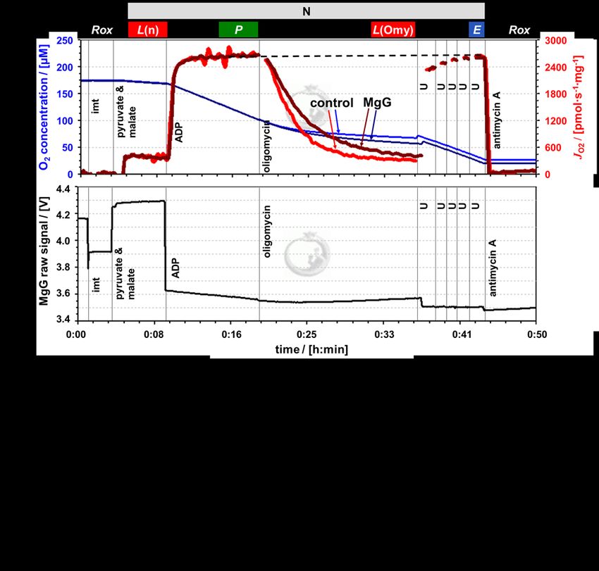
Bioenerg Commun 2021.1 doi:10.26124/bec:2021-0001

(Krumschnabel et al 2014). The effect of ΔΨp+ fluorescent probes can be explained since
they accumulate in the mitochondrial matrix and thus possibly affect mitochondrial
function.
      Amplex UltraRed is frequently employed in mitochondrial physiology studies to
analyze H2O2 production. This dye was shown to affect mitochondrial respiration, even
though the mitochondrial membranes are not permeable to this fluorophore (Makrecka-
Kuka et al 2015). Therefore, it is important to analyze whether MgG affects mitochondrial
respiration, despite the fact that mitochondrial membranes are not permeable to this
fluorophore.
      In the present technical communication, we report the effect of MgG on
mitochondrial respiration, providing an important contribution for the applicability of
the MgG assay for analysis of P»/O2 ratios in mitochondrial preparations. The
experimental MgG concentration was chosen for simultaneous assessment of ATP
production and mitochondrial respiration, which is the gold standard to evaluate
mitochondrial function. Coupling control protocols using NADH- and succinate-linked
substrates showed that mitochondrial respiration was not affected by MgG in LEAK,
OXPHOS- and ET-states, thus making it possible to obtain flux control efficiencies.

2. Materials and methods
2.1. Reagents
      Magnesium Green was purchased from Invitrogen/Thermo Fisher Scientific (cat. Nº
M3733). Antimycin A (cat. Nº A8674), ATP (cat. Nº A2383), CCCP (cat. Nº C2759), malate
(cat. Nº M1000), MgCl2 1 M (cat. Nº M1028), oligomycin (cat. Nº O4876), pyruvate (cat.
Nº P2256), rotenone (cat. Nº R8875), SF 6847 (cat. Nº T182), and succinate (cat. Nº
S2378) were obtained from Sigma Aldrich. ADP was acquired from Millipore (cat. Nº
117105), and carboxyatractyloside from Calbiochem (cat. Nº 216201).
      ADP and ATP were dissolved in deionized H2O without addition of Mg 2+ salts, pH
was adjusted to 6.9 with KOH. Magnesium Green, malate, succinate, carboxyatractyloside
and MgCl2 were dissolved or diluted in deionized H 2O whereas antimycin A, CCCP,
oligomycin, rotenone and SF 6847 were dissolved in ethanol p.a. All solutions were
aliquoted and stored at -20 °C, except pyruvate, which was dissolved in deionized H2O
fresh on the day of each experiment.
2.2. Animals
      Wild-type C57BL/6N adult mice (N=3 per experimental group) were housed in the
animal facility of the Medical University of Innsbruck (maximum 5 mice per cage) and,
maintained at 23±3 °C, relative humidity 45−65 % with a controlled 12 h light/dark cycle
in a conventional animal facility. Mice were fed ad libitum with free access to water. All
procedures were conducted according to the Austrian Animal Experimentation Act in
compliance with the European convention for the protection of vertebrate animals used
for experimental and other scientific purposes (Tierversuchsgesetz 2012; Directive
2010/63/EU; BMWFM-66.011/0128-WF/V/3b/2016).

 www.bioenergetics-communications.org                                                 3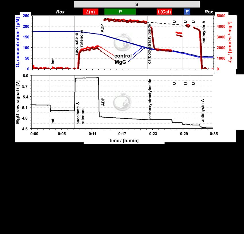
MgG and O 2 flux

2.3. Cardiac mitochondrial isolation and protein concentration determination
       Following cervical dislocation, the hearts were immediately excised and transferred
into ice-cold biopsy preservation solution (BIOPS: 10 mM Ca 2+-EGTA - 0.1 µM free Ca2+,
20 mM imidazole, 20 mM taurine, 50 mM K+-MES, 0.5 mM dithiothreitol, 6.56 mM MgCl2,
5.77 mM ATP, 15 mM phosphocreatine, pH 7.1 adjusted with KOH) for short period of
time (1–2 h; Fontana-Ayoub et al 2016). All procedures were performed on ice (Gnaiger
et al 2000a). Mouse heart mitochondria were isolated following the protocol described
by Fontana-Ayoub and Krumschnabel (2015). The heart (~ 80−120 mg) was washed to
remove blood clots and minced with 1 mL of BIOPS. The tissue was homogenized with 2
mL isolation buffer (IB1: 0.5 M mannitol; 0.5 M sucrose; 0.1 M EGTA; pH 7.4 adjusted with
Tris; 2.5 mg/mL BSA and 0.5 mg/mL subtilisin, the latter two added freshly on the day of
use) in a 10 mL glass-Teflon Potter Elvehjem homogenizer, 6−8 × with about 1000 rpm
mechanical rotation. 3 mL of IB1 was added to the homogenate which was centrifuged at
800 g for 10 min at 4 °C. The supernatant was centrifuged again, at 10 000 g for 10 min at
4 °C. The pellet was resuspended carefully using a 1 mL pipette in 0.5 mL IB2 (IB1 withou t
subtilisin). After addition of 2 mL IB2, the homogenate was centrifuged again at 10 000 g
for 10 min at 4 °C. The pellet was resuspended in 200 µL of IB3 (IB1 without BSA and
subtilisin) and kept on ice until use on the same day within 2 h.
       Protein concentration was used for calculation of mass specific O 2 flux, determined
using the kit DC Protein Assay (Bio-Rad, Hercules, CA, US). Absorbance was measured at
620 nm with a Tecan Infinite TM F200 spectrophotometer (Tecan, Männedorf,
Switzerland), using BSA at different concentrations as standards (Lowry et al 1951).
2.4. High-resolution respirometry
      Oxygen consumption and ATP production measurements were performed
simultaneously at 37 °C in the O2k-FluoRespirometer (O2k, Oroboros Instruments,
Innsbruck, Austria). The O2k includes two Duran® glass chambers with stirring (750
rpm) and controlled temperature for closed-chamber respirometry using polarographic
oxygen sensors (POS). Smart Fluo-Sensors Blue were used, with excitation LED 465 nm
and filters for the LED and photodiode selected for Magnesium Green™). Specific
amperometric emission and detection settings ― fluorescence light intensity of 500 and
gain 100 ― were applied with the software DatLab 7.4 (Oroboros Instruments, Innsbruck,
Austria) with continuous data recording set at 2 s time intervals. Standardized
calibrations and instrumental O2 background tests were performed (Doerrier et al 2018).
The time-derivative of the O2 concentration is calculated real-time by DatLab, providing
traces of O2 flux corrected for the O2 instrumental background (Gnaiger 2001).
      Experiments were run with cardiac isolated mitochondria at protein concentrations
in the range of 0.026−0.049 mg/mL in modified mitochondrial respiration medium
MiR05-MgG (MgCl2 1 mM instead of 3 mM in MiR05, EGTA 500 µM, KH2PO4 10 mM, Hepes
20 mM, lactobionic acid 60 mM, D-sucrose 110 mM, taurine 20 mM, BSA 1 g/L, pH
adjusted with KOH to 7.1). This modification of MiR05 (Gnaiger et al 2000a) was
optimized for measurement of ATP production with MgG.

 4                                               Cardoso et al (2021) Bioenerg Commun 2021.1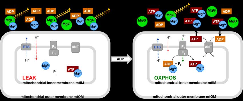
Bioenerg Commun 2021.1 doi:10.26124/bec:2021-0001
2.5. ATP production measurement with MgG
MgG (Magnesium Green™, pentapotassium salt, cell impermeant) does not
permeate biological membranes. Therefore, the plasma membrane barrier function must
be removed, as achieved in mitochondrial preparations − isolated mitochondria, tissue
homogenates, permeabilized tissues and cells. MgG remains outside of the mitochondrial
matrix and fluoresces when bound to Mg 2+. In the phosphorylation reaction
ADP + Pi ATP
reactants and MgG bind Mg 2+ according to their apparent dissociation constants. When
ADP is added to the experimental chamber, there is a fast drop of the fluorescence signal.
If mitochondria and fuel substrates are present, ATP is generated and exchanged with
ADP by the ANT. ATP has a higher affinity to Mg 2+ compared to ADP. As ATP concentration
increases in the medium, the free Mg 2+ concentration declines, less MgG is bound to Mg 2+,
and the fluorescence decreases. After calibration of the fluorescence signal in terms of
free [Mg2+], ATP concentration in the medium is calculated according to Chinopoulos et al
(2009; 2014), taking in account that: (1) the initial concentration of ATP is zero, (2) the
initial concentration of ADP is known, (3) the concentration of Mg 2+ is measured, and (4)
apparent Kd values for ADP and ATP with Mg 2+ are obtained experimentally.
The free Mg2+ concentration was calibrated in MiR05-MgG containing the
mitochondrial sample, fuel substrates, carboxyatractyloside, and oligomycin. MgCl2 was
titrated in 10 steps of 0.1 mM to obtain a non-linear fit for calibration of the amperometric
signal converted from current to voltage U (free [Mg2+] = (a × U2) + (b × U) + c)
(Chinopoulos et al 2014). After calibration, the apparent Kd (Kd’) of ADP and ATP for Mg2+
was determined for each experimental condition by performing multiple titrations with
ADP or ATP. To test for an influence of EGTA on the MgG calibration, similar calibrations
and Kd’ determination curves were performed in the absence of sample, fuel substrates,
and with varying EGTA concentrations in the range of 5 – 500 µM.
2.6. Substrate-uncoupler-inhibitor-titration (SUIT) protocols
Coupling control protocols (SUIT-006) assess different coupling control states ―
LEAK, OXPHOS and ET ― at a constant ET-pathway state (Gnaiger et al 2020). The effect
of MgG on mitochondrial respiration was evaluated by adding 1.1 µM MgG to the
experimental chamber prior to sample addition, but not to the controls which were
maintained at an identical fluorescence light intensity. Since the fluorescent dye was
diluted in water and a volume of only 2 µL was added into the 2 mL chamber, no solvent
addition was performed in the control group without MgG. After addition of mitochondria
into the O2k chambers, residual oxygen consumption Rox was measured in the absence
of substrates. Two coupling-control protocols were used to study simultaneously oxygen
consumption and ATP production with the following titrations: NADH-pathway with 5
mM pyruvate and 2 mM malate, or Succinate-pathway with 0.5 µM rotenone and 10 mM
succinate. First, LEAK respiration was measured in the absence of ADP. Second, OXPHOS
capacity was measured after addition of 2 mM ADP. Oligomycin (7.5−10.0 nM) or
carboxyatractyloside (0.3 – 0.4 mM) were added to induce again a LEAK state. Both
inhibitors have the same function in the context of the present experiments, to induce
www.bioenergetics-communications.org 5MgG and O 2 flux
LEAK respiration by inhibition of the phosphorylation system. This was followed by
stepwise titration of uncouplers up to the optimum concentration, when the maximum O 2
flux was achieved as a measure of ET capacity. CCCP (0.5 µM steps) or SF 6847 (25−50
nM steps) were used. Both protonophores have the same function. Finally, Rox was
measured after addition of the CIII inhibitor antimycin A (2.5 µM). The fluorescence signal
in the control group did not change on the scale used to analyze the MgG data, such that
corrections for changes of autofluorescence were not required.
2.7. Data analysis
The assays were repeated 3 times with independent mitochondrial preparations,
with or without MgG, for each condition tested. Data analysis for O 2 consumption,
calculations of Kd’ and ATP production following Chinopoulos et al 2014, were performed
using the templates provided with the software DatLab 7.4.
3. Results and discussion
To determine ATP production and respiration, a variation of the MiR05 medium was
prepared without EGTA nor MgCl2, which were titrated during the calibrations. EGTA is
used to chelate Ca2+ ions that could affect mitochondrial function. In addition, free Ca 2+
affects the MgG assay since this fluorophore has a higher affinity for Ca 2+ than Mg2+
(Molecular Probes 2005).
The chemicals used to prepare the media may contain contaminations. Titration of
KH2PO4 increased fluorescence of MgG, which was decreased upon addition of EGTA (not
shown). With a low concentration of EGTA (5 µM), it was not possible to chelate all
contaminants, as shown by the lower signal after titration of ADP or ATP (in the presence
of 1 mM MgCl2) compared to the signal before MgCl2 titration, thus making it impossible
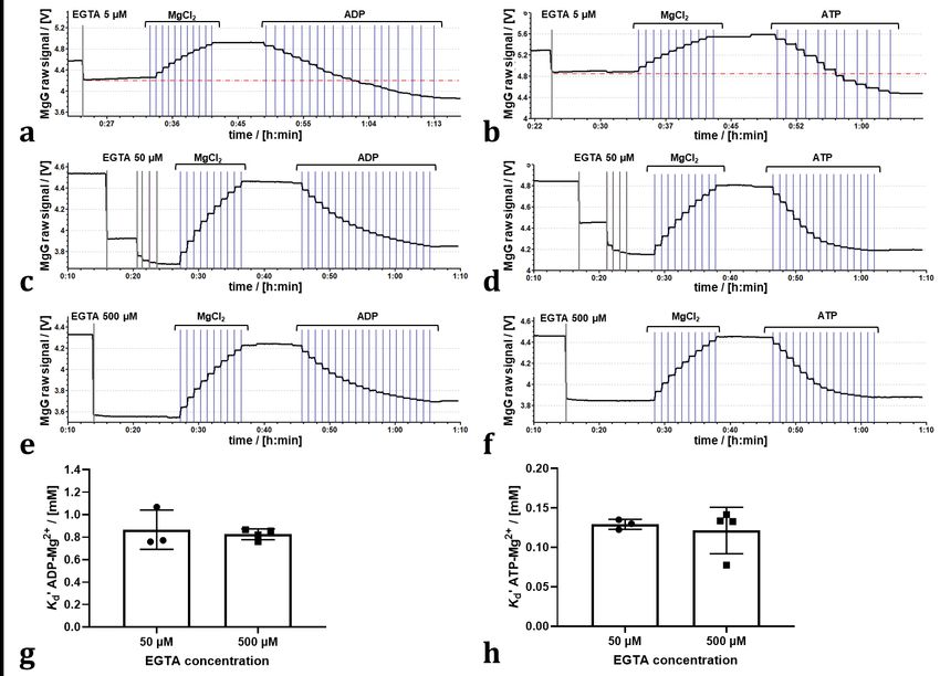
to determine the Kd’ values (Figure 1a and b). 50 µM EGTA was needed (Figure 1c and d),
with similar results at 500 µM EGTA (Figure 1e and f). The Kd’ of ADP3--Mg2+ and ATP4--
Mg2+ determined in the presence of 50 µM or 500 µM EGTA were not different (Figure 1g
and h).
Even though 500 µM EGTA partially chelates Mg 2+ in the medium, this EGTA
concentration did not interfere with the calibrations of MgG (Figure 1e and f). Therefore,
500 µM EGTA was selected to foster a higher capacity for binding contaminating cations
under experimental conditions in the presence of mitochondria. With the modified MiR05
(1 mM MgCl2), titration of 2 mM ADP leads to binding of ~ 60 % of the initial free Mg2+.
Even if 500 µM EGTA is mostly bound as EGTA-Mg2+, still 0.5 mM Mg2+ would be free
before ADP titration, and a minimum of ~ 0.19 mM would be free upon titration of 2 mM
ADP. Under our experimental conditions, the free Mg 2+ was sufficient to monitor the
further decrease in MgG fluorescence over time due to ATP production (Figures 3b and
4b). As shown by Chinopoulos et al (2009), too low concentrations of free Mg 2+ decrease
the signal-to-noise ratio. Media with 500 µM EGTA and 3 mM MgCl 2 have been used to
determine ATP production using MgG (Pham et al 2014, Devaux et al 2019). However,
with 3 mM MgCl2, the decrease in the signal upon titration of ADP or ATP is lower, such
that 1 mM MgCl2 was considered to be optimum.
6 Cardoso et al (2021) Bioenerg Commun 2021.1Bioenerg Commun 2021.1 doi:10.26124/bec:2021-0001
Figure 1. Effect of EGTA concentration on K d’ of ADP3--Mg2+ and ATP4--Mg2+:
Representative traces of calibration in MiR05 (10 titrations of 0.1 mM MgCl2 by TIP2k)
and titrations of (a, c, e) ADP (0.25 mM/titration) or (b, d, f) ATP (0.20 mM/titration), in
the presence of 5, 50 (5 titrations of 10 µM EGTA) or 500 µM EGTA. (g, h) Kd’ for ADP3--
Mg2+ or ATP4--Mg2+ determined in the presence of 50 or 500 µM EGTA. Dots represent
independent measurements; bars represent average and standard deviation. No
statistical difference was found (p > 0.05; Welch’s t-test).
Figures 3a and 4a show superimposed traces of O2 concentration and O2 flux per
mass. Coupling control of mitochondrial respiration was measured in two different
electron-transfer-pathway control states. In the N-protocol, the NADH-linked pathway
through Complex I (CI) was evaluated in the presence of pyruvate and malate which
stimulate dehydrogenases of the TCA cycle, leading to reduction of NAD + to NADH. NADH
is the substrate of CI, with further electron flow into the Q-junction, CIII and CIV (Figure
2). In the S-protocol, CI was inhibited by rotenone to prevent reverse electron transfer
and accumulation of oxaloacetate, which is an inhibitor of succinate dehydrogenase
(Makrecka-Kuka et al 2015; Gnaiger 2020), and respiration was measured supported by
succinate as the substrate of CII (Figure 3).
www.bioenergetics-communications.org 7MgG and O 2 flux
Figure 2. Simultaneous measurement of respiration and ATP production by high-
resolution FluoRespirometry in mitochondria isolated from mouse heart.
Representative traces for coupling control protocol SUIT-006 with NADH-linked
substrates (N-protocol), following additions (respiratory states): isolated mitochondria
imt (ROX), pyruvate & malate (LEAK), ADP (OXPHOS), oligomycin (LEAK), uncoupler U
(ET), and antimycin A (ROX). Experiment 2019-02-07 P5 04: (a) O2 concentration (dark
and lighter blue traces) and O2 flux per mass (dark and lighter red), 1.1 µM MgG versus
control; (b) MgG fluorescence signal; (c) ATP concentration calculated from MgG signal
calibrated as Mg2+ concentration.
In both protocols, LEAK respiration was measured (1) L(n), in the absence of
adenylates and (2) L(Omy) or L(Cat), in the presence of phosphorylation system
inhibitors. Respiration in these two LEAK states was similar, but slightly lower in L(Omy)
with the N-protocol (Figure 2a, Table 1). L(n) stabilized quickly, whereas for L(Omy)
8 Cardoso et al (2021) Bioenerg Commun 2021.1Bioenerg Commun 2021.1 doi:10.26124/bec:2021-0001 inhibition of respiration was slow at the low concentration of 7.5−10.0 nM oligomycin. In the S-protocol with sequential addition of rotenone followed by succinate, L(n) increased for a few minutes until stabilization (Figure 3a). Inhibition by carboxyatractyloside (0.3−0.4 µM) was immediate, and L(Cat) tended to be slightly lower than L(n) (Table 1). Figure 3. Simultaneous measurement of respiration and ATP production by high - resolution FluoRespirometry in mitochondria isolated from mouse heart. Representative traces for coupling control protocol SUIT-006 with succinate as substrate (S-protocol), following additions (respiratory states): isolated mitochondria imt (ROX), succinate & rotenone (LEAK), ADP (OXPHOS), carboxyatractyloside (LEAK), uncoupler U (ET), and antimycin A (ROX). Experiment 2019-03-18 P5-03: (a) O2 concentration (dark and lighter blue traces) and O2 flux per mass (dark and lighter red), 1.1 µM MgG versus control; (b) MgG fluorescence signal; (c) ATP concentration calculated from MgG signal calibrated as Mg2+ concentration. www.bioenergetics-communications.org 9
MgG and O 2 flux

Table 1. Coupling control efficiency (P-L)/P and P»/O2 ratio in absence or presence
of MgG. Average ± SD, N=3. OXPHOS capacity P and LEAK respiration L corrected for
residual oxygen consumption Rox. L(n)/L(inh) ratios: L in the absence of adenylates (n)
over L with an inhibitor (inh) of the phosphorylation system, oligomycin Omy or
carboxyatractyloside Cat for the N- or S-pathway, respectively. L(inh) is used in (P-L)/P.
 Protocol               (P-L)/P           L(n)/L(inh)         P»/O2               P»/O
 N-pathway - MgG       0.90 ± 0.01         1.13 ± 0.05           -                  -
 N-pathway + MgG       0.88 ± 0.02         1.12 ± 0.04        2.33 ± 1.07      1.16 ± 0.53
 S-pathway - MgG       0.62 ± 0.05         1.27 ± 0.16           -                  -
 S-pathway + MgG       0.61 ± 0.05         1.12 ± 0.26        2.78 ± 0.74      1.39 ± 0.37

       OXPHOS capacity P was measured in the presence of a kinetically saturating
concentration of ADP. The optimum uncoupler concentrations to measure maximum ET
capacity E were 6.0−7.0 µM CCCP in the N-protocol, and 0.150−0.175 µM SF 6847 in the
S-protocol. In the N-protocol, P was stable over time and identical to E. However, in the S-
protocol, P showed a slight decrease over time. Extrapolating this trend of declining O 2
flux to the point where ET capacity was measured explains why E appears to be lower
than P (dashed trendline, Figure 3a). In both protocols, therefore, E = P, indicating that
OXPHOS capacity was not limited by the phosphorylation system. This agrees with results
for mouse heart mitochondria on coupling control even in the combined NS-pathway
(Lemieux et al 2017). Parallel measurements were performed in the presence and
absence of 1.1 µM MgG with the N- and S-protocol. This low concentration of MgG is
sufficiently high for calculating ATP production (Figures 3b and c and Figures 4b and c).
       The MgG assay to measure ATP production can be used concomitantly with high -
resolution respirometry, providing information real-time. Other methods are available to
detect ATP production real-time. Spectrophotometric detection of NADPH can be used in
conjunction with the coupled enzyme system hexokinase and glucose-6-phosphate
dehydrogenase (Horgan, 1978). This assay has been adapted for simultaneous detection
of O2 consumption and NADPH (Lark et al 2016). The luciferin/luciferase assay can be
used for continuous measurement of ATP production (Manfredi et al 2002). It is
important to note that luciferase consumes O2, and instruments typically used for
luminometry do not allow monitoring of O2 concentration in parallel.
       Another method for continuous measurement of the P»/O 2 ratio is the steady-state
ADP injection-respirometry (Gnaiger et al 2000b; 2001). The phosphorylation rate is set
by continuous injection of ADP as the rate-limiting step while measuring O2 consumption
stimulated to a constant sub-maximal level. Chance and Williams (1955) originally
described a polarographic ADP pulse-titration method to determine the P»/O2 ratio,
titrating a known concentration of ADP, which leads to a peak of O2 consumption
stimulated by the complete phosphorylation of ADP to ATP. The ADP pulse-titration
method has been extended and critically discussed by Gnaiger (2001).
       End-point assays are available to detect ATP levels, providing discontinuous
measurement of ATP production. These include chromatography (high performance
liquid chromatography, HPLC; thin layer chromatography, TLC); nuclear magnetic

 10                                               Cardoso et al (2021) Bioenerg Commun 2021.1
Bioenerg Commun 2021.1 doi:10.26124/bec:2021-0001 resonance detection of 2-deoxyglucose and its phosphorylated form, and radioactivity measurements using 32P (Menegollo et al 2019; Morciano et al 2017; Fink et al 2017; Sausen et al 2019). Figure 4. O2 consumption in the absence and presence of MgG by mitochondria isolated from mouse heart. The respiratory rates indicated in the abscissa were measured by HRR with two coupling control protocols SUIT -006, with the following respiratory states: ROX, LEAK (in the absence of adenylates), OXPHOS, LEAK (in the presence of inhibitors), ET, and ROX. Sequential titrations are described for (a) N- protocol (experiments 2019-02-05 P3-04, 2019-02-06 P3-03 and 2019-02-07 P5-04) and (b) S-protocol (experiments 2019-03-13 P6-03, 2019-03-14 P3-03 and 2019-03-18 P5- 03). For both graphs the three symbol shapes show independent mitochondrial preparations (prep.), whereas open and closed symbols compare results in controls and in the presence of MgG from the same preparation; bars represent the average. www.bioenergetics-communications.org 11
MgG and O 2 flux

Figure 5. Chemical background signal with MgG. MiR05 (without MgCl2) in the
absence of sample, and titrations as indicated in the figure. Note the increase in
fluorescence upon titration of (a) succinate and (b) pyruvate, despite dilution of the MgG
by 1 % and 0.25% respectively. Titration of mitochondria isolated from mo use heart (imt)
leads to change in optical properties and decreases the signal in the (a, b) presence and
(c) absence of MgG. Calibration with MgCl2 [mM].

     The fluorometric MgG assay applied simultaneously with O 2 consumption by HRR
has been used extensively (Iftikar, Hickey 2013; Goo et al 2013; Chinopoulos et al 2014;
Pham et al 2014; Power et al 2014; Salin et al 2016; Napa et al 2017; Masson et al 2017;

 12                                              Cardoso et al (2021) Bioenerg Commun 2021.1
Bioenerg Commun 2021.1 doi:10.26124/bec:2021-0001
Salin et al 2018; Devaux et al 2019; Salin et al 2019). Understanding whether MgG may
affect respiration is crucial for such studies, particularly for P»/O 2 ratios obtained in
different electron-transfer-pathway states.
It is well established that different dyes commonly applied to measure
mitochondrial membrane potential inhibit OXPHOS capacity, e.g., safranin, rhodamine
123 and its derivatives TMRM and TMRE (Krumschnabel et al 2014; Scaduto, Grotyohann
1999). Surprisingly, Amplex UltraRed used to detect H 2O2 flux impairs respiration despite
not accumulating in the mitochondria (Makrecka-Kuka et al 2015). Therefore, we studied
the effect of MgG on respiration. MgG at 1.1 µM did not affect NADH-linked nor succinate-
linked respiration in any coupling control state (LEAK, OXPHOS and ET) measured in
mitochondria isolated from mouse hearts (Figure 4). In addition, residual oxygen
consumption was not affected by MgG. Similar controls should be applied in studies of
mitochondria from other species and tissues or cells, and under different experimental
conditions including media with different composition and different MgG concentrations.
Addition of succinate and pyruvate in the absence of sample resulted in a chemical
background effect (Figure 5a and b). A similar increase of fluorescence was seen after
titration of succinate or pyruvate in the presence of isolated mitochondria (Figures 4b
and 3b), which is therefore explained by the chemical background. This enforces the
recommendation of obtaining the Kd’ for ATP4--Mg2+ and ADP3--Mg2+ in the presence of
the substrates used in the reaction media, as discussed previously (Chinopoulos et al
2014). The addition of sample leads to a decrease in the amperometric signal both in the
chambers with and without MgG (Figure 5a, b and c), indicating that this effect is at least
partly due to shadowing/blocking the light. The intensity of this signal change varies, as
well as the initial MgG signal might vary. Therefore, it is advisable to perform a calibration
with each sample tested under the same experimental conditions (same batches of media,
chemicals and MgG, in the same instrumental chamber used).
The NADH-pathway has three coupling sites, CI, CIII and CIV, whereas the succinate-
pathway has only the latter two, resulting in a lower P»/O 2 ratio. When dividing ATP flux,
calculated from the increase in ATP concentration over time, by the simultaneously
measured O2 flux, then P»/O2 flux ratios (JP»/JO2) are obtained. The P»/O2 is twice the
classical P»/O (Table 1). P»/O2 obtained for the S-pathway was close to the theoretically
expected value (Gnaiger et al 2020). The result obtained for the N-pathway was lower
than expected. A limitation of the present study is the low number of replicates (N = 3),
with a high variability of P»/O2 ratios. Further experiments are necessary to investigate
and compare P»/O2 ratios, which is beyond the aim of this technical communication.
Coupling control efficiencies are closely related to P»/O 2 ratios. The coupling
control efficiency is defined as (E-L)/E, ranging from 0, at zero coupling, to 1 in a fully
coupled system. In the present case of P = E, the coupling control efficiency is expressed
as the P-L control efficiency, (P-L)/P (Gnaiger 2020). As expected, a higher P-L control
efficiency of 0.89 ± 0.02 was found for the N-pathway than 0.62 ± 0.05 for the S-pathway
(pooled data with and without MgG, average ± standard deviation, N = 6; Table 1). These
correspond to a RCR = P/L of 9.6 ± 1.8 for the N-pathway and 2.6 ± 0.3 for the S-pathway.
www.bioenergetics-communications.org 13MgG and O 2 flux
In summary, MgG did not affect respiration in any of the coupling control states.
These results demonstrate that measurement of O 2 consumption is reliable concomitant
with the MgG assay in SUIT protocols with different pathway states and coupling states .
Acknowledgements
We thank Marco Di Marcello and Manuela Passrugger for expert technical support on
media and chemicals preparation, equipment maintenance and mitochondria isolation ,
Cristiane Cecatto for discussions on autofluorescence corrections, and the BEC reviewers
for helpful suggestions. This work was partially funded by the European Union’s Horizon
2020 research and innovation programme under grant agreement No. 859770, NextGen-
O2k project. An initiative of the MitoEAGLE Task Group of the Mitochondrial Physiology
Society.
Abbreviations
Amp amperometric; ANT adenosine nucleotide translocase; BSA bovine serum albumin;
CI to CIV Complex I to IV; CCCP carbonyl cyanide m-chlorophenyl hydrazone; ΔΨp+ mt-
membrane potential; EGTA ethylene glycol tetraacetic acid; E ET capacity; ETS electron
transfer system; FOF1 ATP synthase; Hepes N-(2-hydroxyethyl)piperazine-N′-(2-
ethanesulfonic acid); HRR high-resolution respirometry; imt isolated mitochondria; JO2 O2
flux; Kd dissociation constant; Kd’ apparent Kd; L LEAK respiration; LED light-emitting
diode; MES 2-(N-morpholino)ethanesulfonic acid hydrate; MgG Magnesium Green; P
OXPHOS capacity; P»/O ADP phosphorylated per atom oxygen consumed; P»/O 2 ADP
phosphorylated per molecular oxygen consumed; Pi inorganic phosphate; RCR
respiratory acceptor control ratio; Rox residual oxygen consumption; SUIT substrate-
uncoupler -inhibitor-titration; TCA tricarboxylic acid; TMRM tetramethylrhodamine
methyl ester; TMRE tetramethylrhodamine ethyl ester; TPP + tetraphenylphosphonium;
Tris 2-amino-2-(hydroxymethyl)-1,3-propanediol; U uncoupler.
References
Budinger GRS, Duranteau J, Chandel NS, Schumacker PT (1998) Hibernation during hypoxia in
cardiomyocytes. Role of mitochondria as the O 2 sensor. J Biol Chem 273:3320-6.
Chance B, Williams GR (1955) Respiratory enzymes in oxidative phosphorylation. I. Kinetics of oxygen
utilization. J Biol Chem 217:383-93.
Chinopoulos C, Vajda S, Csanady L, Mandi M, Mathe K, Adam-Vizi V (2009) A Novel Kinetic Assay of
Mitochondrial ATP-ADP Exchange Rate Mediated by the ANT. Biophys J 96:2490-504.
Chinopoulos C, Kiss G, Kawamata H, Starkov AA (2014) Measurement of ADP-ATP exchange in relation to
mitochondrial transmembrane potential and oxygen consumption. Methods Enzymol 542:333-48.
Devaux JBL, Hedges CP, Birch N, Herbert N, Renshaw GMC, Hickey AJR (2019) Acidosis maintains the
function of brain mitochondria in hypoxia-tolerant triplefin fish: a strategy to survive acute hypoxic
exposure? Front Physiol 9:1941.
Doerrier C, Garcia-Souza LF, Krumschnabel G, Wohlfarter Y, Mészáros AT, Gnaiger E (2018) High-
Resolution FluoRespirometry and OXPHOS protocols for human cells, permeabilized fibers from small
biopsies of muscle, and isolated mitochondria. Methods Mol Biol 1782:31-70.
Fink BD, Bai F, Yu L, Sivitz WI (2017) Regulation of ATP production: dependence on calcium concentrati on
and respiratory state. Am J Physiol Cell Physiol 313:C146-53.
Fontana M, Krumschnabel G (2015) Isolation of mouse heart mitochondria. Mitochondr Physiol Network
20.06(01):1-2.
14 Cardoso et al (2021) Bioenerg Commun 2021.1Bioenerg Commun 2021.1 doi:10.26124/bec:2021-0001
Fontana-Ayoub M, Fasching M, Gnaiger E (2016) Selected media and chemicals for respirometry with
mitochondrial preparations. Mitochondr Physiol Network 03.02(18):1-10.
Gnaiger E, Wyss M (1994) Chemical forces in the cell: Calculation for the ATP system. In: What is Controlling
Life? (Gnaiger E, Gellerich FN, Wyss M, eds) Modern Trends in BioThermoKinetics 3. Innsbruck Univ
Press:207-12.
Gnaiger E (2001) Bioenergetics at low oxygen: dependence of respiration and phosphorylation on oxygen
and adenosine diphosphate supply. Respir Physiol 128:277-97.
Gnaiger E (2020) Mitochondrial pathways and respiratory control. An introduction to OXPHOS analysis. 5th
ed. Bioenerg Commun 2020.2: 112 pp. doi:10.26124/bec:2020-0002.
Gnaiger E et al ― MitoEAGLE Task Group (2020) Mitochondrial physiology. Bioenerg Commun 2020.1.
doi:10.26124/bec:2020-0001.v1.
Gnaiger E, Kuznetsov AV, Schneeberger S, Seiler R, Brandacher G, Steurer W, Margreiter R (2000a)
Mitochondria in the cold. In: Life in the Cold (Heldmaier G, Klingenspor M, eds) Springer, Heidelberg,
Berlin, New York:431-42.
Gnaiger E, Méndez G, Hand SC (2000b) High phosphorylation efficiency and depression of uncoupled
respiration in mitochondria under hypoxia. Proc Natl Acad Sci U S A 97:11080-5.
Goo S, Pham T, Han JC, Nielsen P, Taberner A, Hickey A, Loiselle D (2013) Multiscale measurement of cardiac
energetics. Clin Exp Pharmacol Physiol 40:671-81.
Horgan DJ (1978) A spectrophotometric assay of ATP synthesized by sarcoplasmic reticulum. Aust J Biol Sci
31:21-4.
Iftikar FI, Hickey AJ (2013) Do mitochondria limit hot fish hearts? Understanding the role of mitochondrial
function with heat stress in Notolabrus celidotus. PLoS One 8:e64120.
Krumschnabel G, Eigentler A, Fasching M, Gnaiger E (2014) Use of safranin for the assessment of
mitochondrial membrane potential by high-resolution respirometry and fluorometry. Methods Enzymol
542:163-81.
Lark DS, Torres MJ, Lin CT, Ryan TE, Anderson EJ, Neufer PD (2016) Direct real-time quantification of
mitochondrial oxidative phosphorylation efficiency in permeabilized skeletal muscle myofibers. Am J
Physiol Cell Physiol 311:C239-45.
Lemieux H, Blier PU, Gnaiger E (2017) Remodeling pathway control of mitochondrial respiratory capacity
by temperature in mouse heart: electron flow through the Q-junction in permeabilized fibers. Sci Rep
7:2840, DOI:10.1038/s41598-017-02789-8.
Leyssens A, Nowicky AV, Patterson L, Crompton M, Duchen MR (1996) The relationship between
mitochondrial state, ATP hydrolysis, [Mg2 +] i and [Ca2+] i studied in isolated rat cardiomyocytes. J Physiol
496:111-28.
Lowry OH, Rosebrough NJ, Farr AL, Randall RJ (1951) Protein measurement with the Folin phenol reagent.
J Biol Chem 193:265-275.
Makrecka-Kuka M, Krumschnabel G, Gnaiger E (2015) High-resolution respirometry for simultaneous
measurement of oxygen and hydrogen peroxide fluxes in permeabilized cells, tissue homogenate and
isolated mitochondria. Biomolecules 5:1319-38.
Manfredi G, Yang L, Gajewski CD, Mattiazzi M (2002) Measurements of ATP in mammalian cells. Methods
26:317-26.
Masson SWC, Hedges CP, Devaux JBL, James CS, Hickey AJR (2017) Mitochondrial glycerol 3-phosphate
facilitates bumblebee pre-flight thermogenesis. Sci Rep 7:13107.
Menegollo M, Tessari I, Bubacco L, Szabadkai G (2019) Determination of ATP, ADP, and AMP Levels by
Reversed-Phase High-Performance Liquid Chromatography in Cultured Cells. Methods Mol Biol
1925:223-32.
Molecular Probes. Fluorescent Magnesium Indicators. Revised: 05–May–2005. Available online at:
https://assets.thermofisher.com/TFS-Assets/LSG/manuals/mp01290.pdf
Morciano G, Sarti AC, Marchi S, Missiroli S, Falzoni S, Raffaghello L, Pistoia V, Giorgi C, Di Virgilio F, Pinton P
(2017) Use of luciferase probes to measure ATP in living cells and animals. Nat Protoc 12:1542-62.
Napa K, Baeder AC, Witt JE, Rayburn ST, Miller MG, Dallon BW, Gibbs JL, Wilcox SH, Winden DR, Smith JH,
Reynolds PR, Bikman BT (2017) LPS from P. gingivalis negatively alters gingival cell mitochondrial
bioenergetics. Int J Dent 2017:2697210.
www.bioenergetics-communications.org 15MgG and O 2 flux
Pham T, Loiselle D, Power A, Hickey AJ (2014) Mitochondrial inefficiencies and anoxic ATP hydrolysis
capacities in diabetic rat heart. Am J Physiol 307:C499–507.
Power A, Pearson N, Pham T, Cheung C, Phillips A, Hickey A (2014) Uncoupling of oxidative phosphorylation
and ATP synthase reversal within the hyperthermic heart. Physiol Rep pii:e12138.
Salin K, Villasevil EM, Auer SK, Anderson GJ, Selman C, Metcalfe NB, Chinopoulos C (2016) Simultaneous
measurement of mitochondrial respiration and ATP production in tissue homogenates and calculation
of effective P/O ratios. Physiol Rep 10.14814/phy2.13007.
Salin K, Villasevil EM, Anderson GJ, Selman C, Chinopoulos C, Metcalfe NB (2018) The RCR and ATP/O
indices can give contradictory messages about mitochondrial efficiency. Integr Comp Biol 58:486-94.
Salin K, Villasevil EM, Anderson GJ, Lamarre SG, Melanson CA, McCarthy I, Selman C, Metcalfe NB (2019)
Differences in mitochondrial efficiency explain individual variation in growth performance. Proc Biol Sc i
286:20191466.
Sausen CW, Rogers CM, Bochman ML (2019) Thin-Layer Chromatography and Real-Time Coupled Assays
to Measure ATP Hydrolysis. Methods Mol Biol 1999:245-253.
Scaduto RC Jr, Grotyohann LW (1999) Measurement of mitochondrial membrane potential usi ng
fluorescent rhodamine derivatives. Biophys J 76:469-77.
Copyright © 2021 The authors. This Open Access peer-reviewed communication
is distributed under the terms of the Creative Commons Attribution License,
which permits unrestricted use, distribution, and reproduction in any medium,
provided the original authors and source are credited. © remains with the
authors, who have granted BEC an Open Access publication license in perpetuity.
16 Cardoso et al (2021) Bioenerg Commun 2021.1You can also read