Targeting ADT-Induced Activation of the E3 Ubiquitin Ligase Siah2 to Delay the Occurrence of Castration-Resistant Prostate Cancer
←
→
Page content transcription
If your browser does not render page correctly, please read the page content below
ORIGINAL RESEARCH
                                                                                                                                                published: 16 April 2021
                                                                                                                                        doi: 10.3389/fonc.2021.637040

                                              Targeting ADT-Induced Activation
                                              of the E3 Ubiquitin Ligase Siah2 to
                                              Delay the Occurrence of Castration-
                                              Resistant Prostate Cancer
                           Edited by:
                Matthew K. Summers,           Tingmang Yan 1†‡, Dapeng Zhou 1‡, Youwei Shi 1, Di Cui 1, Juntao Jiang 1, Bangmin Han 1,
             The Ohio State University,       Shujie Xia 1, Zhou Wang 2,3,4,5, Haitao Liu 1*, Wenhuan Guo 6* and Yifeng Jing 1*
                        United States
                                              1 Department of Urology, Shanghai General Hospital, Shanghai Jiao Tong University School of Medicine, Shanghai, China,
                          Reviewed by:
                                              2 Department of Urology, University of Pittsburgh School of Medicine, Pittsburgh, PA, United States, 3 Department of
                      Marco A. Calzado,
          University of Cordoba, Spain        Pharmacology and Chemical Biology, University of Pittsburgh School of Medicine, Pittsburgh, PA, United States,
                                              4 Department of Pathology, University of Pittsburgh School of Medicine, Pittsburgh, PA, United States, 5 UPMC Hillman
                         Feng Liu-Smith,
         University of Tennessee Health       Cancer Center, University of Pittsburgh School of Medicine, Pittsburgh, PA, United States, 6 Pathology Center, Shanghai
              Science Center (UTHSC),         General Hospital/Faculty of Basic Medicine, Shanghai Jiao Tong University School of Medicine, Shanghai, China
                           United States

                  *Correspondence:            Siah2 is an E3 ubiquitin ligase that targets androgen receptor (AR) and plays an important role
                           Yifeng Jing
                    jyf_123@163.com
                                              in the development of castration-resistant prostate cancer (CRPC). However, the regulation of
                        Wenhuan Guo           Siah2 in prostate cancer (PCa) is largely unknown. In this study, we used AR-dependent and
              wenhuan1130@sina.com
                                              -independent cells lines to investigate the cellular roles of AR and androgen deprivation
                            Haitao Liu
                doctorlht69@163.com           therapy (ADT) on Siah2 protein levels and E3 ligase activity using Western blotting and co-
                    †
                  Present address:            immunoprecipitation. We also validated our findings using patient samples taken before and
                      Tingmang Yan,           after ADT. Finally, we used xenograft tumor models to test the effects of ADT combined with
             Department of Urology,
 Hua Shan Hospital, Fudan University
                                              vitamin K3 (Vit K3) on tumor growth in vivo. Our results showed that AR stabilizes Siah2
 School of Medicine, Shanghai, China          protein by attenuating its self-ubiquitination and auto-degradation, likely by blocking its E3
     ‡
      These authors have contributed          ubiquitin ligase activity. Conversely, ADT decreased Siah2 protein expression but enhanced
                 equally to this work
                                              its E3 ligase activity in PCa cells. Notably, the findings that ADT decreasing Siah2 protein
                                              expression were verified in a series of paired PCa samples from the same patient. Additionally,
                     Specialty section:
           This article was submitted to      we found that ADT-induced Siah2 activation could be abolished by Vit K3. Strikingly, ADT
                        Cancer Molecular      combined with Vit K3 treatment delayed the occurrence of CRPC and dramatically inhibited
             Targets and Therapeutics,
                 a section of the journal     the growth of tumor xenografts compared with ADT treatment alone. AR is an inhibitor of
                   Frontiers in Oncology      Siah2 in PCa, and ADT leads to the continuous activation of Siah2, which may contribute to
         Received: 02 December 2020           CRPC. Finally, ADT+Vit K3 may be a potential approach to delay the occurrence of CRPC.
            Accepted: 26 March 2021
             Published: 16 April 2021         Keywords: castration-resistant prostate cancer, androgen receptor, androgen deprivation therapy, Siah2, E3
                                              ubiquitin ligase
                            Citation:
Yan T, Zhou D, Shi Y, Cui D, Jiang J,
Han B, Xia S, Wang Z, Liu H, Guo W
   and Jing Y (2021) Targeting ADT-
         Induced Activation of the E3
                                              INTRODUCTION
     Ubiquitin Ligase Siah2 to Delay
       the Occurrence of Castration-
                                              Prostate cancer (PCa) is the most commonly diagnosed malignancy and the second leading cause of
           Resistant Prostate Cancer.         cancer-related mortality in American men, with an estimated 174,650 new cases and 31,620 deaths
            Front. Oncol. 11:637040.          expected in 2020 (1). Notably, PCa has also become the most common male urogenital malignancy in
    doi: 10.3389/fonc.2021.637040             China (2). Androgen deprivation therapy (ADT) remains the first-line therapy for men with metastatic

Frontiers in Oncology | www.frontiersin.org                                          1                                         April 2021 | Volume 11 | Article 637040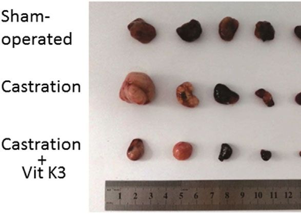
Yan et al.                                                                                                Androgen Deprivation Therapy Activates Siah2

PCa; however, most patients ultimately relapse with castration-              5% antibiotics, and 1% L-glutamine at 37°C with 5% CO2. Cells
resistant prostate cancer (CRPC), which is currently incurable and           were verified as mycoplasma free using PCR. Cells were cultured
accounts for most PCa-associated mortalities (3). Although the               in phenol red-free medium supplied with 5% dextran-coated
mechanisms of CRPC remain unclear, accumulating evidence has                 charcoal-stripped fetal bovine serum (CS-FBS) for 24 h before
shown that androgen receptor (AR) signaling is central to CRPC               treatment with dihydrotestosterone (Sigma-Aldrich, St Louis,
progression (4, 5). Several mechanisms have been suggested to                MO, USA). In some experiments, cells were treated with the
mediate androgen-independent AR signaling, including AR                      protein synthesis inhibitor cycloheximide (Sigma-Aldrich) at 50
mutations, AR gene amplifications or overexpression, expression               mg/ml and/or the proteasome inhibitor MG132 (Sigma-Aldrich)
of specific AR splice variants, intratumoral androgen production,             at 5 mM for various times as described in the figure legends. Cell
and abnormal post-translation modification of AR, such as                     transfection was separately performed with Lipofectamine 2000
phosphorylation, methylation, acetylation, ubiquitination, and               (Invitrogen, Carlsbad, CA, USA) for constructs or siRNAs
SUMOylation (6).                                                             according to the manufacturer’s protocols. The siRNA targeting
    Siah2 is a RING finger type ubiquitin ligase comprising a catalytic       AR was purchased from Thermo Fisher Scientific (Waltham, MA,
RING domain, two zinc fingers, and a C-terminal substrate-binding             USA). The siRNAs targeting Siah2 were as follows: Siah2-1, 5′-
domain (SBD) (7). Many proteins have been identified as substrates            UAUGACUUGCUUUCCUAGGCAAUCCAC-3′; Siah2-2, 5′-
of Siah2, including N-CoR, PHD, Sprouty2, b-catenin, and AR (8–              CCUCCCAUUCCUAACACACUGAUCUAU-3′.
12). Several studies have shown that Siah2 has important roles in
tumorigenesis and metastasis in multiple cancers, including breast           Western Blot Analysis
cancer, lung cancer, pancreatic cancer, melanoma, and PCa (13).
                                                                             and Immunoprecipitation
Importantly, Siah2 has been identified as an E3 ubiquitin ligase of
                                                                             Western blot and immunoprecipitation assays were performed
AR that specifically targets a selective pool of NCOR1-bound,
                                                                             as previously described (14). Primary antibodies against Siah2
repressed AR chromatin complexes for degradation. These
                                                                             (NBP1-19648, Novus Biologicals, Littleton, CO, USA, 1:1000),
complexes are typically involved in lipid metabolism, cell motility,
                                                                             AR (sc-816, Santa Cruz Biotechnology, Dallas, TX, USA, 1:1000),
and proliferation in PCa cells. Additionally, Siah2 is required for
                                                                             Sprouty2 (sc-30049, Santa Cruz Biotechnology, 1:1000), Flag M2
CRPC tumor growth in mice, whereas Siah2 deletion increases the
                                                                             (F1804, Sigma-Aldrich, 1:2000), Myc (MMS-150, Covance,
castration sensitivity of TRAMP mice (12). Thus, Siah2 is a critical
                                                                             Princeton, NJ, USA, 1:2000), HA (MMS-101P, Covance,
player in CRPC development.
                                                                             1:2000), and Tubulin (abs131993, Absin Bioscience, Shanghai,
    Given its role in CRPC, it is important to know how Siah2 is
                                                                             China, 1:5000) were used in the study.
regulated in PCa. We and others have previously reported that
Siah2 is regulated by proteins, such as DHX15 and AKR1C3 in                  In Vivo Ubiquitination Assay
PCa (14, 15). However, the clinical relevance of these studies still         HEK293 cells were transfected with Flag-AR, GFP-AR, and HA-
needs further verification. In this study, we show that AR is a               ubiquitin as indicated for 24 h, and then were treated with 5 mM
substrate of Siah2 that can inhibit Siah2 self-ubiquitination,               MG132 for 16 h before harvest. Cells were lysed in 100 ml RIPA
stabilize Siah2 expression, and decrease its E3 ubiquitin ligase             buffer with 1% SDS to disrupt protein–protein interactions, and
activity in PCa cells. Additionally, ADT significantly reduced                then boiled for 10 min at 95°C. The lysates were diluted 10-fold
Siah2 expression and enhanced its ligase activity. Notably, these            with RIPA buffer and immunoprecipitated with Flag M2 gel for
findings are closely related to clinical PCa samples. Importantly,            3 h followed by incubation with protein A/G Plus-Agarose for
treatment with the specific Siah2 inhibitor, vitamin K3 (Vit K3),             3 h. After three washes, the immunoprecipitates were subjected
delayed LNCaP tumor progression to castration resistance in                  to Western blot analysis.
LNCaP tumors. Therefore, Vit K3 might be an adjuvant that can
be combined with ADT to treat advanced PCa and delay CRPC.                   RT-PCR and Real-Time PCR
                                                                             RT-PCR and real-time PCR were performed as described
                                                                             previously (14). The primer sequences used were as follows:
                                                                             AR, 5′-TGGATGGATAGCTACTCCGG-3′ and 5′-CCCAGA
MATERIALS AND METHODS                                                        AGCTTCATCTCCAC-3′; GAPDH, 5′-CGACCACTTTGT
                                                                             CAAGCTCA-3′ and 5′-AGGGGAGATTCAGTGTGGTG-3′;
Plasmid Constructs                                                           and Siah2, 5′-AGGTTGCCCTCTGCCGATA-3′ and 5′-
All plasmid constructs were created or obtained as previously                ACATAGGTGAGTGGCCAAATCTC-3′.
described (14).
                                                                             Immunohistochemistry
Cell Culture and Transfection                                                Formalin-fixed paraffin-embedded (FFPE) PCa specimens were
The human PCa cell lines LNCaP, 22Rv1, and PC3, and HEK293                   obtained from the surgical pathology archives of Shanghai General
cells were obtained from American Type Culture Collection                    Hospital. Use of these prostate tissues was approved by the Shanghai
(ATCC, Manassas, VA, USA). Cells were maintained in the                      General Hospital Review Board. Immunohistochemistry was
appropriate medium (RPMI-1640 for LNCaP, PC3, and 22Rv1                      performed as previously described (14) using an anti-Siah2
and DMEM for HEK293) supplied with 10% fetal bovine serum,                   antibody (NB110-88113 [24E6H3], Novus Biologicals, 1:500).

Frontiers in Oncology | www.frontiersin.org                              2                                    April 2021 | Volume 11 | Article 637040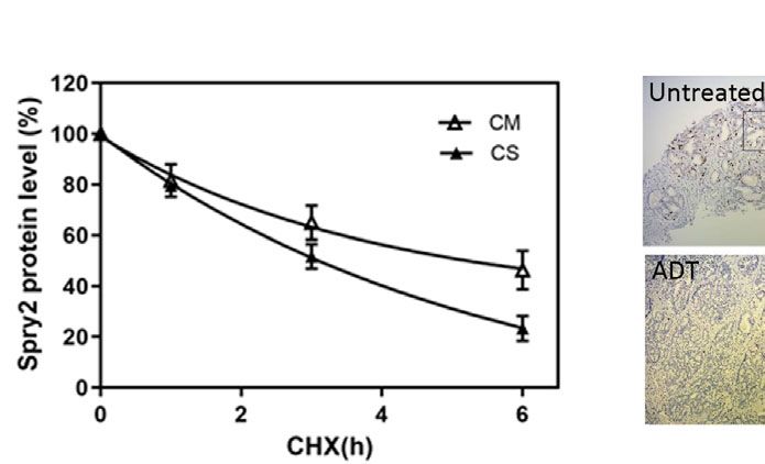
Yan et al.                                                                                           Androgen Deprivation Therapy Activates Siah2

Transwell Migration and BrdU                                            Both siRNAs and flutamide decreased Siah2 protein expression
Incorporation Assays                                                    (Figures 1E, F). Consistently, DHT treatment increases Siah2
The transwell migration and BrdU incorporation assays were              protein level in AR-positive LNCaP cells but not in AR-negative
performed as previously described (14).                                 PC3 cells (Figure S1B). Additionally, co-transfection of HA-
                                                                        Siah2 with different amounts of Flag-AR into HEK293 cells
Animals and Xenograft Tumors                                            showed that Siah2 protein levels increased with increasing
Male athymic BALB/c nude mice (5–6 weeks old) purchased                 amount of exogenous AR expression (Figure 1G). Finally, we
from the Animal Center of the Chinese Academy of Sciences               found AR increased the expression of Wild-type Siah2 (Siah2-
(Shanghai, China) were subcutaneously injected in one flank              WT) but not RING mutant Siah2 (Siah2-RM, which lacks
with 300 ml of LNCaP cells (1 × 10⁶) mixed 1:1 (v:v) with               ubiquitin ligase activity), suggesting that AR stabilizes Siah2 by
Matrigel (Invitrogen). Tumors were measured with calipers twice         inhibiting its ubiquitin ligase activity (Figure 1H). Together,
per week. Tumor volumes were calculated using the formula               these findings demonstrate that AR stabilizes Siah2 protein in
length × width 2 × 0.52. Mice were randomized into three groups         PCa cells.
once the tumor volume reached 0.6 mm3: the sham-operated
(n=5), castration (n=5), and castration+Vit K3(Sigma-Aldrich)           AR Inhibits Siah2 Self-Ubiquitination and
injection groups (n=5). For the latter, a dose of 10 mg/kg Vit K3       Decreases Its E3 Ligase Activity
(dissolved in DMSO at a final concentration of 0.1%) was                 Like other RING finger E3 ubiquitin ligases, Siah2 limits its own
administered via twice weekly intra-peritoneal injections.              expression by self-ubiquitination and auto-degradation, which is a
Tumor growth was monitored twice per week for 7 weeks, at               sign of its ubiquitin-ligase activity. To test the effect of AR
which point the mice were sacrificed and tumors were harvested           stabilization on Siah2 E3 ligase activity, we monitored Siah2-
for Western blot analyses. All animal studies were conducted in         mediated degradation of Sprouty2 (Spry2), one of the classic
accordance with the Shanghai Jiao Tong University Medical               substrates of Siah2 and a marker for Siah2 ligase activity.
School’s Animal Committee guidelines.                                   Overexpressing wide-type Siah2 effectively reduced Spry2 half-
                                                                        life from approximately 5 to 3 h, while co-expression of AR
Statistical Analysis                                                    prolonged Spry2 half-life to approximately 4 h (Figures 2A, B).
Data are presented as mean ± standard error (SEM) or mean ±             RING mutant Siah2 alone or co-expression with AR did not
standard deviation (SD). Statistical analyses were performed with       change the half-life of Spry2 (Figure S2A), which indicated AR
Student’s t-test or one-way ANOVA. P < 0.05 was considered              inhibits Siah2 E3 ligase activity. We next assessed the effect of DHT
statistically significant.                                               on endogenous Spy2 and PHD3, another classic substrate of Siah2.
                                                                        Treatment with 10 nM DHT for 24 h resulted in significantly
                                                                        increased Spry2 and Siah2 levels in both LNCaP and 22Rv1 cells
RESULTS                                                                 (Figure 2C). As expected, DHT treatment increased PHD3
                                                                        expression as well (Figure 2D). These observations suggest that
Androgens Stabilized Siah2 Protein                                      Siah2 ligase activity was inhibited in the presence of AR.
in PCa Cells                                                                We next determined whether AR stabilized Siah2 protein by
Due to the pivotal role of Siah2 in the development of CRPC,            inhibiting its self-ubiquitination. Therefore, we co-expressed
here we asked how Siah2 was regulated during ADT. Therefore,            Flag-Siah2-WT or Flag-Siah2-RM, HA-Ub, and GFP-AR in
we first assessed whether Siah2 expression was altered following         PC3 cells and treated the cells with the proteasome inhibitor
the treatment of PCa cells with androgens. Notably, treatment of        MG132 for 6 h. We then immunoprecipitated Flag-Siah2 using
LNCaP cells with 10 nM dihydrotestosterone (DHT) for 24 h led           anti-Flag M2 beads and performed Western blot analysis with an
to a significant increase in Siah2 protein expression (Figure 1A).       anti-HA antibody to detect Flag-Siah2 ubiquitination levels. As
We next determined the expression of Siah2 in response to DHT           expected, Siah2-WT ubiquitination was decreased in the
in another AR-positive PCa cell line, 22Rv1. Treating 22Rv1 cells       presence of AR, while ubiquitination of Siah2-RM was not
with DHT also dramatically increased Siah2 expression                   changed (Figure 2E). Siah2 comprises three different domains:
(Figure 1A). Furthermore, DHT effect on Siah2 showed a                  N-terminal domain, central RING domain/zinc finger domain,
dose-dependent manner (Figure S1A). Nevertheless, DHT did               and C-terminal SBD. To map the Siah2 domains required for this
not alter Siah2 mRNA levels in either LNCaP or 22Rv1 cells,             AR interaction, we generated truncation mutants of Siah2 (14)
which suggests that DHT regulates Siah2 expression at the post-         and co-transfected them individually with AR into HEK293 cells.
transcriptional level (Figure 1B). To determine whether                 These co-immunoprecipitation assays revealed that both the
androgens altered Siah2 stability, we monitored the half-life of        SBD and the central RING domain/zinc finger domains were
endogenous Siah2 in the presence of the protein synthesis               required for the interaction with AR (Figure 2F).
inhibitor cycloheximide; 10 nM DHT prolonged the half-life of
Siah2 from approximately 1.5 to 5 h in both LNCaP and 22Rv1             ADT Decreased Siah2 Expression and
cells (Figures 1C, D). To further address the role of AR in DHT-        Increased Its Ligase Activity
mediated Siah2 stabilization in PCa cells, siRNAs specifically           Given that AR stabilized Siah2 protein and inhibited its E3 ligase
targeting AR or the antagonist flutamide were used to block AR.          activity, we hypothesized that ADT may increase Siah2 activity in

Frontiers in Oncology | www.frontiersin.org                         3                                     April 2021 | Volume 11 | Article 637040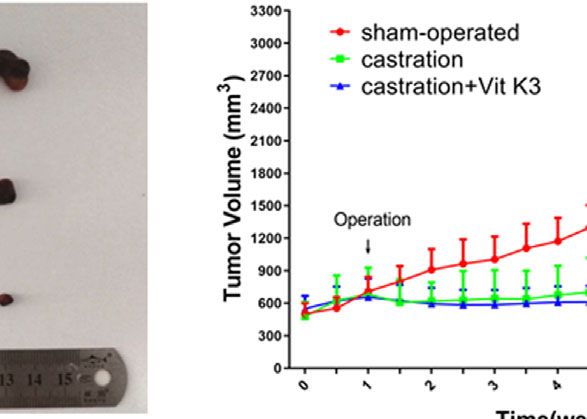
Yan et al.                                                                                                            Androgen Deprivation Therapy Activates Siah2

                         A                                      B

                         C

                         D

                         E                                                           F

                          G                                                          H

 FIGURE 1 | Androgens stabilized Siah2 protein levels. (A) LNCaP and 22Rv1 cells were cultured in CS-FBS medium for 24 h, and then treated with 10 nM DHT or
 vehicle for 24 h. AR and Siah2 protein expression were detected using Western blotting. (B) LNCaP and 22Rv1 cells were treated as described in A, and then AR
 and Siah2 mRNA expression were detected by qPCR. (C) LNCaP and 22Rv1 cells were treated as described in A, and then treated with cycloheximide (CHX, 50
 mg/ml) for 1, 3, and 6 h, after which cell lysates were subjected to Western blotting. (D) Degradation curves of Siah2 by cycloheximide chase experiments in the
 presence or absence of DHT. (E) LNCaP and 22Rv1 cells were treated as described in A, and then treated with flutamide (5 mM) for 24 h. AR and Siah2 protein
 expression were detected using Western blotting. (F) LNCaP and 22Rv1 cells were transfected with two different siRNAs targeting Siah2 or control for 72 h, and
 then subjected to Western blotting to detect Siah2 and AR expression. (G) HEK293 cells were transfected with HA-Siah2 and different doses of Flag-AR for 48 h.
 Cell lysates then were subjected to Western blotting and probed with anti-Flag and anti-HA antibodies. (H) 293 cells were transfected with Flag-AR and HA-Siah2WT
 or HA-Siah2RM for 48 h, cell lysates were subjected to Western blotting and probed with anti-Flag and anti-HA antibodies. NS, non-significant.

PCa. To test the effect of ADT on Siah2 in PCa cells, LNCaP or                      compared with those in complete medium, which was
22Rv1 cells were cultured in medium supplemented with 5% CS-                        consistent with AR expression (Figure 3A). This result
FBS (to mimic ADT conditions) or in complete medium. As                             suggested that ADT treatment decreased Siah2 expression
expected, both Siah2 and Spry2 expressions were significantly                        while enhancing its E3 ligase activity. To further confirm the
decreased when the cells were cultured in CS-FBS medium                             effect of ADT on Siah2 activity, we monitored endogenous Spry2

Frontiers in Oncology | www.frontiersin.org                                     4                                          April 2021 | Volume 11 | Article 637040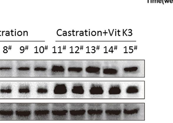
Yan et al. Androgen Deprivation Therapy Activates Siah2
A
B C D
E F
FIGURE 2 | AR inhibited Siah2 auto-ubiquitination and decreased its E3 ligase activity. (A) HEK293 cells were transfected with Flag-Siah2, GFP-AR, and Myc-Spry2
as indicated for 48 h, and then treated with cycloheximide (CHX, 50 mg/ml) for 1, 3, and 6 h. Cell lysates were then subjected to Western blotting. (B) Degradation
curves of Spry2 by cycloheximide chase experiments in the presence or absence of Siah2 or AR. (C) LNCaP and 22Rv1 cells were cultured in CS-FBS medium for
24 h, and subsequently treated with 10 nM DHT or vehicle for 24 h. Spry2, Siah2, and AR protein expression were detected by Western blotting. (D) LNCaP cells
were cultured in CS for 24 h, subsequently treated with 10 nM DHT or vehicle for 24 h. Siah2and PHD3 protein expression were detected by Western blotting.
(E) PC3 cells were transfected with Flag-Siah2-WT or Flag-Siah2-RM, GFP-AR and HA-Ub as indicated for 24 h, and treated with 5 mM MG132 for 16 h. Cell lysates
were immunoprecipitated with Flag antibody and subjected to Western blotting with HA and Flag antibody. (F) GFP-AR was co-transfected with Flag-Siah2
fragments (N, N-terminal region; M-middle region; C, C-terminal region) into HEK293 cells. Cell lysates were immunoprecipitated with Flag antibody and subjected to
Western blotting with Flag and GFP antibodies.
and Siah2 expression in LNCaP cells using the cycloheximide months. Siah2 was detected immunohistochemically both in
chase assay. The Spry2 half-life was reduced from approximately biopsy samples (before ADT) and in radical prostatectomy
4.5 h in cells cultured in complete medium to 3 h when cells samples (after ADT) from the same patient. Siah2 showed a
cultured in CS-FBS medium (Figures 3B, C), which suggests nuclear expression pattern as described previously (14). Notably,
increased Siah2 activity. Siah2 staining was significantly reduced in all patients after ADT
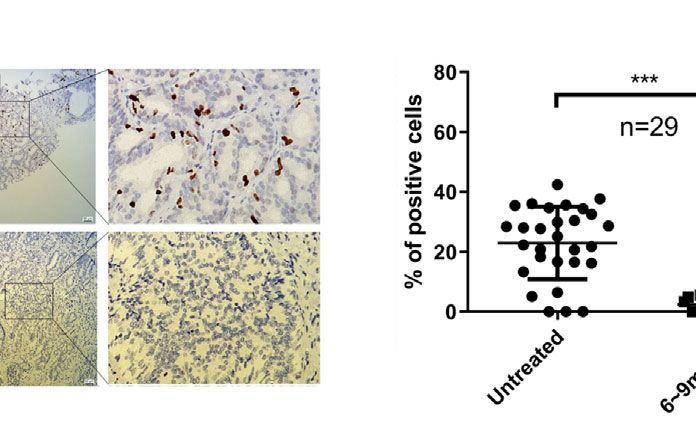
To assess the relevance of our findings in human PCa, we (Figures 3D, E), which was consistent with our in vitro findings.
evaluated Siah2 protein expression in 29 PCa patients who These data suggest that ADT decreases Siah2 protein expression
underwent radical prostatectomy after ADT treatment for 6–9 and enhances its E3 ligase activity in PCa.
Frontiers in Oncology | www.frontiersin.org 5 April 2021 | Volume 11 | Article 637040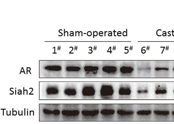
Yan et al.                                                                                                                   Androgen Deprivation Therapy Activates Siah2

                        A                                                  B

                        C                                             D                                         E

 FIGURE 3 | ADT decreased Siah2 expression and increased its ligase activity. (A) LNCaP and 22Rv1 cells were cultured in complete medium for 24 h, and then
 maintained in CS-FBS medium for another 24 h. Spry2, Siah2, and AR protein expression were detected by Western blotting. (B) LNCaP cells were treated as described in
 (A), and then treated with cycloheximide (CHX, 50mg/ml) for 1, 3, and 6 h, after which cell lysates were subjected to Western blotting. (C) Degradation curves of Siah2 by
 cycloheximide chase experiments. (D) Representative images of Siah2 immunohistochemistry staining in PCa specimens from the same patient. (E) Quantification of Siah2
 immunohistochemistry staining in PCa samples from 29 patients who underwent radical prostatectomy after between 6 and 9 months of ADT treatment. ***P < 0.001.

Vit K3 Attenuated the Effects of ADT on                                                  K3 inhibits the growth and motility of PCa cells through a mechanism
Siah2 and Inhibited the Growth and                                                       that involves inhibition of Siah2 activity.
Motility of PCa Cells
Given that Siah2 plays a key role in the development of CRPC (12)                        ADT Combined With Vit K3 Therapy
and that ADT triggers its ligase activation, we next asked whether                       Delayed the Formation of CRPC
Siah2 could be blocked when PCa cells were treated by ADT. The                           Having established that Vit K3 abolished the Siah2 activation
only Siah2-targeting drug described so far is Vit K3 (also known as                      triggered by ADT in PCa cells in vitro, we next sought to
menadione), which was identified as a specific inhibitor of Siah2                          determine whether this mechanism was active in vivo. Therefore,
ubiquitin ligase activity in a screen of U.S. Food and Drug                              we subcutaneously injected LNCaP cells into nude mice and
Administration-approved therapeutic drugs (16). We next verified                          monitored tumor development. Both castration and castration+Vit
whether Vit K3 could attenuate the effect of ADT on Siah2. As shown                      K3 treatment significantly reduced tumor volumes compared with
in Figure 4A, Siah2 and Spry2 expression were decreased in LNCaP                         the sham-treatment group. However, in the castration alone group,
and 22Rv1 cells cultured in CS-FBS medium compared with those in                         tumor volumes started to increase 3 weeks after castration and grew
complete medium, as described previously. Significantly, Vit K3                           extremely fast 4 weeks later, which suggests the formation of CRPC.
treatment increased Siah2 and Spry2 expression in CS-FBS medium                          Strikingly, tumors treated with castration+Vit K3 only grew very
(Figure 4A), which indicates that Vit K3 inhibited Siah2 activity.                       slowly 4 weeks after treatment and still did not show fast growth after
    We next evaluated the physiologic significance of Vit K3 on PCa                       8 weeks (Figures 5A, B). Consistently, tumors derived from the Vit
cells. Treatment with 20 mM Vit K3 for 24 h significantly reduced                         K3 treatment group were much smaller than those from the other
proliferation of AR-positive LNCaP and 22Rv1 cells, but not of AR-                       two groups. Analysis of these tumors revealed significantly decreased
negative PC3 cells (Figure 4B). Given that the effect of Siah2 on PCa                    Siah2 expression in response to castration, whereas Vit K3 treatment
cell proliferation was AR-dependent, we argue that the inhibitory                        dramatically reversed expression Siah2, which was consistent with
effect of Vit K3 on PCa cell growth is Siah2- and AR-dependent. To                       our in vitro findings (Figure 5C). These results strongly suggest that
further verify the role of Siah2, siRNAs targeting Siah2 were                            ADT+Vit K3 treatment may delay CRPC formation.
transfected into LNCaP and 22Rv1 cells. Silencing Siah2 and Vit
K3 treatment independently resulted in reduced PCa cell proliferation,
but Vit K3 treatment in Siah2-knockdown cells did not further reduce                     DISCUSSION
cell proliferation (Figures 4C, D). Similarly, LNCaP cell motility was
inhibited following Vit K3 treatment or Siah2 silencing, but no further                  It is well established that the androgen-AR signaling axis plays a
changes were observed in Siah2-knockdown cells treated with Vit K3                       central role in CRPC. Most of the new drugs for CRPC approved by
(Figures 4E, F). Collectively, these data support a model in which Vit                   the FDA in recent years target AR signaling, including Abiraterone

Frontiers in Oncology | www.frontiersin.org                                          6                                            April 2021 | Volume 11 | Article 637040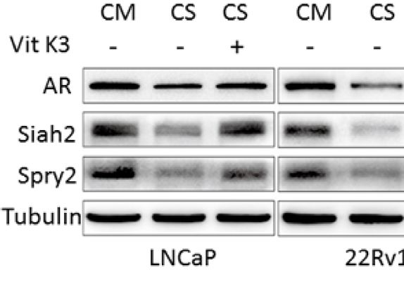
Yan et al.                                                                                                                Androgen Deprivation Therapy Activates Siah2

                          A                                                               B

                          C                                                                D

                          E                                                                   F

 FIGURE 4 | Vit K3 attenuated the effects of ADT on Siah2. (A) LNCaP and 22Rv1 cells were cultured in CS-FBS medium for 24 h, and then treated with Vit K3
 (20 mM) for 24 h. Spry2, Siah2, and AR protein expression were detected by Western blotting. (B) LNCaP, 22Rv1, and PC3 cells were treated with Vit K3 (20 mM)
 for 24 h, and then treated with 10 mM BrdU for 5 h (LNCaP) or 2 h (22Rv1, PC3, and DU145). The BrdU assay was performed as described in the Materials and
 Methods. (C) LNCaP and 22Rv1 cells were transfected with siRNAs targeting Siah2 or control as indicated for 72 h, and then treated with Vit K3 (20 mM) or vehicle
 as indicated for 24 h. BrdU assays were performed as described in (B). (D) The expression of Siah2 protein from cells treated as described for C was detected by
 Western blotting. (E) LNCaP cells were transfected with si-Siah2 or si-control as indicated, and then cultured in transwell chambers, as described in the Materials
 and Methods. Cells were then treated with Vit K3 (20 mM) or vehicle for 24 h, and subsequently were stained with crystal violet, after which the number of cells per
 field was quantified. (F) The expression of Siah2 protein from cells treated as described for E was detected by Western blotting. *P < 0.05; **P < 0.01.

Acetate, an androgen synthesis inhibitor, and Enzalutamide and                         increased Siah2 transcription may not be the primary mechanism
Apalutamide, second-generation AR antagonists. Although these                          underlying the increased Siah2 activity observed in PCa tissues
second-generation antiandrogens show an overall survival benefit                        (15). Siah2 is very unstable because of self-ubiquitination and
for advanced PCa, 20% to 40% of patients still do not respond to                       auto-degradation. Like other Ring finger E3 ubiquitin ligases, the
these therapies, and among the patients that do respond, resistance                    ligase activity of Siah2 is reflected by its protein stability. Several
will eventually occur (17–19). Thus, the mechanisms of castration                      factors, including the deubiquitinating enzymes USP13, AKR1C3,
resistance need to be fully elucidated, as this will identify additional               and DHX15 have been reported to stabilize Siah2 expression, by
targets to treat and/or prevent CRPC.                                                  inhibiting its E3 ligase activity (14, 15, 20).
    Siah2 is an E3 ubiquitin ligase that targets AR and is thought to                      Here, we report that AR significantly stabilizes Siah2 protein
play an important role in the development of CRPC by targeting a                       expression and decreases its ligase activity in PCa cells. Notably,
select pool of chromatin-bound ARs that control the growth,                            androgens extended the half-life of endogenous Siah2 to 5 h, which
survival, and tumorigenic capacity of PCa cells, especially under                      is much longer than the half-life in the presence of any of the other
conditions of androgen deprivation (12). Interestingly, only small                     factors that have been reported to stabilize Siah2 (15, 20). These
sets of metastatic PCa or CRPC showed a moderate 1.5- to 2-fold                        findings suggest that AR is a strong inhibitor of Siah2 ligase activity
increase in Siah2 mRNA (12) These observations suggest that                            in PCa cells. As expected, ADT decreased Siah2 expression and

Frontiers in Oncology | www.frontiersin.org                                        7                                           April 2021 | Volume 11 | Article 637040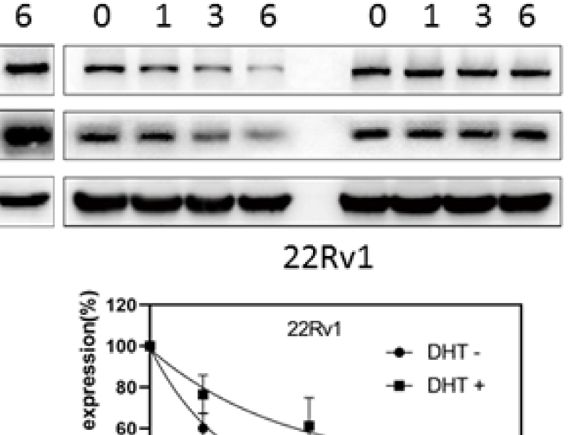
Yan et al.                                                                                                            Androgen Deprivation Therapy Activates Siah2

                          A                                                           B

                          C

 FIGURE 5 | ADT combined with Vit K3 therapy delayed the formation of CRPC. (A) LNCaP cells were subcutaneously injected into BALB/c nude mice, and the
 animals were treated as described in the Materials and Methods. The tumor xenografts with different treatments are shown as indicated. (B) The growth curve of
 tumor xenografts. (C) Xenograft tumor tissue lysates were analyzed by Western blotting to detect AR and Siah2 expression in the sham-operated, castration, and
 castration+Vit K3 groups. *P < 0.05, ****P < 0.0001.

enhanced its ligase activity. Importantly, we tested Siah2 expression                MAPK pathway and the hypoxic response pathway independent
in clinical PCa samples from the same patients before and after                      of reactive oxygen species. In this study, we showed that Vit K3
ADT, and found remarkably reduced Siah2 expression in response                       could abolish ADT-triggered Siah2 activation in PCa cells.
to ADT. These data indicate that during ADT, Siah2 is continuously                   Interestingly, we found that Vit K3 inhibited the growth of AR-
activated in PCa. Based on these findings, we conclude that AR and                    positive LNCaP and 22Rv1 cells, but not AR-negative PC3 cells,
Siah2 potentially form a positive regulatory loop in PCa, in which                   which is consistent with its role of inhibiting Siah2 on PCa cells
Siah2 mediates the ubiquitination-proteasomal degradation of a                       (12). Additionally, Vit K3 did not inhibit the growth or migration
select pool of AR, whereas AR inhibits Siah2 ligase activity. ADT                    of Siah2-KD cells. Thus, we conclude that Vit K3 blocks PCa cell
breaks the balance of these two proteins, which results in                           proliferation and motility at least partially by inhibiting Siah2.
continuous Siah2 activation, which subsequently leads to CRPC.                           Given that Siah2 plays a pivotal role in CRPC and that ADT
    Siah2 has three likely sites for intervention—interfering with its               triggers Siah2 activation in PCa cells, we hypothesized that ADT
E3 ubiquitin ligase activity, the SBD domain, and the Siah–Siah                      combined with a Siah2 inhibitor could block or delay the
dimerization domain (13). We demonstrated that AR reduced                            occurrence of CRPC. Strikingly, we demonstrated in vivo that
Siah2 auto-ubiquitination and increased Spry2 expression, a                          ADT+Vit K3 treatment delayed the formation of CRPC and
classic Siah2 substrate, which indicates that the mechanism                          dramatically inhibited the growth of tumor xenografts compared
through which AR stabilizes Siah2 is by blocking its E3 ligase                       with ADT alone. Importantly, analyses of these tumors suggested
activity. Co-immunoprecipitation studies revealed two domains of                     that Vit K3 delayed CRPC by inhibiting Siah2 activation.
Siah2—the SBD and central RING domain/zinc finger domains—                                In summary, this study provides new insights into the regulation
interacted with AR, consistent with the results of Qi et al. (12). We                of Siah2 in PCa. AR was identified as an inhibitor of Siah2. Because
hypothesize that AR binding to the SBD is degraded as substrate,                     Siah2 is an E3 ubiquitin ligase for AR, we conclude that AR and
whereas AR binding to the central domain blocks the E3 ubiquitin                     Siah2 form a positive regulatory loop in PCa. ADT inhibits AR
ligase activity of Siah2. However, future structural studies will                    signaling, resulting in continuous Siah2 activation, which
enable better assessment of the precise effects of AR on Siah2.                      contributes to CRPC. Thus, ADT combined with Vit K3 may be
    Vit K3 is a quinone used with cancer chemotherapeutics. Vit                      a potential novel approach to delay the occurrence of CRPC.
K3 and its analogs have been showed anticancer activities in
several types of cancer including prostate cancer, breast cancer,
melanoma and liver cancer in vitro and in vivo (16, 21, 22).                         DATA AVAILABILITY STATEMENT
Although the main biological effects on cancers are attributed to its
role in the redox cycle and arylating nucleophilic substrates, Vit K3                The original contributions presented in the study are included in
has been identified as a specific inhibitor of Siah2 that inhibits                     the article/Supplementary Material. Further inquiries can be
both arms of the Siah2 downstream signaling network, the Ras/                        directed to the corresponding authors.

Frontiers in Oncology | www.frontiersin.org                                      8                                          April 2021 | Volume 11 | Article 637040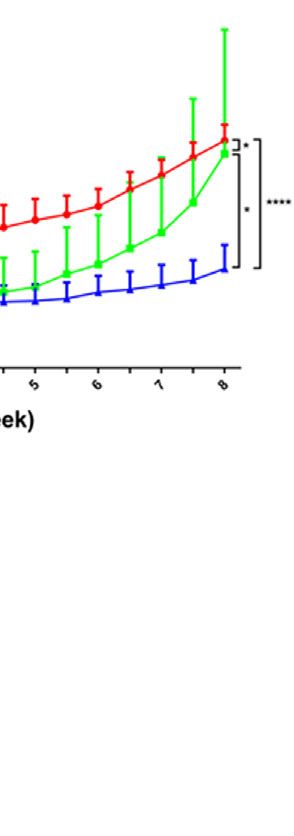
Yan et al.                                                                                                                        Androgen Deprivation Therapy Activates Siah2

AUTHOR CONTRIBUTIONS                                                                         ACKNOWLEDGMENTS
YJ: conceptualization, investigation, supervision, writing the                               We thank James P. Mahaffey, PhD, from Liwen Bianji, Edanz
original draft, reviewing, and editing the manuscript. WG:                                   Editing China (www.liwenbianji.cn/ac), for editing the English
methodology, investigation, reviewing, and editing the                                       text of a draft of this manuscript.
manuscript. TY: methodology, software, investigation, and
writing the original draft. DZ: formal analysis, software,
investigation, reviewing, and editing the manuscript. YS, DC,                                SUPPLEMENTARY MATERIAL
JJ, BH, HL, and SX: formal analysis, reviewing, and editing the
manuscript. ZW: reviewing and editing the manuscript.                                        The Supplementary Material for this article can be found online
All authors contributed to the article and approved the                                      at: https://www.frontiersin.org/articles/10.3389/fonc.2021.
submitted version.                                                                           637040/full#supplementary-material
                                                                                             Supplementary Figure 1 | Androgens stabilize Siah2 protein. (A) LNCaP cells
                                                                                             were cultured in CS for 24 h, subsequently treated with different dose of DHT or
                                                                                             flutamide (5mM) as indicated for another 24 h, and AR and Siah2 protein expression
FUNDING                                                                                      were detected by Western blotting. (B) LNCaP and PC3 cells were cultured in CS
                                                                                             for 24 h, subsequently treated with 10 nM DHT or vehicle for 24 h. AR and Siah2
This work was supported by the National Natural Science                                      protein expression were detected by Western blotting.
Foundation of China (81872098), the National Natural Science
                                                                                             Supplementary Figure 2 | AR inhibits Siah2 self-ubiquitination and decreases its
Foundation of China (81502212), and the Science and                                          E3 ligase activity. (A) 293 cells were transfected with Flag-Siah2, GFP-AR and Myc-
Technology Commission of Shanghai Municipality,                                              Spry2 as indicated for 48 h and then treated with cycloheximide (CHX, 50 mg/ml) for
China (18ZR1430600).                                                                         1, 3, 6 h and cell lysates were subjected to Western blotting.

REFERENCES                                                                                         ubiquitination of androgen receptor. Oncogene (2018) 37:638–50.
                                                                                                   doi: 10.1038/onc.2017.371
 1. Siegel RL, Miller KD, Jemal A. Cancer statistics, 2020. CA Cancer J Clin (2020)          15.   Fan L, Peng G, Hussain A, Fazli L, Guns E, Gleave M, et al. The Steroidogenic
    70:7–30. doi: 10.3322/caac.21590                                                               Enzyme AKR1C3 Regulates Stability of the Ubiquitin Ligase Siah2 in Prostate
 2. Ye D, Zhu Y. Epidemiology of prostate cancer in China: an overview and                         Cancer Cells. J Biol Chem (2015) 290:20865–79. doi: 10.1074/
    clinical implication. Chin J Surg (2015) 53:249–52.                                            jbc.M115.662155
 3. Hellerstedt BA, Pienta KJP. The cturrent state of hormonal therapy for                   16.   Shah M, Stebbins JL, Dewing A, Qi J, Pellecchia M, Ronai ZA. Inhibition of
    prostate cancer. CA Cancer J Clin (2002) 52:154–79. doi: 10.3322/                              Siah2 ubiquitin ligase by vitamin K3 (menadione) attenuates hypoxia and
    canjclin.52.3.154                                                                              MAPK signaling and blocks melanoma tumorigenesis. Pigment Cell
 4. Chan SC, Dehm SM. Constitutive activity of the androgen receptor. Adv                          Melanoma Res (2009) 22:799–808. doi: 10.1111/j.1755-148X.2009.00628.x
    Pharmacol (2014) 70:327–66. doi: 10.1016/B978-0-12-417197-8.00011-0                      17.   Ryan CJ, Smith MR, de Bono JS, Molina A, Logothetis CJ, de Souza P, et al.
 5. Kobayashi T, Inoue T, Kamba T, Ogawa O. Experimental evidence of                               Abiraterone in metastatic prostate cancer without previous chemotherapy.
     persistent androgen-receptor-dependency in castration-resistant prostate                      N Engl J Med (2013) 368:138–48. doi: 10.1056/NEJMoa1209096
     cancer. Int J Mol Sci (2013) 14:15615–35. doi: 10.3390/ijms140815615                    18.   Scher HI, Fizazi K, Saad F, Taplin M-E, Sternberg CN, Miller K, et al.
 6. Tsao CK, Galsky MD, Small AC, Yee T, Oh WK. Targeting the androgen                             Increased survival with enzalutamide in prostate cancer after chemotherapy.
    receptor signalling axis in castration-resistant prostate cancer (CRPC). BJU                   N Engl J Med (2012) 367:1187–97. doi: 10.1056/NEJMoa1207506
    Int (2012) 110:1580–8. doi: 10.1111/j.1464-410X.2012.11445.x                             19.   Fizazi K, Shore N, Tammela TL, Ulys A, Vjaters E, Polyakov S, et al.
 7. Reed JC, Ely KR. Degrading liaisons: Siah structure revealed. Nat Struct Biol                  Darolutamide in Nonmetastatic, Castration-Resistant Prostate Cancer.
    (2002) 9:8–10. doi: 10.1038/nsb0102-8                                                          N Engl J Med (2019) 380:1235–46. doi: 10.1056/NEJMoa1815671
 8. Zhang J, Guenther MG, Carthew RW, Lazar MA. Proteasomal regulation of                    20.   Scortegagna M, Subtil T, Qi J, Kim H, Zhao W, Gu W, et al. USP13 enzyme
    nuclear receptor corepressor-mediated repression. Genes Dev (1998) 12:1775–                    regulates Siah2 ligase stability and activity via noncatalytic ubiquitin-binding
    80. doi: 10.1101/gad.12.12.1775                                                                domains. J Biol Chem (2011) 286:27333–41. doi: 10.1074/jbc.M111.218214
 9. Nakayama K, Frew IJ, Hagensen M, Skals M, Habelhah H, Bhoumik A, et al.                  21.   Wellington KW, Hlatshwayo V, Kolesnikova NI, Saha ST, Kaur M, Motadi
    Siah2 regulates stability of prolyl-hydroxylases, controls HIF1alpha                           LR. Anticancer activities of vitamin K3 analogues. Invest New Drugs (2020)
    abundance, and modulates physiological responses to hypoxia. Cell (2004)                       38:378–91. doi: 10.1007/s10637-019-00855-8
    117:941–52. doi: 10.1016/j.cell.2004.06.001                                              22.   Jamison JM, Gilloteaux J, Taper HS, Summers JL. Evaluation of the in vitro
10. Nadeau RJ, Toher JL, Yang X, Kovalenko D, Friesel R. Regulation of Sprouty2                    and in vivo antitumor activities of vitamin C and K-3 combinations against
    stability by mammalian Seven-in-Absentia homolog 2. J Cell Biochem (2007)                      human prostate cancer. J Nutr (2001) 131:158–60. doi: 10.1093/jn/131.1.158S
    100:151–60. doi: 10.1002/jcb.21040
11. Matsuzawa SI, Reed JC. Siah-1, SIP, and Ebi collaborate in a novel pathway for           Conflict of Interest: The authors declare that the research was conducted in the
    beta-catenin degradation linked to p53 responses. Mol Cell (2001) 7:915–26.              absence of any commercial or financial relationships that could be construed as a
    doi: 10.1016/s1097-2765(01)00242-8                                                       potential conflict of interest.
12. Qi J, Tripathi M, Mishra R, Sahgal N, Fazli L, Ettinger S, et al. The E3 ubiquitin
    ligase Siah2 contributes to castration-resistant prostate cancer by regulation of        Copyright © 2021 Yan, Zhou, Shi, Cui, Jiang, Han, Xia, Wang, Liu, Guo and Jing.
    androgen receptor transcriptional activity. Cancer Cell (2013) 23:332–46.                This is an open-access article distributed under the terms of the Creative Commons
    doi: 10.1016/j.ccr.2013.02.016                                                           Attribution License (CC BY). The use, distribution or reproduction in other forums is
13. Wong CS, Möller A. Siah: a promising anticancer target. Cancer Res (2013)                permitted, provided the original author(s) and the copyright owner(s) are credited and
    73:2400–6. doi: 10.1158/0008-5472.CAN-12-4348                                            that the original publication in this journal is cited, in accordance with accepted
14. Jing Y, Nguyen MM, Wang D, Pascal LE, Guo W, Xu Y, et al. DHX15                          academic practice. No use, distribution or reproduction is permitted which does not
    promotes prostate cancer progression by stimulating Siah2-mediated                       comply with these terms.

Frontiers in Oncology | www.frontiersin.org                                              9                                              April 2021 | Volume 11 | Article 637040
You can also read