ISOLATION OF MOUSE EPIDERMAL KERATINOCYTES AND THEIR IN VITRO CLONOGENIC CULTURE - JOVE
←
→
Page content transcription
If your browser does not render page correctly, please read the page content below
Journal of Visualized Experiments                                                                                           www.jove.com

Video Article
Isolation of Mouse Epidermal Keratinocytes and Their In Vitro Clonogenic
Culture
                    1                 1              1                1             1                 1               1                   1
Rebecca J. Morris , Nyssa Readio , Kelsey Boland , Kelly Johnson , Sonali Lad , Anupama Singh , Ashok Singh , Stephanie Holtorf ,
                1
Samantha Skaar
1
Hormel Institute, University of Minnesota

Correspondence to: Rebecca J. Morris at morri877@umn.edu

URL: https://www.jove.com/video/58701
DOI: doi:10.3791/58701
Keywords: Biology, Issue 150, epidermal keratinocytes, cell culture, adult mouse epidermis, clonal culture, keratinocyte stem cells, 3T3 fibroblast
Date Published: 8/10/2019
Citation: Morris, R.J., Readio, N., Boland, K., Johnson, K., Lad, S., Singh, A., Singh, A., Holtorf, S., Skaar, S. Isolation of Mouse Epidermal
Keratinocytes and Their In Vitro Clonogenic Culture. J. Vis. Exp. (150), e58701, doi:10.3791/58701 (2019).

Abstract
    The protocol described here is a reliable method of harvesting primary keratinocytes from adult female mice (54 ± 2 days old) yielding
                            6
    approximately 30 x 10 viable cells per mouse. Primary adult mouse keratinocytes are harvested from the dorsal skin of female mice. Male mice
    (~6 weeks old) can be used for keratinocyte harvesting depending on the requirements of the experiment. Euthanized mice are shaved and
    sterilized with serial washes in povidone iodine and ethanol solutions (70% alcohol). After disinfecting the mice, the dorsal skin is removed and
    the subcutaneous fat and muscle are removed with a scalpel and discarded. The skins are cut into small pieces and treated with a mild, low
    temperature trypsinization to detach the lower dermis from the epidermis. The scraped epidermises are stirred at low speed, filtered to remove
    the hairs, counted, and re-suspended in culture medium. This method provides an excellent single cell suspension of highly culturable cells for
    many downstream applications.

Video Link
    The video component of this article can be found at https://www.jove.com/video/58701/

Introduction
    Mammalian skin covers the entire body and serves a multitude of functions (e.g., a primary physical barrier, temperature regulation, and moisture
    retention). Over the last five decades, basic research on mouse skin has yielded new information on the structure and function of the skin. The
    mouse skin model has greatly helped understanding of the basic biology behind the complex molecular mechanisms of chemical or ultraviolet
    radiation-induced carcinogenesis. These mouse skin models provided a significant contribution towards understanding of human skin diseases
    and their mitigation. To explore further molecular dissection of the primary keratinocytes in hair follicle developmental biology, stem cell research,
    carcinogenesis, and genetic exploration of epidermis, it is frequently desirable to isolate and culture primary epithelial keratinocytes from mice
    skin to use in parallel with in vivo experiments. The motivating factor in developing this method was the need for an in vitro assay for clonogenic
                                           1,2
    epidermal and hair follicle stem cells . There was also an urgent need for single cell suspensions of epidermal cells suitable for clonogenic
            3                                                         3,4
    assays , fluorescence activated cell sorting, and flow cytometry . The advantages of this method are a robust harvest increase an order of
                                      5,6
    magnitude over other methods , inclusion of the epithelial portion of the hair follicles, and the high culturability of the harvested cells. In the
    1980s, there were no known methods that could meet these requirements. In subsequent years, this method was refined to increase cell yield.
                                                                                                                                                 6
    The principal limitation of this method would be masking of some trypsin sensitive antibodies that would compromise immunoreactivity .

    The mice received husbandry according to the Specific Pathogen Free guidelines. One to five female mice 54 ± 2 days of age were obtained.
    In older mice, the viability of keratinocytes will be reduced due to anagen phase (growth) of the hair cycle. Also, the process of trypsinization is
    more difficult compared to young mice. The harvesting procedure has been successfully optimized for the thinner skin of female mice compared
    to male. Male mice are generally not used for keratinocyte harvests as they have a tendency to fight with one another during housing and
    therefore may leave scratches or wounds on the skin.

Protocol
    All vertebrate animal use protocols have been approved by the University of Minnesota Institutional Animal Care and Use Committee in
    accordance with recommended NIH and government guidelines.

1. Feeder Layer Initialization and Subculturing
    1. Thaw one cell vial containing frozen Swiss mouse 3T3 from the liquid nitrogen tank by placing the vial in a 37 °C water bath for 1-2 min. The
                                                          6
       number of cells in the vial is approximately 1 x 10 .

Copyright © 2019 Creative Commons Attribution-NonCommercial-NoDerivs 3.0 Unported                             August 2019 | 150 | e58701 | Page 1 of 7
License
Journal of Visualized Experiments                                                                                           www.jove.com

  2. Remove the 3T3 cell vial when the final sliver of ice has melted. Wipe the tube with a 70% alcohol swab. Then, transfer the vial to a biosafety
     cabinet.
  3. Transfer the cells slowly into a T-150 flask, and rinse the 3T3 vial with 1 mL of CGM medium. Slowly add 30 mL of pre-warmed 3T3 Complete
     Growth Medium (CGM) to the cells. Gently rock the flask to evenly spread the 3T3 fibroblast cells. Label the flask with cell line details, date,
     and passage number appropriately.
  4. Incubate the cells at 37 °C with 5% CO2 and 95% humidity.
     NOTE: According to the ATCC, the doubling time of Swiss mouse 3T3 is 18 h; when confluent (contact inhibited), the density is about 40,000
                  2
     cells per cm . We use a particular line of 3T3 within 10-15 passages after initialization. More rapid growth or the presence of spindle-shaped
     cells can occur at higher passages and is usually a sign that the 3T3 will not perform as well as feeder cells for adult mouse keratinocytes. If
     rapid growth occurs, it is better to initiate a new line from lower passage cells.
  5. Change media after 24 h to remove dead cells and remaining DMSO (cryopreservation agent), and twice weekly thereafter. Allow cells to
     proliferate around 80% confluence and use T-150 flasks for culture initiation. Use T-225 flasks for subculture after initialization.
  6. To subculture 3T3 fibroblast cells, remove the flask from the cell culture incubator and wash twice with cold sterile PBS (1x) with
     gentamycin. Pipet 10 mL of pre-warmed (37 °C water bath) trypsin solution (0.25%) into each T-225 flask.
       1. Place the flask on a warming plate for 3-8 min. Remove the flask and gently use palms to tap the walls of the flask to detach cells and
            confirm cell detachment under inverted microscope. Trypsin solution may appear cloudy with detached 3T3 cells.

  7. Pipet the trypsin solution 3–4x to wash the cell growth surface of the flask and transfer the trypsinized cell to 30 mL of CGM in a 50 mL
     conical tube. Rewash the flask 3–5x with 10 mL of the CGM-trypsin-cell mixture from the 50 mL conical tube.
     NOTE: The contents of two flask contents can be added to a 50 mL tube; if only one flask is used, then place 10 mL of trypsin/cells into 20
     mL of CGM in a 50 mL tube.
  8. Centrifuge the 50 mL conical tube at 160 x g for 7 min at 4 °C.
  9. Now aspirate the supernatant and resuspend the cell pellet with 5 mL of CGM, pipetting gently ~10x to completely disrupt the cell
     pellet. Add 10 mL of CGM to bring the total volume of the 50 mL conical tube to 15 mL.
       1. Mix again ~10x and place 5 mL of the cell suspension into 3 separate T-150 flasks. The subculture ratio is 1:3 in T-225 flasks or 1:4 in
            T-150 flasks. Add 30 mL of pre-warmed 3T3 CGM to each of the flasks.

  10. Label the flask with the cell line name, the date of cell seeding, and the passage number.
  11. To freeze down 3T3 cells following centrifugation in the subculture step, remove the 50 mL tube from the centrifuge and place on an ice
      bucket. Aspirate the supernatant and resuspend the cell pellet with 1 mL of 5% DMSO-DMEM mixture (see Table 1) for each flask harvested.
  12. Place 1 mL of DMSO-DMEM mixture with cell suspension into each cryovial (one cryovial per 3T3 flask harvested). Label the vial with the
      passage number, the cell line name (3T3), and the date that the cells were frozen.
  13. Place these 3T3 cell vials into cell freezer jar (see Table of Materials) and transfer to -70 °C freezer for >5 h. Careful remove and transfer the
      vials from the cell freezer jar and place in a liquid nitrogen storage tank for long-term storage until next use. Enter information about the cell
      line name, the location in tank and the date in the laboratory liquid nitrogen inventory sheet.

2. Preparation of 3T3 Feeder Layer for X-ray Irradiation and Seeding
  1. Prepare 3T3 cells one week before their irradiation and allow them to grow ~100% confluence. The 3T3 cells are typically within the
     passages of 120 to 130.
  2. For successful clonogenic assays of primary keratinocytes, before seeding 3T3 cells, surface coat the 60 mm Petri dishes with a
     collagen.
       1. Pipet around 2–3 mL of the collagen-coating solution into 60 mm Petri dishes to coat the bottom surface and remove the excess
           collagen-coating liquid (see Table 1). Transfer the Petri dishes to a 37 °C incubator with 5% CO2 for a minimum of 1 h. Do not let the
           dishes dry in incubator.

  3. X-ray irradiation step: Follow steps 1.4–1.6. Use 50 mL of fresh CGM to re-suspend the cell pellet, close the conical tube and seal with
     paraffin sealing film. To avoid any contamination, use plastic bags to double bag the paraffin-sealed cell containing tube and place on ice. A
                                                                                       7
     mitomycin based alternative feeder layer can be used for keratinocyte culture .
  4. Irradiate fibroblast 3T3 cells in the 50 mL conical tube with a 5,000 rads (50 Cgy) dose with a biological x-ray machine. Irradiate
     these cells along with the CGM media.
        1. Following X-ray irradiation, centrifuge cells at 160 x g for 7 min at 4 °C. Aspirate the supernatant media and gently resuspend cell pellet
            with 5 mL of CGM triturating gently ~10x. Now bring the final volume to 30 mL with additional 25 mL of medium and triturate 10x. These
            triturating steps are crucial for uniform 3T3 fibroblast layer for clonogenicity assay.

  5. Viable Cell counting: Count the keratinocytes using a regular glass hemocytometer. Take approximately 0.5 mL of the cells and place into a
     1.5 mL tube. Remove 200 µL of cell mixture and add an equal volume of 0.4% Trypan blue solution and mixture 3–4x. Transfer cells with a
     hemocytometer and count all nucleated cells (small gold and pink cells) as viable cells. Calculate the final concentration of viable gold cells.
                   6                               5
  6. Pipette 1 x 10 3T3 cells (a minimum of 7 x 10 ) cells may be used) into 60 mm collagen-coated Petri dishes (step 2.2) and gently add 4 mL
     of modified high-calcium Williams E media (see Table 1). Allow freshly seeded 3T3 cells to attach for 24 h. On the next day, harvest the fresh
     primary keratinocyte and seed them on top of attached 3T3 fibroblast layer for clonogenic assay as indicated below.

3. Primary Keratinocyte Harvesting and Seeding
  1. Euthanize four to five adult female mice via CO2 inhalation according to approved animal facility/IACUC standards. However, this
     protocol may also be used for single female/male mice.
                                   2
       1. Clip approximately 9 cm of the dorsal fur with an electric animal clipper and place all clipped mice into a 500 mL jar with enough
          povidone iodine antiseptic solution (not scrub) to cover them for 1–2 min. Shake the jar well to evenly coat the antiseptic solution over
          the mice.

Copyright © 2019 Creative Commons Attribution-NonCommercial-NoDerivs 3.0 Unported                           August 2019 | 150 | e58701 | Page 2 of 7
License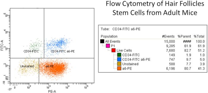
Journal of Visualized Experiments                                                                                          www.jove.com

        2. Pour off the solution and rinse with clean water until the liquid runs clear. Repeat the same antiseptic wash followed by a water rinse.
           After the antiseptic wash with iodine solution, soak all the mice in 70% ethanol for 5–10 min.
           NOTE: Mice with light fur (e.g., BALB/c) will retain a slight yellowish color from the antiseptic iodine washes, while darker mice (e.g.,
           C57BL/6) will not. Notably, cell viability or culture growth is not affected due to the yellow color.

  2. Carefully remove the clipped dorsal skin using thumb forceps and scissors in a laminar flow hood. Place the excised dorsal skin in conical
     tube filled with PBS/2x gentamycin. Avoid using ventral side skin to prevent mammary cell contamination in culture.
  3. Using autoclaved forceps and a scalpel, place one dorsal skin at a time hairy side down onto a thin Petri dish. Start scraping all
     subcutaneous tissue including fat from the ventral side of the skin tissue until it is semi-translucent. Try to remove maximum
     traces of subcutaneous tissue without tearing the skin (hair follicle part). Also, do not scrape for a long time and avoid skin drying.
        1. Keep scraped skin in PBS solution until all other remaining skins are processed. Using a scalpel, slice skins into 0.5 cm x 1–1.5 cm
            strips and place the hairy side up onto a sterile Petri dish.

  4. Carefully pipette 20 mL of PBS/2x Gentamicin solution mixed with trypsin (0.25%) into a 100 mm x 20 mm plastic Petri dish. Using
     sterile forceps, transfer the skins and float them hairy side up on the surface of trypsin solution for 2 h in a 32 °C incubator.
     NOTE: Trypsinization time and temperature are crucial for good yield of highly culturable primary keratinocytes. Although other methods may
     provide good yields of viable cells, the culturability of the keratinocytes has been less satisfactory.
       1. During this 2 h incubation time, coat dishes with collagen coating and place them at 37 °C for 1 h (see step 3.1.2). For clonogenic
            assays, the 60 mm Petri dishes have already been coated 24 h prior to keratinocyte harvesting to allow the irradiated 3T3 feeder layer
            to attach to bottom and spread evenly.
            NOTE: Coating of culture dishes for clonogenic culture is very important for proper attachment, colony formation, and ultimate growth/
            proliferation of mice epidermal keratinocytes.

  5. Epidermal scraping step: Arrange a sterile plastic square Petri dish and create a surface at a 30° incline for proper epidermal
     scraping with 15 mL of harvesting medium (see Table 1). Carefully remove a floating skin strip from the trypsin solution with curved
     forceps. With a new scalpel blade, scrape off the epidermis and hairs into the medium.
       1. Do not use excessive force, but use sufficient force to scrape the epidermis, or it will affect the cell viability.
       2. During epidermis scraping, keep the blade perpendicular to the skin piece. If the blade is angled toward the motion of the blade,
           there are more chances of skin tearing. If the blade is angled away from the blade movement, there are more chances of insufficient
           epidermis removal.

  6. Carefully pour the harvesting medium containing scraped epidermal cells into a sterile 60 mL jar (autoclavable and reusable) with a 1.5 inch
     magnetic stir bar. Rinse the Petri dish with additional harvesting medium to collect remaining epidermal cells and bring the final volume to
     30 mL. Use a magnetic stirrer and stir at 100 rpm for 20 min at room temperature. This process will remove the trapped epidermal cells from
     hairs.
  7. Place a sterile 70 µm cell strainer on the top of a 50 mL conical tube. The cell strainer fits on top of a 50 mL conical tube.
       1. Take the jar inside a biosafety hood. Remove the stir bar and pour the contents into strainer filter attached to a 50 mL conical tube.
            Strain out undesired hair and sheets of stratum corneum.
       2. Press the hairs along with stratum corneum materials within the strainer and manipulate them to release the trapped hair cells. Use
            5 mL of harvesting medium to allow remaining trapped cells to release and flow into the tube. Bring the total volume up to 50 mL in
            conical tube.

  8. Cap the 50 mL conical tube with cell filtrate and centrifuge at 160 x g for 7 min at 4 °C. Next aspirate the supernatant and add 5 mL
     of harvesting medium. Resuspend the cell pellet by triturating gently ~20x with a 5 mL pipet.
       1. At this step, keep the cells in an icebox to avoid any aggregation. Add 25 mL of harvesting medium and triturate again (20–25x).
       2. To ensure an accurate keratinocyte count at this step, take 1 mL of cell suspension and add 19 mL harvesting medium. Now the cell
           dilution is 1:20 in 50 mL conical tube. If keratinocytes are harvested from a single mouse, adjust the volume of the cell suspension.
           Instead of 30 mL, use 15 mL and make a 1:10 dilution for counting the cells.

  9. Pipet 0.5 mL of diluted cells from the 50 mL conical tube (1:20 dilution) and transfer to a 1.5 mL autoclaved microcentrifuge tube.
     Remove 0.2 mL (200 µL) of cell mix to another 1.5 mL microcentrifuge tube and add an equal volume (200 µL) of 0.4% Trypan blue
     solution. Gently mix this solution 3x and transfer cells to a hemocytometer for counting nucleated keratinocytes.
       1. Score all dark blue cells positive for Trypan blue as non-viable dead cells, and score small gold and pinkish cells as viable cells. The
           final keratinocyte yield per mouse should be around 30 million viable cells.

  10. Centrifuge the original 50 mL conical tube (from step 3.8) for 7 min at 160 x g at 4 °C. At this step, resuspend the cells in desired
      medium and make the required dilution for seeding cells.
        1. For mass culture, seed 2 to 4 million viable gold cells in each 35 mm Petri dish in cell culture medium (KGM-type medium +
            supplements for monolayer cultures) (see Table 1). For a clonogenic (colony formation) assay, seed 1,000 cells per 60 mm Petri dish
            on X-ray irradiated Swiss mouse 3T3 feeder layers with collagen-coating and modified William’s E medium with supplements and
            serum.
        2. Use a total of 2 to 4 mL medium for 35 mm and 60 mm Petri dishes, respectively. Also, formulate DMEM and Williams E medium
            procured from ATCC with reduced levels of sodium bicarbonate for use at 5% CO2.

  11. Grow the culture cells (mass or clonogenic) at 32 °C, 95% humidity with 5% CO2. Change the media one day after initial seeding for mass
      culture, and then 3x weekly thereafter. However, for clonal assays, the first medium change is 2 days after cell seeding and 3x weekly
      thereafter.
  12. For clonal culture, cultivate cells at two- and four-week intervals. After completion of clonal cultures (2 or 4 weeks), aspirate the
      medium. Fix the colonies in 10% buffered formalin overnight at room temperature and stain with 0.5% rhodamine B in autoclave
      water for 1 h. Then, remove the rhodamine B stain with a pipette from the edge of the dishes.

Copyright © 2019 Creative Commons Attribution-NonCommercial-NoDerivs 3.0 Unported                          August 2019 | 150 | e58701 | Page 3 of 7
License
Journal of Visualized Experiments www.jove.com
1. Rinse the dishes in cold autoclaved water until the dishes run clear. Put the dishes inclined on the lid of the dishes and let them dry for
final colony counting. Notably, when performing clonogenic culture assay, never pipetJournal of Visualized Experiments www.jove.com
Figure 2: Representative example of flow cytometry of hair follicle stem cells from adult mice. Briefly, the isolated primary keratinocytes
were isolated from dorsal mice skin and filtered for cellular debris using cell filter. The isolated keratinocyte suspension was labeled with CD49f
or α6-integrin (PE) and CD34 (FITC) antibodies along with live dead dye and other controls. FITC and PE isotype control antibodies (Rat
IgG2akappa) were used for compensation purpose (see Table of Materials). Stem cells were selected by means of CD49f (PE channel), which
recognized the external component of the hemi-desmosomes found on basal epidermal and hair follicle cells, and CD34 (FITC channel), which
+ +
recognized the hair follicle stem cells (i.e., CD34-FITC and a6-PE (upper right column, total 5%)). The right side column shows the different
keratinocyte populations namely CD34-FITC only, CD34-FITC and a6-PE cells (double positive stem cell population), a6-PE only, and unstained
+ 6,14
cells. Note that there are two CD34 stem cell populations as have been reported . Please click here to view a larger version of this figure.
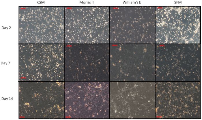
Figure 3: Comparison of four different media on primary cultures of epidermal cells from adult female mice. Primary keratinocytes were
isolated from the dorsal mice skin and counted for seeding using four different media (KGM, SFM, Willem's-E and Morris-2). Equal numbers
of keratinocytes were seeded and followed for their general morphology and growth pattern. Note that the proliferating keratinocytes have
slightly different morphologies. KGM, SFM, and Morris-2 all of which are reduced calcium media have the most robust growth after fourteen
days. In contrast, the cells do not persist when cultured in Williams E medium that has 1.4 mM calcium and a high ratio of potassium to sodium.
Surprisingly, Williams E medium with supplements and twenty percent fetal bovine serum supports keratinocyte clonal cultures far better than
any other medium tested, probably because the enriched Williams E medium helps the 3T3 feeder cells to "feed" better. Please click here to view
a larger version of this figure.
Copyright © 2019 Creative Commons Attribution-NonCommercial-NoDerivs 3.0 Unported August 2019 | 150 | e58701 | Page 5 of 7
LicenseJournal of Visualized Experiments www.jove.com Solutions and Media Comments Harvesting Solutions 2.5% trypsin (5 mL) Dulbecco’s PBS (500 mL) Gentamycin (2 mL) PBS with 2x gentamycin (45 mL) Phosphate-buffered saline (PBS) with 2x gentamycin Trypsin solution Cell Culture Solutions Purecol-fibronectin dish-coating solution: 1 M HEPES (1 mL) 116 mM CaCl2 (1 mL) Bovine serum albumin 1.0 mg/mL (10 mL) Cell culture medium (100 mL) Fibronectin (1 mg) Purecol collagen (1 mL) Cell freezing solution DMSO (2 mL) DMEM with 10% BCS and pen-strep Harvesting Medium Needs to be without calcium 2x gentamycin (1 mL) FBS (50 mL) SMEM (500 mL) High-Calcium Williams' E Media with: EGF (5 µg/mL) 1 mL Glutamine 14.5 mL Hydrocortisone (1 mg/mL) 0.5 mL Insulin 1 mL LinoAcid-BSA (0.1 mg/mL) 0.5 mL MEM Ess AminoAcids 4 mL MEM Vitamins 5 mL Penicillin-Streptomycin 5 mL Transferrin (5mg/mL) 1 mL Vit A (1 mg/1,000 µL) 57.5 µL Vit E 1 mg/mL (4°C) 3.5 µL Vit D2 (10 mg/mL) 50 µL 3T3 fibroblast complete growth medium (CGM) BCS (100 mL) DMEM (900 mL) Penicillin-streptomycin (10 mL) KGM Medium used for mass culture is a reduced calcium medium Morris 2 medium with the same supplements as Williams' E A reduced calcium variant of Williams E with supplements Table 1: Solution and media recipes. Copyright © 2019 Creative Commons Attribution-NonCommercial-NoDerivs 3.0 Unported August 2019 | 150 | e58701 | Page 6 of 7 License
Journal of Visualized Experiments                                                                                               www.jove.com

Discussion
  The keratinocyte harvesting method described here was developed to produce high yields of highly culturable primary epidermal keratinocytes
  from the back of adult female mice. These keratinocytes are suitable for flow cytometry, molecular biology application, and primary cell culture,
  including the clonogenic assay reported here. This keratinocyte harvesting method also has been useful for studying other downstream aspects
                                                              1,13
  of cutaneous chemical carcinogenesis and tumor promotion .

  There are several critical steps in the protocol. First, for rigor and reproducibility, female mice of age 54 ± 2 days were used. This has enabled
  us to perform sensitive and quantitative colony assays from multiple strains of mice and to use them as a phenotype in linkage analysis leading
                                                                  10,11
  to identification of least one new stem cell regulatory gene         . Second, it is helpful to cut the skins into smaller pieces, as the trypsinization is
  more effective than trying to keep the skin whole. Third, the time and the temperature of the trypsin flotation is very important to the subsequent
  culturability. Moreover, one needs to "poke considerably" at the hairs remaining on the filter to dislodge the attached cells. This step is important
  to obtain high yields of cells. Furthermore, the 32 °C temperature of the incubator is important for the longer-term cultivation of keratinocytes from
                                                                                                      1
  adult mice. Finally, in this protocol, the parallel readouts of in vitro and in vivo experiments are based on primary cultures rather than subcultures
  or cell lines. Notably, if harvesting primary keratinocytes for the first time using this protocol, keep the remaining dermis after scraping to confirm
  the full removal of hair follicles along with epidermis by histopathological observation.

  The principal strength of this protocol for harvesting adult mouse keratinocytes is that it produces high yields of culturable cells that are
  suitable for many downstream applications. The chief limitation is that some cell surface epitopes may be sensitive to trypsinization such that
                                                                                                                                       6                5
  immunostaining may be compromised. Therefore, when testing a new cell surface antibody, it may be prudent to use a dispase or thermolysin
  based method for harvesting for comparison. The significance of this protocol is that it has been rigorously tested, quantified, and applied to such
                                                            1,9,10,11                                   8                                        3,4
  diverse applications as assays for clonogenic stem cells           , for mass cultures in biochemistry , for FACS sorting in stem cell analysis , for
                    3,12
  molecular biology , and for ongoing RNA and DNA sequencing.

Disclosures
  The authors have nothing to disclose.

Acknowledgments
  The authors have no acknowledgments.

References

  1. Morris, R. J., Tacker, K. C., Fischer, S. M., Slaga, T. J. Quantitation of primary in vitro clonogenic keratinocytes from normal adult murine
      epidermis, following initiation, and during promotion of epidermal tumors. Cancer Research. 48 (22), 6285-6290 (1988).
  2. Tani, H., Morris, R. J., Kaur, P. Enrichment for murine keratinocyte stem cells based on cell surface phenotype. Proceedings of the National
      Academy of Sciences of the United States of America. 97 (20), 10960-10965 (2000).
  3. Morris, R. J. et al. Capturing and profiling adult hair follicle stem cells. Nature Biotechnology. 22 (4), 411-417 (2004).
  4. Trempus, C. S. et al. Enrichment for living murine keratinocytes from the hair follicle bulge with the cell surface marker CD34. Journal of
      Investigative Dermatology. 120 (4), 501-511 (2003).
  5. Redvers, R. P.. Kaur, P. Serial cultivation of primary adult murine keratinocytes. Methods in Molecular Biology. 289, 15-22 (2005).
  6. Jensen, K. B., Driskell, R. R.. Watt, F. M. Assaying proliferation and differentiation capacity of stem cells using disaggregated adult mouse
      epidermis. Nature Protocols. 5 (5), 898-911 (2010).
  7. Blacker, K. L., Williams, M. L.. Goldyne, M. Mitomycin C-treated 3T3 fibroblasts used as feeder layers for human keratinocyte culture retain
      the capacity to generate eicosanoids. Journal of Investigative Dermatology. 89 (6), 536-539 (1987).
  8. Morris, R. J. Procedure for harvesting epidermal cells from the dorsal epidermis of adult mice for primary cell culture in “high calcium” defined
      medium. Cambridge University Press. (1994).
  9. Wu, W. Y.. Morris, R. J. Method for the harvest and assay of in vitro clonogenic keratinocytes stem cells from mice. Methods in Molecular
      Biology. 289 79-86 (2005).
  10. Popova, N. V., Teti, K. A., Wu, K. Q.. Morris, R. J. Identification of two keratinocyte stem cell regulatory loci implicated in skin carcinogenesis.
      Carcinogenesis. 24 (3), 417-425 (2003).
  11. Popova, N. V., Tryson, K. A., Wu, K. Q.. Morris, R. J. Evidence that the keratinocyte colony number is genetically controlled. Experimental
      Dermatology. 11 (6), 503-508 (2002).
  12. Trempus, C. S. et al. Comprehensive microarray transcriptome profiling of CD34-enriched mouse keratinocyte stem cells. Journal of
      Investigative Dermatology. 127 (12), 2904-2907 (2007).
  13. Baer-Dubowska, W., Morris, R. J., Gill, R. D.. DiGiovanni, J. Distribution of covalent DNA adducts in mouse epidermal subpopulations after
      topical application of benzo(a)pyrene and 7,12-dimethylbenz(a)anthracene. Cancer Research. 50 (10), 3048-3054 (1990).
  14. Blanpain, C., Lowry, W. E., Geoghegan, A., Polak, L.. Fuchs, E. Self-renewal, multipotency, and the existence of two cell populations within
      an epithelial stem cell niche. Cell. 118 (5), 635-648 (2004).

Copyright © 2019 Creative Commons Attribution-NonCommercial-NoDerivs 3.0 Unported                              August 2019 | 150 | e58701 | Page 7 of 7
License
You can also read