Adipose tissue derived stem cells modulate immune function in vivo and promote long term hematopoiesis - Spandidos Publications
←
→
Page content transcription
If your browser does not render page correctly, please read the page content below
EXPERIMENTAL AND THERAPEUTIC MEDICINE 19: 1725-1732, 2020

                Adipose tissue‑derived stem cells modulate immune
               function in vivo and promote long‑term hematopoiesis
                          in vitro using the aGVHD model
                MING JIANG1,2*, XIAOJUAN BI2*, XIANLIN DUAN1, NANNAN PANG1, HONGBO WANG2,
                                HAILONG YUAN1, RONGYAO ZHANG1 and LEI CUI1,3

                      1
                       Hematologic Disease Center, The First Affiliated Hospital of Xinjiang Medical University,
           Xinjiang Uygur Autonomous Region Research Institute of Hematology; 2State Key Laboratory of Pathogenesis,
                  Prevention and Treatment of High Incidence Diseases in Central Asia, Clinical Medicine Institute,
         The First Affiliated Hospital of Xinjiang Medical University, Urumqi, Xinjiang Uygur Autonomous Region 830054;
                                3
                                  School of Medicine, Tongji University, Shanghai 200092, P.R. China

                                     Received February 9, 2018; Accepted November 6, 2019

                                                   DOI: 10.3892/etm.2020.8430

Abstract. The present study was designed to investigate the         reconstitution in aGVHD rats. ADSCs decreased aGVHD
effect of adipose‑derived stem cells (ADSCs) on acute graft vs.     severity by immunomodulation. ADSCs support the prolif-
host disease (aGVHD) and hematopoietic recovery after               eration of hematopoietic stem/progenitor cells in vitro. The
allogeneic hematopoietic stem cell transplantation. ADSCs,          present study demonstrated that ADSCs may reduce aGVHD
bone marrow‑derived stem cells (BMSCs) and fibroblasts              by influencing the balance of IL‑4 and INF‑γ and can promote
were cultured. ADSCs were cocultured with hematopoietic             long‑term hematopoiesis.
stem/progenitor cells. Then, ADSCs were infused into the
aGVHD rat model. The survival of the rats was recorded.             Introduction
Livers and small intestines were obtained from sacrificed rats
for pathological examinations. Expression of the Sry gene in        Acute graft vs. host disease (aGVHD) is the most common
recipient rats that survived longer than 21 days was examined by    complication after allogeneic hematopoietic stem cell trans-
real‑time PCR to detect the presence of donor Y chromosome.         plantation (allo‑HSCT). It is reported that the incidence
Expression of serum interferon (INF)‑γ and interleukin (IL)‑4       of aGVHD has reached 32±3% in several transplantation
was detected by ELISA at 0, 7, 14, 21 and 50 days after trans-      centers (1), and severe aGVHD is the most prevalent cause of
plantation. Transplantation of ADSCs improved the survival of       death after transplantation. The first choice of treatment for
aGVHD rats. Survived ADSCs participated in hematopoietic            aGVHD is steroids. When poor clinical outcome is shown,
                                                                    a combination of steroids with immunosuppressive drugs is
                                                                    recommended. Given that some aGVHD cases persist even
                                                                    after treatment with this combination, novel approaches to
Correspondence       to: Professor Ming Jiang, Hematologic          overcome aGVHD have been explored in recent years.
Disease Center, The First Affiliated Hospital of Xinjiang Medical       Mesenchymal stem cells (MSCs) are multipotent cells that
University, Xinjiang Uygur Autonomous Region Research Institute     can self‑renew and differentiate into various somatic lineages.
of Hematology, 137 Liyushan Road, Urumqi, Xinjiang Uygur            MSCs have also been reported to regulate immunological reac-
Autonomous Region 830054, P.R. China                                tions by secreting soluble cytokines and/or by direct contact
E‑mail: jiangmingyy@126.com                                         with lymphocytes. MSCs were first identified and isolated from
                                                                    bone marrow (2), and were subsequently confirmed to exist in
*
    Contributed equally                                             a variety of tissues such as adipose, muscle, tendon, umbilical
                                                                    cord blood and amniotic fluid. Le Blanc et al (3) reported that
Abbreviations: ADSCs, adipose‑derived stem cells; aGVHD,            infusion of bone marrow‑derived MSCs (BMSCs) systemi-
acute graft vs. host disease; BMSCs, bone marrow‑derived stem
                                                                    cally attenuated aGVHD in a mouse model, indicating the
cells; MSCs, mesenchymal stem cells; SD, Sprague‑Dawley;
BMMNCs, bone marrow mononuclear cells; HSCs, hematopoietic
                                                                    therapeutic potential of MSCs in amelioration of aGVHD.
stem cells; IL‑4, interleukin 4; INF‑γ, interferon‑γ                    Adipose‑derived mesenchymal stem cells (ADSCs), which
                                                                    were first identified by Zuk et al in 2001 (4), share similar
Key words: adipose‑derived stem cells, hematopoiesis support,       biological characteristics and immunological phenotype with
immunomodulation, acute graft vs. host disease, bone marrow stem    BMSCs. It is believed that ADSCs confer more advantages
cells                                                               in terms of proliferation and cause reduced damages than
                                                                    BMSCs (5). The present study was designed to ascertain
                                                                    whether ADSCs alleviate the incidence and severity of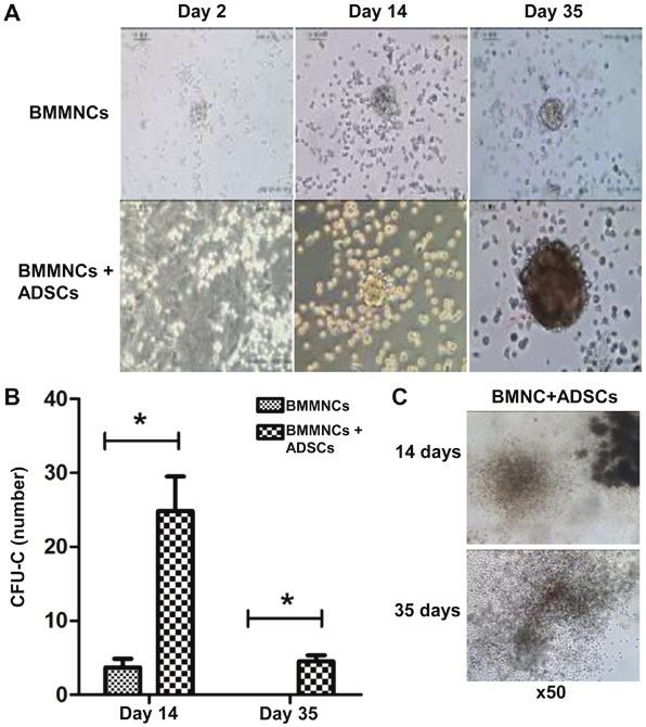
1726                                  JIANG et al: ADSCs MODULATE HEMOPOIESIS IN aGVHD

aGVHD in a rat model. Hemopoiesis after treatment with             (cat. #119307; Biolegend), HCAM‑FITC (cat. #203906;
ADSCs was also observed.                                           Biolegend), CD106 ‑PE (cat. #20 0403; Biolegend),
                                                                   CD49‑d‑FITC (cat. #200103; Biolegend), and CD29‑PE
Materials and methods                                              (cat. #102207; Biolegend). At 4˚C, the sample was incubated for
                                                                   30 min in the dark before flow cytometry using the CytoFLEX
Animals. Specific‑pathogen‑free Sprague‑Dawley (SD) and            V2‑B4‑R0 Flow Cytometer (C02944; Beckman Coulter).
Wistar rats (n=10 for each type of rat) were provided by the       Then, EXPOTM32 MultiCOMP Software (Beckman Coulter,
Animal Center, Xinjiang Medical University, China [license         Inc.) was used for data analysis. Adipogenic and osteogenic
no. SCXK (Xin) 2003‑001]. The study was approved by the            differentiation of cells was identified by Oil red staining and
Ethics Committee of The First Affiliated Hospital of Xinjiang      Alizarin red staining, respectively. Fibroblasts were obtained
Medical University, China (lot no. 20080701017). The rats          from rat dermis and cultured according to previously described
were housed in a specific‑pathogen‑free laboratory as approved     methods (7).
by the US Association for Assessment and Accreditation of              For Oil red staining, ADSCs and BMSCs in logarithmic
Laboratory Animal Care (AAALAC; https://www.aaalac.org).           growth were mixed with low‑glucose DMEM containing 10%
Donor rats were male SD rats and recipient rats were female        FBS, 0.1 µmol/l dexamethasone, 200 µmol/l indometacin,
Wistar rats aged 6‑8 weeks and weighing 180‑210 g. Before          and 0.5 mmol/l 3‑isobutyl‑1‑methylxanthine. After culture
sacrifice, all rats were given acidified water containing eryth-   for 8‑10 days, transparent lipid droplets appeared, and the
romycin (250 mg/l) and gentamicin (pH 3.0‑3.5) for bowel           cells were fixed with 4% paraformaldehyde before washing
cleansing. Animal experiments were conducted in the Animal         with PBS twice. Then, the cells were stained with Oil red for
Experimental Center of Clinical Research Institute of the First    10 min before observation.
Affiliated Hospital of Xinjiang Medical University. All animal         For Alizarin red staining, ADSCs and BMSCs in
experiments were performed following the US Guidelines for         logarithmic growth were mixed with low‑glucose DMEM
the Use and Management of Laboratory Animals (6). After            osteogenic induction medium containing 10% FBS, 0.1 µmol/l
intraperitoneal anesthesia with 10% chloral hydrate at a dose      dexamethasone, 50 µmol/l ascorbic acid, and 10 mmol/l
of 300 mg/kg body weight, the animals were sacrificed by           sodium β‑glycerophosphate. After culture for 15 days, black
neck dislocation after losing consciousness. No rats developed     sediments appeared, and the cells were fixed with 4% para-
peritonitis due to the use of chloral hydrate. Then, tissues and   formaldehyde before washing with PBS twice. Then, the cells
blood samples were obtained.                                       were stained with 1% Alizarin red (pH 4.2) for 3 min.

Culture and identification of ADSCs, BMSCs and fibroblasts.        Coculture of ADSCs with hematopoietic stem/progenitor cells.
After intraperitoneal anesthesia with 10% chloral hydrate at       Bone marrow mononuclear cells (BMMNCs) were obtained by
a dose of 300 mg/kg body weight, the rats were sacrificed by       density gradient centrifugation using rat lymphocyte separa-
neck dislocation. Then, the rats were soaked in 75% ethanol for    tion medium (1.083 g/cm3). After centrifugation, mononuclear
15 min. Bilateral inguinal skin was cut, bilateral inguinal fat,   cells were resuspended and centrifuged at 290 x g for 5 min.
femur and tibia were isolated and obtained, and the required       Then, the cells were collected and cocultured with ADSCs,
cells were obtained according to the experimental method           BMSCs or fibroblasts, respectively, which had been treated
described below.                                                   with mitomycin C (0.5 µg/ml) for 24 h. The colony‑forming
    Bilateral inguinal fat was aseptically obtained, washed        ability of non‑adherent mononuclear cells cocultured with
with phosphate‑buffered saline (PBS, pH 7.4) and cut into          ADSCs, BMSCs or fibroblasts was determined after 14 and
small pieces. Following digestion with 0.1% type I collagenase     35 days using methylcellulose colony‑forming assay (Stem
(Worthington Biochemical Corp.) for 30 min, the samples            Cell Technologies, Canada). The number and morphological
were centrifuged at 1,200 x g for 10 min and the superna-          characteristics of hematopoietic colony‑forming units (CFUs)
tant was discarded. Cells were resuspended in low‑glucose          were observed under an inverted microscope (x100 magni-
Dulbecco's modified Eagle's medium (DMEM) supplemented             fication; DMI4000B; Leica Microsystems). Each test was
with 100 U/ml penicillin, 100 mg/ml streptomycin and 10%           performed 3 times.
fetal bovine serum (FBS) (Gibco; Thermo Fisher Scientific,
Inc.), and plated at a density of 4x10⁴ cells/cm 2 in 100‑mm       Giemsa staining. The cells on the slides were fixed with
culture dishes (Falcon, USA).                                      methanol for 5 min, and Giemsa staining working solution
    BMSCs were harvested from bone marrow in the femur             (Sangon) was added onto the slides before incubation at room
and tibia by flushing with 5 ml low‑glucose DMEM using a           temperature for 8 min. Then, the slides were washed with
21G syringe. Cells were incubated at a density of 6‑8x10⁶/ml       distilled water and dried in the air before observation under
for 48 h to allow adhesion. When reaching 70‑80% confluency,       a microscope.
the cells were passaged and BMSCs before the 4th passage
were used in subsequent studies.                                   Infusion of ADSCs in the aGVHD rat model. After sacrifice of the
    ADCSs and BMSCs at passage 3 were prepared into a              rats, splenic lymphocytes were obtained by grinding the spleen
single‑cell suspension after trypsinization with 0.25% trypsin.    into a single‑cell suspension. Then, inguinal adipose tissue was
After centrifugation at 1,000 x g for 10 min, the supernatant      obtained for isolation and culture of ADSCs. The femur of male
was removed before washing with PBS twice. Cells (1x10 6)          rats was extracted, the epiphysis was cut off, and the bone marrow
were bound with monoclonal antibodies (100 µl system, the          cells of all the donors were washed out with complete culture
antibody was 0.25 µg). The antibodies included: CD34‑PE            medium. Then, the single‑cell suspension of bone marrow cells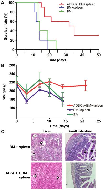
EXPERIMENTAL AND THERAPEUTIC MEDICINE 19: 1725-1732, 2020 1727
was formed by passing the cells through 200 meshes of metal
mesh. Allogeneic hematopoietic stem cells (HSCs) were obtained.
According to previous reports (8,9), infusion of allogeneic HSCs
alone is not able to induce aGVHD. Therefore, the present study
established the model of aGVHD by infusing a mixture of allo-
geneic HSCs and spleen lymphocytes. Briefly, after receiving a
total of 6 Gy body irradiation, the rats were transplanted with a
mixture of BMMNCs (2x10⁸ cells/kg) and spleen cells (3x10⁸/kg)
(BM+spleen group) by infusion via caudal vein of the tail.
To observe the suppressive effects of ADSCs on aGVHD,
ADSCs were infused at a dose of 1x10⁷/kg, in combination
with either the mixture of BMMNCs and spleen lymphocytes
(ADSCs+BM+spleen group) or BMMNCs alone (BM group),
through the caudal vein of rats at 4‑6 h after irradiation.
Occurrence of aGVHD was characterized by clinical mani-
festations such as weight loss, abnormal hunched posture,
decreased motion, hair loss and skin ulcers (10). Livers
and small intestines were obtained from sacrificed rats for
pathological examinations. Life span exceeding 50 days was
considered a long‑period of survival. Expression of the Sry
gene in recipient rats that survived longer than 21 days was
examined by real‑time PCR to detect the presence of donor
Y chromosome. Briefly, the Blood & Cell Culture DNA Midi Figure 1. Morphology and the differentiation potential of ADSCs at the third
Kit (cat. no. 13343; Qiagen) was used to extract DNA. Using passage. (A) Morphology of ADSCs at the third passage observed under an
inverted microscope. Scale bar, 200 µm. (B) Giemsa's staining of ADSCs.
the primers 5'‑GAGGGTTATACTTTGCAGCGTGAA‑3' and
Scale bar, 200 µm. (C) Oil red O staining of ADSCs on days 5‑8 after
5'‑CTG CTGT TTCTG CTGTAGTGG GT‑3', the Sry gene on adipogenic induction. Scale bar, 100 µm. (D) Alizarin red staining of ADSCs
the Y chromosome of the rats was determined. The reaction on day 14 after osteogenic induction. Scale bar, 200 µm. ADSCs, adipose
system (25 µl) was composed of 12.5 µl PCR mix, 0.5 µl tissue‑derived stem cells.
upstream primer, 0.5 µl downstream primer, 1 µl cDNA and
10.5 µl ddH2O. PCR condition consisted of initial denaturation
at 95˚C for 60 sec; 40 cycles of denaturation at 95˚C for 15 sec, and spleen lymphocytes (BM+spleen group) (14 days) was
annealing at 58˚C for 15 sec and elongation at 72˚C for 45 sec. the same with that of rats that received BMMNCs alone (BM
The reaction product underwent agarose gel electrophoresis. group) (14 days). After transplanting ADSCs into the aGVHD
Simultaneously, expression of serum interferon‑γ (IFN‑γ; model, the median survival time of rats (ADSCs+BM+spleen
eBioscience; Thermo Fisher Scientific, Inc.) and interleukin‑4 group) was significantly extended to 33.5 days, indicating that
(IL‑4; eBioscience; Thermo Fisher Scientific, Inc.) was ADSCs improved survival of the aGVHD rats (Fig. 3A). After
detected by ELISA at 0, 7, 14, 21 and 50 days after transplanta- transplanting BMMNCs and spleen lymphocytes, the rats
tion. showed typical aGVHD manifestations, including hunched
arched posture, hair loss and decreased movement range of
Statistical analysis. Data were analyzed using SPSS 13.0 motion after 14 days. Body weight changes of rats from each
software (IBM, Corp.), and are expressed as means ± standard group at different time points after transplantation showed that
deviations. For analysis of the survival rate, Kaplan‑Meier ADSCs significantly reduced the weight loss of the recipient
method and the log‑rank method were used. For analysis of rats on day 14 after transplantation, and no difference between
IL‑4/INF‑γ levels and cell colonies, Student's t‑test was used the BM+spleen group and BM group was observed (Fig. 3B).
for analysis of the data. For analysis of body weight and white Moreover, pathological changes in liver and intestinal
blood cell count, one‑way ANOVA followed by Newman‑Keuls track such as sinusoidal dilation and congestion, periportal
test was utilized. A value of P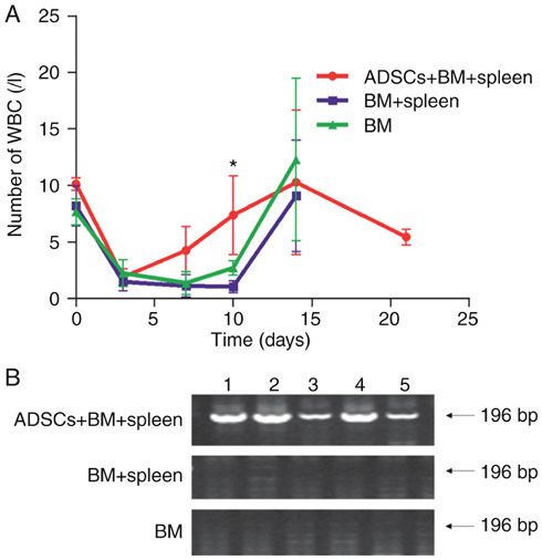
1728                                           JIANG et al: ADSCs MODULATE HEMOPOIESIS IN aGVHD

Figure 2. Immunophenotype of ADSCs and BMSCs. The third passage cells were treated with FITC‑ or PE‑labeled antibodies, followed by flow cytometry.
Mesenchymal cell markers included CD49d, CD106, CD29 and HCAM. ADSCs, adipose tissue‑derived stem cells; BMSCs, bone marrow‑derived stem cells.

                                                                                  Figure 4. ADSCs promotes hematopoietic reconstitution. (A) Leukocyte
                                                                                  count of aGVHD model rats in the BM group, BM+spleen group and
                                                                                  ADSCs+BM+spleen group. n=10. *P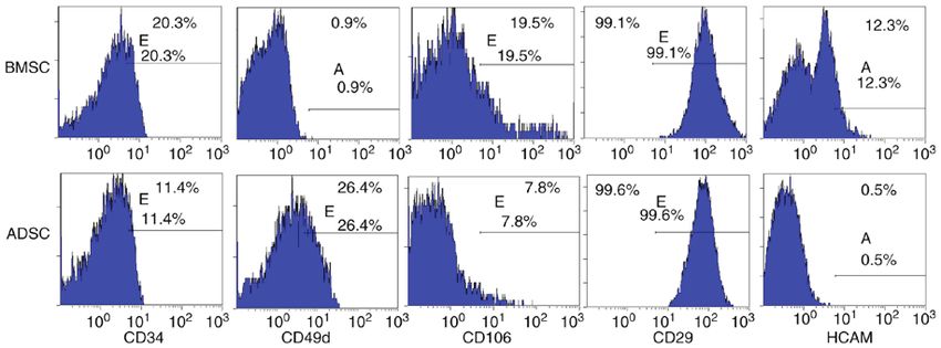
EXPERIMENTAL AND THERAPEUTIC MEDICINE 19: 1725-1732, 2020                                                      1729

Figure 5. Immune‑related factor expression during the restoration of aGVHD in rats. (A) IL‑4 levels on day 7 (n=4, *P0.05); (C) IL‑4 levels on day 14 (n=3, *P
1730                                       JIANG et al: ADSCs MODULATE HEMOPOIESIS IN aGVHD

Figure 6. Methylcellulose analysis exhibited colony‑forming units after coculture of BMMNCs with ADSCs. (A) Changes in cell morphology at 2, 14 and
35 days. Magnification, x100. (B) With prolonged time, methylcellulose analysis exhibited colony‑forming units in the BMMNCs + ADSCs group on days 14
and 35. No colonies were visible in the BMMNCs group (*P
EXPERIMENTAL AND THERAPEUTIC MEDICINE 19: 1725-1732, 2020                                               1731

in vivo. Further in‑depth research concerning these features of          5. Mirlashari MJ, Josefsen D, Landsverk K, Hasvold G, Gullestad H
                                                                            and Kvalheim G: Culturing of human adipose derived mesen-
ADSCs will be carried out in the future.                                    chymal stem cells (ADSCS) under hypoxic conditions affects
                                                                            production of wound healing cytokines and growth factors.
Acknowledgements                                                            Cytotherapy 16: S93, 2014.
                                                                         6. Institute of Laboratory Animal Resources (US). Committee on
                                                                            Care and Use of Laboratory Animals. Guide for the care and
Not applicable.                                                             use of laboratory animals. US Department of Health and Human
                                                                            Services, Public Health Service, National Institutes of Health,
                                                                            1986.
Funding                                                                  7. Frazier K, Williams S, Kothapalli D, Klapper H and
                                                                            Grotendorst GR: Stimulation of fibroblast cell growth, matrix
The study was supported by the National Natural Science                     production, and granulation tissue formation by connective tissue
                                                                            growth factor. J Invest Dermatol 107: 404‑411, 1996.
Foundation of China (no. 81460023) and Natural Science                   8. Hu J, Yan M, Jiang M, Shi W, Xu Y, Zhang D and Li J:
Foundation of Xinjiang Uygur Autonomous Region                              Establishment of a rat model of acute graft‑versus‑host disease
(no. 200821113).                                                            after allogeneic bone marrow transplantation. Chin J Com
                                                                            Med 17: 581‑584, 2007.
                                                                         9. Vogelsang GB, Hess AD, Gordon G and Santos GW: Treatment
Availability of data and materials                                          and prevention of acute graft‑versus‑host disease with thalido-
                                                                            mide in a rat model. Transplantation 41: 644‑647, 1986.
                                                                        10. Renkonen R and Häyry P: Bone marrow transplantation in the rat.
The datasets used and/or analyzed during the current study                  I. Histologic correlations and quantitation of cellular infiltrates in
are available from the corresponding author on reasonable                   acute graft‑versus‑host disease. Am J Pathol 117: 462‑470, 1984.
request.                                                                11. Ju XP, Xu B, Xiao ZP, Li JY, Chen L, Lu SQ and Huang ZX:
                                                                            Cytokine expression during acute graft‑versus‑host disease after
                                                                            allogeneic peripheral stem cell transplantation. Bone Marrow
Authors' contributions                                                      Transplant 35: 1179‑1186, 2005.
                                                                        12. Dong Y, Zhu T, Xia R, Li Y, Ge X, Zeng Q, Ni J and Li Q:
                                                                            Mechanism of bone marrow mesenchymal stem cells for treating
MJ and LC designed and directed the experiment. XB, HW                      model mice with aplastic anemia. J Clin Rehabil Tissue Eng
and RZ performed the experiments. XD, NP and HY collected                   Res 13: 7138‑7142, 2009.
the data and performed the statistical analysis. XB wrote the           13. Wu KH, Tsai C, Wu HP, Sieber M, Peng CT and Chao YH:
                                                                            Human application of ex vivo expanded umbilical cord‑derived
manuscript. LC and XD reviewed and edited the manuscript.                   mesenchymal stem cells: Enhance hematopoiesis after cord
All authors read and approved the manuscript and agree to be                blood transplantation. Cell Transplant 22: 2041‑2051, 2013.
accountable for all aspects of the research in ensuring that the        14. Kern S, Eichler H, Stoeve J, Klüter H and Bieback K: Comparative
                                                                            analysis of mesenchymal stem cells from bone marrow, umbilical
accuracy or integrity of any part of the work are appropriately             cord blood, or adipose tissue. Stem Cells 24: 1294‑1301, 2006.
investigated and resolved.                                              15. Le Blanc K, Frassoni F, Ball L, Locatelli F, Roelofs H, Lewis I,
                                                                            Lanino E, Sundberg B, Bernardo ME, Remberger M, et al:
                                                                            Mesenchymal stem cells for treatment of steroid‑resistant, severe,
Ethics approval and consent to participate                                  acute graft‑versus‑host disease: A phase II study. Lancet 371:
                                                                            1579‑1586, 2008.
Ethical approval for the study was granted from the Ethics              16. Te Boome LC, Mansilla C, van der Wagen LE, Lindemans CA,
                                                                            Petersen EJ, Spierings E, Thus KA, Westinga K, Plantinga M,
Committee of the First Affiliated Hospital of Xinjiang Medical              Bierings M, et al: Biomarker profiling of steroid‑resistant acute
University (Urumqi, China).                                                 GVHD in patients after infusion of mesenchymal stromal cells.
                                                                            Leukemia 29: 1839‑1846, 2015.
                                                                        17. Jitschin R, Mougiakakos D, Von Bahr L, Völkl S, Moll G,
Patient consent for publication                                             Ringden O, Kiessling R, Linder S and Le Blanc K: Alterations
                                                                            in the cellular immune compartment of patients treated with
Not applicable.                                                             third‑party mesenchymal stromal cells following alloge-
                                                                            neic hematopoietic stem cell transplantation. Stem Cells 31:
                                                                            1715‑1725, 2013.
Competing interests                                                     18. Galipeau J: The mesenchymal stromal cells dilemma‑does a
                                                                            negative phase III trial of random donor mesenchymal stromal
                                                                            cells in steroid‑resistant graft‑versus‑host disease represent a
The authors declare that they have no competing interests.                  death knell or a bump in the road? Cytotherapy 15: 2‑8, 2013.
                                                                        19. Bora P and Majumdar AS: Adipose tissue‑derived stromal
References                                                                  vascular fraction in regenerative medicine: A brief review on
                                                                            biology and translation. Stem Cell Res Ther 8: 145, 2017.
                                                                        20. Schäff ler A and Büchler C: Concise review: Adipose
 1. Piemontese S, Ciceri F, Labopin M, Bacigalupo A, Huang H,               tissue‑derived stromal cells‑basic and clinical implications for
    Santarone S, Gorin NC, Koc Y, Wu D, Beelen D, et al: A survey           novel cell‑based therapies. Stem Cells 25: 818‑827, 2007.
    on unmanipulated haploidentical hematopoietic stem cell             21. Zhang Y, Zhu Y, Li Y, Cao J, Zhang H, Chen M, Wang L and
    transplantation in adults with acute leukemia. Leukemia 29:             Zhang C: Long‑term engraftment of myogenic progenitors from
    1069‑1075, 2015.                                                        adipose‑derived stem cells and muscle regeneration in dystrophic
 2. Murray IR, West CC, Hardy WR, James AW, Park TS, Nguyen A,              mice. Hum Mol Genet 24: 6029‑6040, 2015.
    Tawonsawatruk T, Lazzari L, Soo C and Péault B: Natural history     22. Bai W, Jiang M, Cui L and Yi S: An experimental study for the
    of mesenchymal stem cells, from vessel walls to culture vessels.        immunological properties of human adipose‑derived stem cells
    Cell Mol Life Sci 71: 1353‑1374, 2014.                                  after expansion. J Tissue Eng Reconstr Sur 4: 312‑314, 2008.
 3. Le Blanc K, Rasmusson I, Sundberg B, Götherström C,                 23. Cui L, Yin S, Liu W, Li N, Zhang W and Cao Y: Expanded
    Hassan M, Uzunel M and Ringdén O: Treatment of severe acute             adipose‑derived stem cells suppress mixed lymphocyte reaction
    graft‑versus‑host disease with third party haploidentical mesen-        by secretion of prostaglandin E2. Tissue Eng 13: 1185‑1195, 2007.
    chymal stem cells. Lancet 363: 1439‑1441, 2004.                     24. Yañez R, Lamana ML, García‑Castro J, Colmenero I, Ramírez M
 4. Zuk PA, Zhu M, Mizuno H, Huang J, Futrell JW, Katz AJ,                  and Bueren JA: Adipose tissue‑derived mesenchymal stem
    Benhaim P, Lorenz HP and Hedrick MH: Multilineage cells                 cells have in vivo immunosuppressive properties applicable
    from human adipose tissue: Implications for cell‑based therapies.       for the control of the graft‑versus‑host disease. Stem Cells 24:
    Tissue Eng 7: 211‑228, 2001.                                            2582‑2591, 2006.
1732 JIANG et al: ADSCs MODULATE HEMOPOIESIS IN aGVHD
25. Yao Y, Song X, Cheng H, Tang G, Hu X, Zhou H and 29. Arthur A, Cakouros D, Cooper L, Nguyen T, Isenmann S,
Wang J: Dysfunction of bone marrow vascular niche in acute Zannettino AC, Glackin CA and Gronthos S: Twist‑1 enhances
graft-versus-host disease after MHC-haploidentical bone marrow bone marrow mesenchymal stromal cell support of hematopoi-
transplantation. PLoS One 9: e104607, 2014. esis by modulating CXCL12 expression. Stem Cells 34: 504-509,
26. Nasef A, Chapel A, Mazurier C, Bouchet S, Lopez M, Mathieu N, 2016.
Sensebé L, Zhang Y, Gorin NC, Thierry D and Fouillard L: 30. Sun HP, Zhang X, Chen XH, Zhang C, Gao L, Feng YM,
Identification of IL‑10 and TGF‑beta transcripts involved in the Peng XG and Gao L: Human umbilical cord blood-derived
inhibition of T-lymphocyte proliferation during cell contact with stromal cells are superior to human umbilical cord blood-derived
human mesenchymal stem cells. Gene Expr 13: 217‑226, 2007. mesenchymal stem cells in inducing myeloid lineage differentia-
27. Najar M, Rouas R, Raicevic G, Bouf ker HI, Lewalle P, tion in vitro. Stem Cells Dev 21: 1429-1440, 2012.
Meuleman N, Bron D, Toungouz M, Martiat P and Lagneaux L: 31. Zhang R, Bi X, Ma Y, Duan X and Jiang M: Biological charac‑
Mesenchymal stromal cells promote or suppress the prolif- terization of C57 mouse bone marrow mesenchymal stem cells
eration of T lymphocytes from cord blood and peripheral blood: using a whole bone marrow adherent culture technique. Chin J
The importance of low cell ratio and role of interleukin‑6. Tissue Eng Res 18: 45‑50, 2014.
Cytotherapy 11: 570‑583, 2009.
28. de Barros AP, Takiya CM, Garzoni LR, Leal-Ferreira ML, This work is licensed under a Creative Commons
Dutra HS, Chiarini LB, Meirelles MN, Borojevic R and Attribution-NonCommercial-NoDerivatives 4.0
Rossi MI: Osteoblasts and bone marrow mesenchymal stromal International (CC BY-NC-ND 4.0) License.
cells control hematopoietic stem cell migration and proliferation
in 3D in vitro model. PLoS One 5: e9093, 2010.You can also read