Experimental Toxoplasmosis in Pigeons (Columba livia)
←
→
Page content transcription
If your browser does not render page correctly, please read the page content below
Asian Journal of Research in Infectious Diseases

                               3(1): 16-26, 2020; Article no.AJRID.52343
                               ISSN: 2582-3221

                               Experimental Toxoplasmosis in Pigeons
                                                     (Columba livia)
                               Ayhan Atasever1a, Görkem Ekebaş1b, Duygu Yaman Gram1*c,
                                                        Cahit Babür2 and Abdullah Inci3d
                   1
                  Department of Pathology, Faculty of Veterinary Medicine, University of Erciyes, 38280,
                                                                                  Talas/Kayseri, Turkey.
             2
              Department of Biological Products, Ministry of Health General Directorate of Public Health,
      Microbiology Reference Laboratory and Parasitology Laboratory, 06800, Cankaya/Ankara, Turkey.
               3
                Department of Parasitology, Faculty of Veterinary Medicine, University of Erciyes, 38280,
                                                                                  Talas/Kayseri, Turkey.

                                                                                                  Authors’ contributions

    This work was carried out in collaboration among all authors. Authors AA, GE, DYG and AI designed
           the study, performed the statistical analysis, wrote the protocol and wrote the first draft of the
     manuscript. Authors CB and AI managed the analyses of the study. Authors GE and DYG managed
                             the literature searches. All authors read and approved the final manuscript.

                                                                                                       Article Information

                                                                                         DOI: 10.9734/AJRID/2020/v3i130117
                                                                                                                       Editor(s):
          (1) Dr. Shweta Sharma, Department of Microbiology, Dr. Ram Manohar Lohia Hospital and PGIMER, New Delhi, India.
          (2) Dr. Win Myint Oo, Associate Professor, Department of Physical Medicine and Rehabilitation, Sibu Clinical Campus,
                                                                                                     SEGi University, Malaysia.
                                                                                                                    Reviewers:
                                                                         (1) Hassan Yahaya, Bayero University, Kano, Nigeria.
                                                                    (2) Saw Bawm, University of Veterinary Science, Myanmar.
                                                                                    (3) Marco Antonio Santillan Flores, Mexico.
                                                 Complete Peer review History: http://www.sdiarticle4.com/review-history/52343

                                                                                          Received 16 September 2019
                                                                                          Accepted 21 November 2019
    Original Research Article
                                                                                           Published 06 January 2020

ABSTRACT

    The purpose of the present study was to establish experimental model of toxoplasmosis in pigeons,
    to investigate pathogenesis and compare tissue lesions by clinical, histopathological, serological
    and bioassay techniques. Total of 60 unknown aged pigeons (Columba livia), 21 males and 39
    females were used. They were divided into groups as oral (Group I and II) and parenteral (Group III,
    IV, V and VI) infection groups (Table 1). While some pigeons in Group IV showed acute infection
    signs such as anorexia, weight loss, pale cockscomb, bend of head and neck and partial paralyze;
    chronic infection signs such as anorexia, weakness, weight loss, diarrhea, difficulties in breathing,
_____________________________________________________________________________________________________

*Corresponding author: E-mail: dyamangram@gmail.com;
a
 ORCID: 0000-0002-6327-1604; bORCID: 0000-0003-2404-0384; cORCID: 0000-0001-9094-677X;
d
 ORCID: 0000-0003-1614-0756.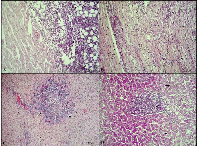
Atasever et al.; AJRID, 3(1): 16-26, 2020; Article no.AJRID.52343

 and conjunctivitis were seen in Group IV, V, and VI. In necropsy, the pigeons in Group IV had
 hyperemia and focal hemorrhages in the meninges and brain; the pigeons in Groups V and VI had
 yellowish color of the liver, the pigeons in Group V had the pale chest muscles, pericardial
 thickening and opaqueness. There were no macroscopic findings in pigeons in Group I and III.
 Histopathological examination revealed nonsuppurative meningoencephalitis and tachyzoites and
 bradyzoite cysts formation of T. gondii in brain tissue, lymphoid cell infiltration and necrotic focal
 hepatitis and nephritis in Group IV. While pigeons in Group V had nonsuppurative focal myositis,
 myocarditis, hepatitis, gastritis, enteritis, pneumonitis, and necrotic pancreatitis, one of them had
 toxoplasma bradyzoite cyst in the sinusoid in the liver. In group VI, nonsuppurative focal hepatitis,
 myocarditis, nephritis and necrotic pancreatitis were detected in pigeons. Bioassay tests were
 performed with tissue samples taken from seropositive pigeons and parasitic tachyzoites were
 isolated from the peritoneal fluid of the mice. Seropositivity in the oral and parenteral groups was
 determined by Sabin-Feldman Dye Test (SFDT) and Indirect Hemagglutination Assay (IHA). As a
 result; in similar studies that will be performed investigating pathogenesis of Toxoplasmosis and
 subclinical cases that may be overlooked, serologic tests and bioassay applications should be used
 together for the diagnosis of toxoplasmosis.

Keywords: Bioassay; histopathology; pigeon; serology; tachyzoites.

1. INTRODUCTION                                             5.9% [2,20-25,28,29]. Immunohistochemistry and
                                                            histopathological examination are important to
Toxoplasmosis is an important zoonotic infection            confirm the diagnosis of toxoplasmosis [22,30].
that caused by Toxoplasma gondii and the                    Immunohistochemistry is more appropriate in
disease distributed worldwide and can affect                cases in which toxoplasmosis cannot be
mammals including human, domestic and wild                  diagnosed      in      routine   histopathological
animals and also wild and domestic avian                    examination. For this reason, microscopic
species [1-6]. T. gondii infection is subclinical in        findings are very important in the diagnosis of
many bird species [1,4,5,7-11]. In addition to              toxoplasmosis [31]. The prevalence of T. gondii
natural toxoplasmosis cases in domestic birds,              has been different in naturally [1,6,11,20-22,24-
experimental studies have been conducted in                 26,28-30,32-37] and experimentally [2,4,7-
many poultry species such as white quails,                  10,12,27] infected poultry. The serological
Japanese quails, chicken, broiler, pigeon, turkey           diagnosis of toxoplasmosis is usually made by
and pheasants [4,5,7-15]. T. gondii infection has           detecting specific antibodies by indirect
been reported in numerous domestic and wild                 fluorescent antibody test (IFAT) and Modified
avian species by serologic [16-19] and                      agglutination     test     (MAT)    [26,27,37-40].
experimental studies [2,5,8,12,14,15] in Turkey.            Additionally, bioassay tests are thought to be
Pigeons have been shown to be susceptible to                more useful if brain and heart are both used
toxoplasmosis by natural studies [20-26] or                 [1,11,14,41-44].
experimental studies [2,3,27], and morbidity and
mortality in pigeons have been reported to be               The objective of this study was to establish
higher than in other birds. T. gondii oocyte-               experimental model of toxoplasmosis in free-
contaminated soil, water and cats play an                   living pigeons in rocky areas and cities using oral
important role in the spread of toxoplasmosis               and parenteral (intracerebral, intramuscular,
[1,2,4,6,11,20]. Experimental studies have shown            intravenous) infection, and also to diagnose the
that the pigeons that are infected with T. gondii           disease     using     clinical, histopathological,
can spread the disease with their faeces and                bioassay, and serological methods and to obtain
also pigeons living freely in rocky areas and               information about pathogenesis.
cities have a risk of spreading the disease
[2,20,21,23,25]. In addition, since pigeons are             2. MATERIALS AND METHODS
used in Chinese cuisine [19] and in the racing
industry in Taiwan the disease has gained                   Animal material: This study was conducted with
zoonotic importance. Seroprevalence studies for             unknown aged pigeons (Columba livia), 21 males
toxoplasmosis have been performed all over the              and 39 females and all pigeons were obtained
world and it has been reported to be between 4-             from a commercial farm in Kayseri/Turkey.

                                                       17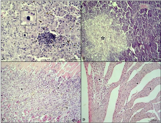
Atasever et al.; AJRID, 3(1): 16-26, 2020; Article no.AJRID.52343

         Table 1. Experimental groups and inoculum doses that were used in the study
                  5                                                              5
    Oral group (10 tachyzoites)                        Parenteral group (10 tachyzoites)
Control group     Infection group   Control group   Intracerebral group Intramuscular    Intravenous group
(I. Grup)         (II. Grup)        (III. Grup)     (IV. Grup)           group (V. Grup) (VI. Grup)

Experimental plan: T. gondii RH strain was                  Bioassay: After necropsy, tissues (brain, heart,
isolated from human and was provided from the               and muscles of chest and legs) of the pigeons
Ankara Refik Saydam Hifzisihha Research                     were bioassayed [1,11,23,26,33]. As reported
Centre. The required concentration of inoculums             previously, specific pathogen free (SPF) 3- to 6-
    5
(10 tachyzoites/0,5 ml) was obtained from                   wk-old Swiss Albino mice (20-25 g) were injected
tachyzoites. Before administrating T. gondii,               intraperitoneally with 0.5 ml suspension that was
bloods of five pigeons in each group were tested            prepared from the brain, heart and skeletal
for sero-negativity. Sabin-Feldman Dye Test                 muscles of each pigeon. Brain, heart, lung,
(SFDT) and Indirect Hemagglutination Assay                  skeletal muscle and other tissues were examined
(IHA) were used in serological detection of                 histopathologically for toxoplasmosis in mice
toxoplasmosis.     Experimental     groups     and          which died between days of 5 and 11. Peritoneal
inoculum doses that were used in the study are              liquid of each mouse was microscopically
shown in Table 1. While Group I was used as a               examined for the presence of tachyzoites
control group for Group II, Group III was served            [1,11,16,17,23,26].
as a control group (parallel concurrent) for Group
IV, Group V and Group VI.                                   3. RESULTS

Necropsy and histopathological examination:                 Clinical findings: While the pigeons that had
Following inoculation among the pigeons under               neurological symptoms on day 13 in Group IV (n:
the clinical observation, two of them died on the           6) showed acute infection signs such as
5th and 9th days from the group II; two pigeons             anorexia, weight loss, pale cockscomb, bend of
died on the 6th and 19th days in Group VI and               head and neck and partial paralyze; chronic
one pigeon died on the fifth day in Group IV.               infection signs such as anorexia, weakness,
Tissue samples (brain, heart, skeletal muscle,              weight loss, diarrhea, difficulties in breathing, and
ovary, testis, gizzard, crop, liver, kidney, spleen,        conjunctivitis were seen in Group IV (n: 2), Group
pancreas, and intestine) that were taken from               V (n: 10) and Group VI (n: 7). No clinical signs
pigeons died from acute toxoplasmosis in Group              were observed in pigeons of groups I and III.
IV (n: 1) and non-specific reasons (n: 4) were              Serological findings: Serum samples of
fixed in neutral formalin solution. On the 13th day         pigeons were serologically examined with SFDT
of post inoculation, six of the pigeons in Group IV         and IHA. Results are shown in Table 2 according
that were showed neurological signs and all the             to the experimental groups. The difference
remaining pigeons (n = 49) on the 45th day were             between SFDT and IHA was not statistically
anesthetized with 0.015 ml %2 xylazine [29] and             significant in all routes of administration (P>0.05)
then blood samples were collected through                   (Table 3).
cardiac     puncture     and     necropsies    were
performed. All the samples were fixed in 10%                Necropsy findings: The pigeon died from acute
neutral formalin. Followed by routine procedure             toxoplasmosis in Group IV (n: 1) showed
the samples embedded in paraffin and sectioned              hyperemia and hemorrhages of meninges and
at 5 μm. After staining with haematoxylin and               brain. No pathological findings were observed
eosin (HxE), sections were examined with light              during necropsy in pigeons which died due to
microscope. Also some tissue samples were                   housing conditions and nonspecific reasons (n:
collected from brain, heart, chest and leg                  4) in Group II and Group VI. On the 13th day of
muscles for bioassay.                                       postinoculation, six of the pigeons in Group IV
                                                            that had neurological symptoms were showed
Serological      examination:     Serums  were              hyperemia and focal hemorrhages of meninges
obtained from blood samples during necropsy.                and brain. Necropsies were performed on the
Seropositivity was determined by SFDT and IHA.              45th day of postinoculation all the remaining
Fisher Exact test and SPSS 20.0 package                     pigeons (n: 49). The pigeons had yellowish
program were used for statistical analysis of               colored liver in Groups V and VI. In group V,
serological test results of SFDT and IHA.                   pigeons had pale chest muscles, and additionaly,

                                                       18
Atasever et al.; AJRID, 3(1): 16-26, 2020; Article no.AJRID.52343
Table 2. Serological and bioassay results of oral and parenteral groups. * Noninfectious death (housing conditions)
Oral Group Parenteral Group
Serological Results Bioassay Results Serological Results (Dilutions) Bioassay Results
I. Group II. Group I. Group II. Group III. Group IV. Group V. Group VI. Group III. Group IV. Group V. Group VI.
Group
SFDT IHA SFDT IHA SFDT IHA SFDT IHA SFDT IHA SFDT İHA
1 - - 1/64 1/80 - - - - 1/64 1/160 1/1024 1/160 1/64 1/160 - + + +
2 - - 1/16 1/80 - - - - 1/256 - 1/16 - -* -* - + + -*
3 - - -* -* - -* - - 1/16 1/80 1/256 1/160 1/256 1/160 - + + +
4 - - 1/64 - - - - - -* -* 1/1024 1/160 1/16 1/80 - -* + +
5 - - 1/64 1/80 - - - - 1/64 1/160 1/64 1/80 1/16 - - + + +
6 - - -* -* - -* - - 1/64 1/160 1/256 1/160 1/16 1/80 - + + +
7 - - 1/256 1/160 - - - - 1/64 - 1/1024 1/160 1/64 1/160 - + + +
8 - - 1/64 1/80 - - - - 1/256 1/160 1/64 1/80 1/16 - - + + +
9 - - 1/16 - - - - - 1/16 - 1/64 1/80 -* -* - + + -*
10 - - 1/16 1/160 - - - - 1/64 1/80 1/256 - 1/16 1/80 - + + +
19Atasever et al.; AJRID, 3(1): 16-26, 2020; Article no.AJRID.52343
Table 3. The difference between SFDT and IHA was not statistically significant in all routes of
administration (P>0,05)
Group names Tests Positive Negative P value
Group II (oral) SFDT 8 0 P= 0,467
IHA 6 2
Group IV (intracerebral) SFDT 9 0 P= 0,206
IHA 6 3
Group V (intramuscular) SFDT 10 0 P= 0,211
IHA 7 3
Group VI (intravenous) SFDT 8 0 P= 0,467
IHA 6 2
four pigeons had an opaque appearance and interglandular region of glandular stomach of
thickening of the pericardium in the heart. another one. Furthermore, lymphoid cell
No pathologic findings were noted on infiltration was detected in intestine of one pigeon
macroscopic examination in Groups I, III and and lungs of another two pigeons. Almost all
IV. pigeons in group V had hyperemia, focal
hemorrhages, necrosis and focal lymphoid cell
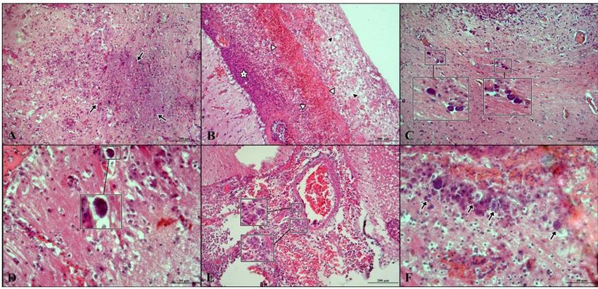
Histopathological findings: Nonspecific infiltration foci in the kidneys. One of the pigeons
hyperemia and focal hemorrhages were detected had bradyzoite cyst in the sinusoid of the liver
in some of the tissue sections (liver, lung and adjacent to the focal lymphoid cell infiltration area
intestine) that were prepared from the pigeons in (Fig. 2A). Focal necrosis and lymphoid cell
Group I and III. However, no lesion associated infiltrations was detected in the pancreas of two
with toxoplasmosis was found in these sections. pigeons (Fig. 2B). Multifocal necrosis and
There were no pathological findings of lymphocyte-rich mononuclear cell infiltration in
toxoplasmosis in pigeons in Group II. All pigeons both centre and periphery of necrotic area were
in Group IV showed severe hyperemia and focal seen in skeletal muscles of all pigeons (Fig. 2C)
hemorrhages with necrosis in brain sections (Fig. and heart muscles of four pigeons (Fig. 2D). The
1A). Nuclear material consisting of fibrin and cell appearance of the lesions in muscles was similar
debris, lymphoid cell infiltrations and in Group VI. Hyperemia and focal hemorrhages
haemorrhages were observed in the meninges were also observed in heart muscles of two
(Fig. 1B). A few strawberry-like bradyzoites pigeons and skeletal muscles of three pigeons in
containing crescent shaped tachyzoites in a Group VI (Fig. 3A-B). There were also hyperemia
macrophage cytoplasm were observed in the and small, round and sharp-sided lipid vacuoles
meninges. There was a large area of necrosis in in the cytoplasm of hepatocytes in the liver of
the occipital and temporal lobes of the brain four pigeons in this group. In addition, focal
where there were disrupted nuclear chromatins, necrotic areas and lymphoid cell infiltrations were
glia cells, macrophages, lymphocytes, and a observed in liver (Fig. 3C). Focal hemorrhages
mass of fibrins. A few strawberry-like bradyzoites and focal lymphoid cell infiltration areas were
containing crescent shaped tachyzoites in a observed in the kidneys. Focal necrosis and
macrophage cytoplasm were perivascularly lymphoid cell infiltrations was detected in the
observed in the peripheral necrotic areas in the pancreas of a pigeon in Group VI (Fig. 3D).
meninges and substansia grisea (Fig. 1C, D).
Additionally, free tachyzoites were also noted in Bioassay findings: Bioassay was performed in
paranchyma (Fig. 1E, F). There were also mice from mixture suspension prepared from the
hyperemia and small, round and sharp-sided lipid tissues (brain, heart, chest and leg muscles) of
vacuoles in the cytoplasm of hepatocytes in the pigeons. Results of the bioassay are given in
liver of some pigeons in this group. In addition, Table 2. In addition, when mice died on 5th- 11th
focal necrotic areas and lymphoid cell infiltrations days, mice tissues (brain, heart, lung, liver etc.)
were observed. Whereas only one pigeon were histopathologically tested for
showed focal necrotic areas and lymphoid cell toxoplasmosis, and peritoneal fluid was tested for
infiltrations in the kidney; some of the pigeons T. gondii tachyzoites. While no tachyzoites in
showed similar lesions in the liver. histopathological examinations were detected in
the organs of dead mice in sections, presence of
In group V, the focal lymphoid cell infiltration was tachyzoites were determined in the examination
seen in the brain of the one pigeon and in the of peritoneal liquid.
20Atasever et al.; AJRID, 3(1): 16-26, 2020; Article no.AJRID.52343
Fig. 1. Large areas of necrosis (arrows) is characterized by nonsuppurative inflammation in the
brain of pigeons (A); The appearance of nuclear material consisting of lymphoid cell
infiltrations (white star), hemorrhage (white arrowheads), fibrin mass and cell debris (black
arrowheads) (B); The appearance of strawberry shaped bradyzoit tissue cysts containing
crescent shaped tachyzoites located perineuronal and perivascular at the periphery of necrotic
areas in substantia grisea in the brain of pigeons (C, D). The appearance of free tachyzoites
(arrows) in substantia grisea, in the brain of pigeons (E, F). (Group IV), H&E stain
Fig. 2. One of the pigeons in Group V had a bradyzoit cyst structure within the sinusoid and
the focal lymphoid cell infiltration (arrows) in the liver (A); The apperance of focal necrotic
areas (star) in the pancreas of pigeons (B); In the skeletal (C) and heart (D) muscles of the
pigeons; they had multifocal necrosis in muscle fibers and lymphocyte-rich mononuclear cell
infiltration (arrowheads) in the necrotic areas. (Group V), H&E stain
21Atasever et al.; AJRID, 3(1): 16-26, 2020; Article no.AJRID.52343
Fig. 3. In the heart (A) and skeletal (B) muscles of the pigeons; they had multifocal necrosis in
muscle fibers and lymphocyte-rich mononuclear (arrowheads) cell infiltration in the necrotic
areas; they also had focal necrotic areas and lymphoid cell infiltration (arrows) in liver (C);
Focal necrosis (arrows) and (D)lymphoid cell infiltration (arrowheads) were observed in one
pancreas of pigeons. (Group VI), H&E stain
4. DISCUSSION after inoculation have been reported in poultry [7-
11]. In the present study similar results were
In the literature, T. gondii oocysts were given observed. Deaths without infection (falling into
orally in most of the studies [2,4,5,7-11,27,40, the water and similar reasons) were observed in
44-46]. Furthermore, in another study [12], two pigeons in Group II, one pigeon in Group IV
oocysts and bradyzoites were inoculated orally, and three pigeons in Group VI. While six pigeons
and tachyzoites were inoculated intravenously. In in Group IV had neurological symptoms on the
addition, there are some studies in which the 13th day of inoculation, necropsy examination
toxoplasma tachyzoites in quail, white turkeys revealed that hyperemia in the meninges and
and broilers are administered as oral, brain and focal hemorrhages were attributed to
intramuscular, intravenous, intraperitoneal, acute toxoplasmosis in this pigeons.
cloacal and intracerebral [14,15,46]. There is
also a study [3] in which pigeons were Rare clinical findings have been reported in
administered subcutaneously, intramuscularly experimental toxoplasmosis in poultry [6,7,12-15,
and intravenously with T. gondii tachyzoites. 27,40,44,46-48]. In this study, neural symptoms
In the present study, in addition to oral and characterized by torticollis, ataxia and tremor
intravenous administration, T. gondii was given were detected in six pigeons that given T. gondii
intramuscular and intracerebral routes similar tachyzoites in Group IV. Weakness and weight
with Atasever et al. [14,15]. In this study, it was loss were found in three pigeons in the same
investigated that if pigeons had infection despite group and in all other groups except for those
some of the tachyzoites are destroyed because given orally. Although most of the T. gondii
of the protective properties of gastric secretions. infections in poultry are subclinical, this study has
shown that there are 6 pigeons with acute
Acute toxoplasmosis-related deaths in neurological findings unlike other researchers.
experimental infections and unexplained deaths Necrosis of the visceral organs, muscular
22Atasever et al.; AJRID, 3(1): 16-26, 2020; Article no.AJRID.52343
dystrophy, pericardial and myocardial thickening, in other groups were tested and found
gastric ulceration, lung hepatization, seropositive using the both methods (SFDT and
hepatosplenomegaly and enteritis have been IHA) and results were not statistically significant
reported as the prominent lesions for (P>0.05).
toxoplasmosis in poultry [1,6-15,40,46,47,49,50].
Similar macroscopic findings were observed in In the diagnosis of toxoplasmosis in poultry as in
the present study in pigeons given tachyzoites other animal species, tachyzoites were obtained
through intramuscular, intracerebral and by bioassay method from mice. Natural
intravenous routes. [11,31,41,42,43,49,50] and experimental
toxoplasmosis [2,5,7-10,12,15,27,44] cases have
Unlike other researchers, large areas of necrosis been confirmed using bioassay in many poultry
in the pancreas were detected in two pigeons in species. In the present study, unlike other
Group V and one in Group VI. It was reported researchers [7-10,12,15,41-43,49,50] a single
that histological features of experimental suspension has been prepared and bioassay
toxoplasmosis consist of necrotic hepatitis, were done in mice using samples (brain, heart,
splenitis, nephritis, pneumonia, non-suppurative chest and skeletal muscles) from all pigeons that
encephalitis, pancreatitis, typhlitis, colitis, were seropositive. Tachyzoites of the parasite
adrenalitis, oesophagitis, and gastric were detected in mouse peritoneal fluid and the
inflammation [7-13,40,45-47]. However, in the bioassay results obtained in this study were in
present study, necrotizing inflammation was parallel with those reported in the literature.
observed only in the liver, brain, lungs, and However, it has not been possible to comment
spleen. on which of the tissues in the suspension given
to the mice had the causative agent.
Natural [3,11,20,28,32,33,48,50] and
experimental [2,4,5,7-10,12-15,27,40,45] In the present study, all groups were seropositive
toxoplasmosis have been serologically tested except Group I and Group III. No pathological
and found seropositive in many poultry species. findings except lymphoid cell infiltration in kidney,
Seroprevalence of toxoplasmosis has been pancreas, heart muscle and liver were observed
determined in owls, pigeons, turkeys and ducks in studies conducted in poultry with intravenous
using MAT [11,21,22,28,32,33,48], in laying hens tachyzoites administration [12,14,16] was similar
using MAT, IHA, IFAT, SFDT, ELISA and latex with the presented study which probably
agglutination test (LAT) [36-39,46] and in suggests that it may be related to the inactivation
pigeons using MAT, PCR, LAT, IHA, SFDT [21- of the causative agent by circulating macrophage
26]. In addition, toxoplasmosis has been cells.
determined in pet birds using LAT and MAT
[11,40,50]. Limited numbers of studies were The absence of a positive result
conducted on toxoplasmosis in domestic and (histopathological and bioassay) other than
wild poultry in Turkey [16-19]. Seroprevalence of serology in Group II is consistent with destruction
experimental toxoplasmosis in poultry has been of the agent by various enzymes released from
detected using different serologic tests [2,7-10, the stomach and intestines.
14,15]. Researchers tested toxoplasmosis using
MAT, LAT, IHA, and SFDT and concluded that As a result; nonsuppurative meningoencephalitis
MAT is more sensitive in detecting experimental was detected microscopically and free
toxoplasmosis in bobwhite quail, Japanese tachyzoites and bradyzoites cyst formations
quails, turkey and pheasant [8,9,11,15]. In related to toxoplasmosis were observed
addition, MAT, ELISA, SFDT, LAT and IHA tests microscopically in brain tissues of pigeons in
were used in experimental toxoplasmosis in Group IV. Pigeons in this group also had
turkeys and pheasants and MAT and ELISA microscopic findings of focal nonpurulent
were reported to be more sensitive compared hepatitis and nephritis with focal foci of lymphoid
with others [7,10,14]. Similarly, ELISA was used cell infiltration and necrosis. One of the pigeons
for serological detection of T. gondii infection in had bradyzoite cyst in the sinusoid of the liver, as
experimentally infected chickens and pigeons well as nonsuppurative focal myositis,
[2,34,37]. In the present study, SFDT and IHA myocarditis, hepatitis, gastritis, enteritis,
were used in serological detection of pneumonia in other pigeons. Nonsuppurative
toxoplasmosis. T. gondii specific antibody was focal hepatitis, myocarditis, nephritis and necrotic
5
detected in pigeons orally given 10 tachyzoites pancreatitis lesions were observed
using the both tests (SFDT and IHA). All pigeons microscopically in pigeons in group VI.
23Atasever et al.; AJRID, 3(1): 16-26, 2020; Article no.AJRID.52343
5. CONCLUSION 8. Dubey JP, Ruff MD, Kwok OCH, Shen SK,
Wilkins GC, Thulliez P. Experimental
This study concludes that the intramuscular toxoplasmosis in Bobwhite quail (Colinus
administration was the most effective route Virgmiaus). J Parasitol. 1993;79:935-936.
followed by intracerebral administration. For 9. Dubey JP, Goodwin MA, Ruff MD, Kwok
future seropositivity testing of subclinical cases OCH, Shen SK, Wilkins GC, Thulliez P.
the histopathological examination and bioassay Experimental toxoplasmosis in Japanese
applications should be performed together in quail. J Vet Diagn Invest. 1994a;6:216-
order to determine T. gondii tachyzoites and 221.
bradizoites which will make differential diagnosis 10. Dubey JP, Ruff MD, Wilkins GC, Shen SK,
of dead and live animals. Kwok OCH. Experimental toxoplasmosis in
Pheasants (Phasianus Colchicus). J Wildl
CONSENT Dis. 1994b;30:40-45.
11. Dubey JP. A review of toxoplasmosis in
It is not applicable. wild birds. Vet Parasitol. 2002;106:121-
153.
ETHICAL APPROVAL 12. Kaneto CN, Costa AJ, Paulillo AC, Moraes
FR, Murakami TO, Meireles MV.
The experiments were carried out in accordance Experimental toxoplasmosis in broiler
with the Guidelines for Animal Experimentation chicks. Vet Parasitol. 1997;69:203-210.
approved by Erciyes University, Experimental 13. Kinjo T. Studies on experimental
Animal Ethics Committee (permit no: toxoplasmosis in chickens. Jap J Vet Res.
15/02/2008). 1961;9:125-126.
14. Atasever A, Cahit Babür, Abdullah İnci,
COMPETING INTERESTS Görkem Ekebaş, Anıl İça. Beyaz
Hindilerde Deneysel Toksoplazmozis.
Authors have declared that no competing İstanbul Üniv Vet Fak Derg. 2017;43(2):
interests exist. 123-131.
DOI: 10.16988/iuvfd.321873
REFERENCES 15. Atasever A, Cahit Babür, Abdullah İnci,
Görkem Ekebaş, Anıl İça. Japon
1. Dubey JP. Toxoplasmosis of animals and bıldırcınlarında (Coturnix coturnix japónica)
nd
humans. 2 Ed. CRC Press, Taylor & deneysel toksoplazmozis. Türkiye Parazitol
Francis Group, Maryland, USA. 2009;1-18. Derg. 2017;41:62-70.
2. Biancifiori F, Rondini C, Grelloni V, DOI: 10.5152/tpd.2017.5117
Frescura T. Avian toxoplasmosis: 16. Altınöz F, Babür C, Kılıç S. Konya
Experimental infection of chicken and Yöresinde Yumurta Tavuklarında Sabin-
pigeon. Comp Immunol Microbiol Infect Feldman boya testi ile Toxoplasma gondii
Dis. 1986;9(4):337-346. (Nicolle ve Manceaux, 1908)
3. Jacobs L, Melton ML, Cook MK. seropozitifliğinin araştırılması. Türkiye
Experimental toxoplasmosis in pigeons. Parazitol Derg. 2007;31:4-6.
Exp Parasitol. 1953;2(4):403-416. 17. Babür C, Gıcık Y, İnci A. Ankara’da
4. Bickford AA, Saunders JR. Experimental güvercinlerde Sabin-Feldman boya testi ile
toxoplasmosis in chicken. Am J Vet Res. anti-Toxoplasma gondii antikorlarının
1966;27:308-318. araştırılması. Türkiye Parazitol Derg.
5. Boch J, Rommel M, Weiland G, Janitschke 1998;22:308-10.
K, Sommer R. Experimentelle toxoplasma- 18. İnci A, Babür C, Çam Y, İça A. Kayseri
infectionen bei legehennen. Berl Meunch yöresinde bazı yırtıcı kuşlarda Sabin-
Tieraerztl Wochenschr. 1966;79:352-355. Feldman boya testi Toxoplasma gondii
6. Boch J, Supperer R. Veterinarmedizinische (Nicole Ve Manceaux, 1908)
th
parasitologie. 4 Ed. Verlag Paul Parey, seropozitifliğinin araştırılması. Fırat Üniv
Berlin und Hamburg, Germany. 1983;76- Sağ Bil Derg. 2002a;16:177-179.
81. 19. İnci A, Babür C, İşcan KM, İça A.
7. Dubey JP, Camargo ME, Ruff MD, Wilkins Bıldırcınlarda (Coturnix Coturnix Japonica)
GC, Shen SK, Kwok OCH, Thulliez P. Toxoplasma gondii (Nicolle Ve Manceaux,
Experimental toxoplasmosis in Turkeys. J 1908) spesifik antikorlarının Sabin-
Parasitol.1993;79:949-952. Feldman boya testi ile araştırılması.
24Atasever et al.; AJRID, 3(1): 16-26, 2020; Article no.AJRID.52343
Türkiye Parazitol Derg. 2002b;26:20- barn-owls (Tyto alba) and pigeons
22. (Columba livia) in New Jersey. Vet
20. Ibrahim HM, Osman GY, Mohamed AH, Al- Parasitol. 1990;36(1-2):177-180.
Selwi AG, Nishikawa Y, Abdel-Ghaffar F. 29. Mushi EZ, Binta MG, Chabo RG, Ndebele
Toxoplasma gondii: Prevalence of natural R, Panzirah R. Seroprevalence of
infection in pigeons and ducks from middle Toxoplasma gondii and Chlamydia psittaci
and upper Egypt using serological, in domestic pigeons (Columba livia
histopathological, and immunohisto domestica) at Sebele, Gaborone,
chemical diagnostic methods. Vet Parasitol Botswana. Onderstepoort J Vet Res.
Reg Stud Reports. 2018;13:45-49. 2001;68(2):159-161.
DOI: 10.1016/j.vprsr.2018.04.002 30. Mc Culloch WF. Toxoplasmosis review and
th
21. Yan C, Yue CL, Qiu SB, Li HL, Zhang H, assesment, in 11 Annu. Meet. American
Song HQ, Zhu XQ. Seroprevalence of Assoc. Veterinary Laboratory Diagnosis,
Toxoplasma gondii infection in domestic New Orleans, Louisiana, October 6 to 11.
pigeons (Columba livia) in Guangdong 1968;503-504.
Province of southern China. Vet Parasitol. 31. Dubey JP, Webb DM, Sundar N,
2011;177(3):371-373. Velmurugan GV, Bandini LA, Kwok OCH,
DOI: 10.1016/j.vetpar.2010.12.004 Su C. Endemic avian toxoplasmosis on a
22. Salant H, Landau DY, Baneth G. A cross- farm in Illinois: Clinical disease, diagnosis,
sectional survey of Toxoplasma gondii biologic and genetic characteristics of
antibodies in Israeli pigeons. Vet Parasitol. Toxoplasma gondii isolates from chickens
2009;165:145–149. (Gallus domesticus) and a goose (Anser
DOI: 10.1016/j.vetpar.2009.06.031 anser). Vet Parasitol. 2007;148(3-4):207-
23. Waap H, Vilares A, Rebelo E, Gomes S, 212.
Ângelo H. Epidemiological and genetic DOI: 10.1016/j.vetpar.2007.06.033
characterization of Toxoplasma gondii in 32. Devada K, Anandan R, Dubey JP.
urban pigeons from the area of Lisbon Serologic prevalence of Toxoplasma gondii
(Portugal). Vet Parasitol. 2008;157(3):306- in chickens in Madras, India. J Parasitol.
309. 1998;84:621-622.
DOI: 10.1016/j.vetpar.2008.07.017 33. El-Massary A, Mahdy OA, El-Ghaysh A,
24. Mushi EZ, Binta MG, Chabo RG, Ndebele Dubey JP. Prevalence of Toxoplasma
R, Panzirah R. Parasites of domestic gondii antibodies in sera of Turkeys,
pigeons (Columba livia domestica) in chickens, and ducks from Egypt. J
Sebele, Gaborone, Botswana. J S Afr Vet Parasitol. 2000;86:627-628.
Assoc. 2000;71:249-250. DOI:10.1645/0022-3395(2000)086[0627:
25. Tsai YJ, Chung WC, Lei HH, Wu YI. POTGAI]2.0.CO;2
Prevalence of antibodies to Toxoplasma 34. Dubey JP. Toxoplasma gondii infections in
gondii in pigeons (Columba livia) in chickens (Gallus domesticus): Prevalence,
Taiwan. J Parasitol. 2006;92:871. clinical disease, diagnosis and public
DOI: 10.1645/GE-716R2.1 health significance. Zoonoses and Public
26. Karatepe M, Kılıç S, Karatepe B, Babür C. Health. 2010;57(1):60-73.
Prevalence of Toxoplasma gondii DOI: 10.1111/j.1863-2378.2009.01274.x
antibodies in domestic (Columba livia 35. Chumpolbanchorn K, Anankeatikul P,
domestica) and wild (Columba livia livia) Ratanasak W, Wiengcharoen J, Andrew
pigeons in Niğde region, Turkey. Turkiye Thompson RC, Sukthana Y. Prevalence of
Parazitol Derg. 2011;35(1):23-26. Toxoplasma gondii indirect fluorescent
DOI: 10.5152/tpd.2011.06 antibodies in naturally- and experimentally-
27. Godoi FSLD, Nishi SM, Pena HFDJ, infected chickens (Gallus domesticus) in
Gennari SM. Toxoplasma gondii: Thailand. Acta Parasitol. 2009;54:194-196.
Diagnosis of experimental and natural DOI: 10.2478/s11686-009-0034-2
infection in pigeons (Columba livia) by 36. Magalhaes FJ, da Silva JG, Ribeiro-
serological, biological and molecular Andrade M, Pinherio Júnior JW, Mota RA.
techniques. Rev Bras Parasitol Vet. High prevalence of toxoplasmosis in free-
2010;19(4):237-243. range chicken of the Fernando de Noronha
DOI: 10.1590/S1984-29612010000400009 Archipelago, Brazil. Acta Tropica.
28. Kirkpatrick CE, Colvin BA, Dubey JP. 2006;159:58-61.
Toxoplasma gondii antibodies in common DOI: 10.1016/j.actatropica.2016.03.034
25Atasever et al.; AJRID, 3(1): 16-26, 2020; Article no.AJRID.52343
37. Muhammad FZ, Garedaghi Y. 44. Bangoura B, Zöller B, Koethe M, Ludewig
Seroprevalence of toxoplasmosis in free M, Pott S, Fehlhaber K, Daugschies A.
range chickens in Tabriz area of Iran by Experimental Toxoplasma gondii oocyst
using ELISA test. Natural Science and infections in turkeys (Meleagris gallopavo).
Discovery. 2016;2(1):20-3. Vet Parasitol. 2013;196(3-4):272-277.
DOI: 10.20863/nsd.11240 DOI: 10.1016/j.vetpar.2013.03.032
38. Dubey JP, Levy MZ, Sreekumar C, Kwok 45. Miller NL, Frenkel JK, Dubey JP. Oral
OH, Shen SK, Dahl E, Lehmann T. Tissue infections with Toxoplasma cyst and
distribution and molecular characterization oocysts in felines, other mammals, and in
of chicken isolates of Toxoplasma gondii birds. J Parasitol. 1972;58:928-931.
from Peru. J Parasitol. 2004;90(5):1015- 46. Derakhshanfar A, Hatam G, Sohrabi K,
1018. Mirzaei M. Clinical, serological and
DOI: 10.1645/GE-329R histopathological signs of toxoplasmosis in
39. Bartova E, Sedlák K, Literák I. Serologic broiler chickens (Gallus domesticus) after
survey for toxoplasmosis in domestic birds experimental infection. Comp Clin Path.
from the Czech Republic. Avian Pathol. 2012;21(6):1379-1382.
2009;38(4):317-320. DOI: 10.1007/s00580-011-1300-x
DOI: 10.1080/03079450903055405 47. Parenti E, Cerruti Sola S, Turilli C,
40. Martínez-Carrasco C, Bernabé A, Ortiz JM, Corazzola S. Spontaneous toxoplasmosis
Alonso FD. Experimental toxoplasmosis in in canaries (Serius canaria) and other
red-legged partridges (Alectoris rufa) fed small passerine cage birds. Avian Pathol.
Toxoplasma gondii oocysts. Vet Parasitol. 1986;15:154-158:.
2005;130(1-2):55-60. 48. Quist CF, Dubey JP, Lutrell MP, Davidson
DOI: 10.1016/j.vetpar.2005.03.003 WR. Toxoplasmosis in wild turkeys: A case
41. Foster BG, Forrest RG, Blanco. Isolation of report and serologic survey. J Wildl Dis.
Toxoplasma gondii from naturally infected 1995;31:255-8.
chickens. Tex J Sei. 1969;20:323-324. 49. Vickers MC, Hartley WJ, Mason RW,
42. Jacobs L, Melton ML. Toxoplasmosis in Dubey JP, Schollam L. Blindness
chickens. J Parasitol. 1966;52:1158- associated with toxoplasmosis in canaries.
1162. JAVMA. 1992;200:1723-1725.
43. Mason RW, Hartley WJ, Dubey JP. Lethal 50. Williams MS, Fulton RM, Render JA,
toxoplasmosis in a Little Penguin Mansfield L, Bouldin M. Ocular
(Eudyptula minor) from Tasmania. J encephalitic toxoplasmosis in canaries.
Parasitol. 1991;72:328-329. Avian Dis. 2001;45:262-267.
_________________________________________________________________________________
© 2020 Atasever et al.; This is an Open Access article distributed under the terms of the Creative Commons Attribution License
(http://creativecommons.org/licenses/by/4.0), which permits unrestricted use, distribution, and reproduction in any medium,
provided the original work is properly cited.
Peer-review history:
The peer review history for this paper can be accessed here:
http://www.sdiarticle4.com/review-history/52343
26You can also read