Development of a rabbit's urethral sphincter deficiency animal model for anatomical-functional evaluation
←
→
Page content transcription
If your browser does not render page correctly, please read the page content below
Vol. 38 (1): 17-24, January - February, 2012
   original article

Development of a rabbit’s urethral sphincter deficiency
animal model for anatomical-functional evaluation
_______________________________________________
M. Skaff, E.R.S. Pinto, K.R.M. Leite, F.G. Almeida

Department of Urology (MS, ERSP, FGA), School of Medicine at Federal University of Sao Paulo, Sao
Paulo, Brazil and Laboratory of Medical Investigation - Department of Urology (KRML), School of
Medicine at State University of Sao Paulo, Sao Paulo, Brazil

ABSTRACT									ARTICLE INFO
_______________________________________________________________                       _____________________
Objective: The aim of the study was to develop a new durable animal model (using      Key words:
rabbits) for anatomical-functional evaluation of urethral sphincter deficiency.       Stem cells; tissue engineering;
Materials and Methods: A total of 40 New Zealand male rabbits, weighting 2.500        urinary incontinence;
kg to 3.100 kg, were evaluated to develop an incontinent animal model. Thirty-two     animal model; transabdominal
animals underwent urethrolysis and 8 animals received sham operation. Before          urethrolysis; urethral sphincter
and at 2, 4, 8 and 12 weeks after urethrolysis or sham operation, it was perfor-      deficiency
med cystometry and leak point pressure (LPP) evaluation with different bladder
distension volumes (10, 20, 30 mL). In each time point, 10 animals (8 from the        Int Braz J Urol. 2012; 38: 17-24
study group and 2 from the sham group) were sacrificed to harvest the bladder and     ________________
urethra. The samples were evaluated by H&E and Masson’s Trichrome to determine
urethral morphology and collagen/smooth muscle density.                               Submitted for publication:
Results: Twelve weeks after urethrolysis, it was observed a significant decrease      October 27, 2010
in LPP regardless the bladder volume (from 33.7 ± 6.6 to 12.8 ± 2.2 cmH2O). The
                                                                                      ________________
histological analysis evidenced a decrease of 22% in smooth muscle density with a
proportional increase in the collagen, vessels and elastin density (p < 0.01).        Accepted after revision:
Conclusions: Transabdominal urethrolysis develops urethral sphincter insufficiency    May 16, 2011
in rabbits, with significant decrease in LPP associated with decrease of smooth
muscle fibers and increase of collagen density. This animal model can be used to
test autologous cell therapy for stress urinary incontinence treatment.

INTRODUCTION                                                50% of the continence mechanism and inconti-
                                                            nence occurs as a consequence of decreased ac-
        Stress urinary incontinence (SUI) is a high         tivity of the smooth muscle of the intrinsic urethra.
prevalent condition with great economic and qual-                    In the past, different approaches have been
ity of life impact (1). Loss of adequate anatomic           described to improve the urethral continence. One
urethral support and intrinsic sphincter deficiency         minimally invasive alternative was the injection
(ISD) are the two major components related to the           of bulking agents (2). Several materials were tested
development of stress urinary incontinence (SUI).           with different success rates (3,4). However, none
Surgical techniques are effective to repair the ana-        of them sustained significant results according to
tomic support defect. However, the treatment of             time (5). The lack of an ideal substance to use as
ISD component is a much more complex issue.                 bulking agent and the potential to urethral func-
The intrinsic sphincter accounts for approximately          tion regeneration by means of cell therapy and

                                                       17
IBJU | Animal model for urethral sphincter deficiency

tissue engineering lead to studies based on cells        3.100 kg were used to develop an incontinent ani-
transplantation into the urethral wall and rhab-         mal model to test cell therapy.
dosphincter (6-8). However, such therapies are as-                The study was designed to determine if
sociated with major potential risks that should be       a standardized peri-urethral lesion (urethrolysis)
extensively evaluated before clinical application.       would determine urethral functional and morpho-
        One of the biggest challenges to develop         logical changes, as previously described in rats
new SUI treatments is the lack of an ideal animal        (10). Thirty two animals underwent urethrolysis
model to test such treatments. This is particularly      and 8 rabbits were kept as a sham group. Animals
true when planning to develop a successful cell          underwent cystometry and LPP determination,
transplantation therapy for a non life-threating         before surgical procedure and at 2, 4, 8 and 12
disease. An animal model to test cell injections         weeks after intervention. In each of the previous
should allow: reproducible injection in an ade-          mentioned time point, 10 animals (8 animals from
quate thick urethral wall, an easy and reproduc-         the study group and 2 animals from the sham
ible evaluation of functional and morphological          group) were sacrificed to harvest the bladder and
regeneration/recuperation, an autologous trans-          urethra.
plantation, a mid and long term follow-up and
the evaluation of possible cell migration teratoge-      Anesthesia, analgesics and antibiotic prophylaxis
nicity and mutagenicity.
        The majority of SUI animal models were                    For the cystometric and LPP evaluation,
developed in rats. However, low bladder capacity,        the rabbits were anesthetized using 20 mg/kg of
presence of early detrusor contraction, small ure-       intramuscular (IM) Ketamine, 10 minutes prior
thral wall thickness and small diameter make the         the study. Transabdominal urethrolysis was per-
cells injections and functional analysis of such         formed after the first cystometric evaluation, us-
animals very complicated with several possible           ing 4 mg/kg of additional intramuscular Xylazine.
biases. Furthermore, the urethral cell injections        Whenever necessary, we used a maintenance dose
described in the literature (9) were performed ei-       of 5 mg/kg of Ketamine and 1 mg/kg of Xylazine
ther with isogenic or immunossupressed animals           (IM) during the surgical procedure.
and it is much more feasible to perform autolo-                   Antimicrobial prophylaxis was carried out
gous cell transplantation in rabbits than in rats.       with 100 mg of sodium Cefalotine (IM), in a single
On the other hand, rabbits are bigger animals,           dose, one hour before the surgery. Postoperative
with larger bladder capacity, shorter relative func-     analgesia was carried out with 100 mg of sodium
tional urethra, thicker urethra wall, larger urethral    Dipirona (IM), every 8 hours, for 2 days.
lumen, stable bladder and allow autologous cells                  To harvest bladder and urethra, the ani-
transplantation. In the present study, we exam-          mals were anesthetized similarly to the urethroly-
ined the possibility to develop a urethral sphincter     sis, being sacrificed by an intravenous high anes-
deficiency animal model in rabbits that would be         thetic dose.
suitable for the study of urethral cell injections.
                                                         Cystometric evaluation and LPP determination
MATERIALS AND METHODS
                                                                  A 4 Fr. catheter was placed inside the blad-
        The present study was conducted after ap-        der through the urethra. The bladder was emptied
proval and following all requirements from the           and the catheter was connected to a three-way
Ethical Committee of our institutions.                   stopcock that allowed connection to the infusion
                                                         pump (Harvard Apparatus model P-22) and to the
Developing urinary incontinence model                    pressure transducer (Viotti 5600/ Urosystem DS-
                                                         5600 version 4.52- Viotti & Assoc. - Sao Paulo -
      After the initial evaluation, a total of 40        Brazil). The bladder was filled out at a continuous
New Zealand male rabbits, weighting 2.500 kg to          rate of 3 ml per minute (about 10% of the blad-

                                                        18
IBJU | Animal model for urethral sphincter deficiency
der capacity), with methylene blue diluted in sa- with Hematoxilin-Eosin (H&E) and Masson’s Tri-
line (1:1000) to facilitate determination of urinary chrome. Stereologic evaluation was performed to
leakage (10). The bladder capacity was determined evaluate collagen and smooth muscle fibers den-
by filling it out until bladder contraction and mic- sity. Stereology is a method that uses systematic
turition ocurred. and randomized samples, counting the collagen
During the filling phase, abdominal pres- and smooth muscle fibers density. The collagen
sure was slowly increased by gentle and continu- and smooth muscle fibers were analyzed and
ous pressure with 2 fingers placed on the lower counted in 100 random sterology microscopi-
abdomen directly over the bladder until one drop cal fields under great magnification (400 X), of
was observed through urethral meatus (10). As the urethra cross-section for each animal using
soon as the leak was observed, the pressure was Olympus microscope BX-51, with differential in-
withdrawed and the bladder pressure rapidly re- terference contrast. Only one person analyzed the
turned to baseline, demonstrating that leakage histological slides.
was not associated to bladder contraction.
The pressure in the bladder at the exact Statistics evaluation
moment that a drop was visualized on the ure-
thral meatus was considered as LPP. This measure The results were analyzed considering the
was performed three times (the average value type, distribution and nature of the variables using
was used to statistical analysis) in three different parametric or nonparametric tests. It was applied
moments of the filling phase; after infusion of the Wilcoxon’s or Friedman’s analysis of variance
10, 20, and 30 mL. to compare pre and postoperatively LPP values
in different timepoints for each animal. The ste-
Transabdominal urethrolysis reological data were evaluated by Kruskal-Wallis’
test. Statistical significance was determined at p
The urethrolysis was performed as previ- values < 0.05.
ous described in rats (10). Briefly, after anesthesia
and adequate antiseptic preparation, a suprapubic RESULTS
midline incision of 4 cm was performed in order to
expose the bladder urethra. In the sham group, the The average functional bladder capacity
incision was closed after complete exposure of the pre and 12 weeks post urethrolysis was 34.5 ± 6.4
bladder and urethra. To perform the urethrolysis, mL and 36.2 ± 7.4 mL, respectively (p = 0.185).
we identified the ureter bilaterally and carried out After urethrolysis, there was a progressive de-
the dissection of the entire urethra. Careful was crease in the ALPP. It becomes more evident with
taken to avoid vesical arteries and ureteral injury. significant decrease (p < 0.05) at 2 weeks after
To totally release the urethra, we opened the en- urethrolysis (Figure-1). The pre and post urethrol-
dopelvic fascia and sectioned the pubic-urethral ysis LPP evaluation with different bladder disten-
ligaments on both sides, dissecting the urethra cir- sion and at different time points are shown in
cumferentially from its proximal to its distal point. Figure-1. The bladder volume did not interfere in
The incision was closed in 2 separated layers using the LPP value as shown in Figure-1. There was no
Vycril 4.0 in the abdominal wall and nylon 4.0 in significant difference between the control groups
the skin. at various stages of study (Figure-1).
At 4 weeks, the LPP was similar to the
Histological analysis LPP at 8 and 12 weeks (p > 0.05 - Figure-1). In
the same way, there was a progressive increase
After 2, 4, 8 and 12 weeks, the animals in collagen and decrease in smooth muscle in the
were sacrificed, the urethra and bladder were for- urethra wall. The control group presented un-
malin fixed, paraffin embedded and cross-sec- changed collagen/smooth muscle distribution in
tioned in 5 μm sections. The slides were stained the urethra wall. The animals submitted to ure-
19IBJU | Animal model for urethral sphincter deficiency Figure 1 – Average ALPP after filling the bladder with 10, 20 and 30 mL at different timepoints: pre-urethrolysis 2,4,8 and 12 weeks after urethrolysis. Sham group with 20 ml bladder distension *p
IBJU | Animal model for urethral sphincter deficiency
Figure 2 - Muscle and collagen distribuition (%) in the wall at different time points.
PO = post operative in each timepoint - 2, 4, 8 and 12 weeks.
* Kruskal Wallis – p< 0.05 – compared with control group.
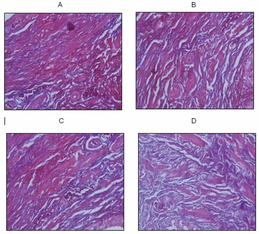
Figure 3 - Masson´s Tricrome staining at the different time points (Magnification = 400X).
A = Control Group at 8 weeks; B =2 weeks; C = 4 weeks; D = 8 weeks after urethrolysis.
Stained in red = muscle.
Stained in purple = collagen and elastin.
21IBJU | Animal model for urethral sphincter deficiency
determined by a positive sneeze test performed 4 occlusion pressure. The authors hypothesized a
weeks following vaginal distension, only one third probable spontaneous regeneration of the nervous
of the rats were incontinent (15). In the present tissue (21).
study, we observed an easily reproducible periure- The main reason to develop an animal
thral lesion. Once we standardized the periurethral model is to allow safety tests before human use.
lesion technique, all animals developed a signifi- In the past years, it has been proposed cell therapy
cant and consistent decrease in LPP. injection as an alternative for urinary inconti-
Another method described to determine nence treatment. Human use has been described
urethral resistance in rodents is the tilting table based mainly on tests performed in isogenic or
test (18). The rat is mounted on a vertical tilting imunocompromised rats (9). We believe that such
table to simulate the orientation of the human pel- therapy should be tested using autologous cell
vis with respect to gravity and the spinal cord of transplantation before human use. The lack of uri-
the rat is transected at the T8-T9 level to eliminate nary incontinence animal model that allows au-
bladder interference, while maintaining the ure- tologous cell harvesting restricts such tests. Our
thral closure mechanisms intact (18). animal model can test autologous cellular therapy
Other studies with female rats were de- injection in the urethra wall.
scribed injuring the pudendal nerve (19). The The major difficulty to create a urinary
main problem with pudendal nerve crush models incontinent animal model is related to function-
is that they create a recoverable localized dener- al-anatomic characteristics that distinguish man
vation of the striated muscle component and the from other animals. This difficulty in getting an
whole pudendal innervation. To study stem cells animal model has led to surgical approaches and
effect on urethra, the urethral lesions should not therapies for humans without being suitably tested
heal spontaneously, because we need long term in animal trials. The mostly used urinary inconti-
follow-up. In addition, it should include the stri- nence and sphincter deficiency animal models to
ated and smooth urethral sphincter component to test injectable cell therapy have been developed
better reproduce the clinical aspects. in female rats. The main problems with the use of
In 2004, an animal model of intrinsic such animals are: the difficulty to perform autolo-
sphincter dysfunction has been described by elec- gous tests, the high regenerative capacity leading
trocoagulation of the urethral surrounding tissues to recovery of the urethral function after a certain
(20); this model created a long lasting decrease period of time, the small bladder capacity (around
in urethral resistance and more significant tis- 1.5 mL), difficulty to determine the leak pressure
sue damage than childbirth simulations. Another due to bladder interference (bladder contraction
study developed a urethral damage by perform- during functional evaluation), a relatively longer
ing a transabdominal urethrolysis in female rats. urethral length with a small diameter and thin
The authors observed a significant decrease in LPP wall.
and retrograde urethral perfusion pressure up to Rabbits present reasonable anesthetic-sur-
24 weeks. An histological analysis demonstrated a gical resistance, allowing multiple surgical inter-
reduction of muscle fibers and an increase of the ventions and autologous cell harvesting; present a
conjunctive tissue (10). In our study, we performed urethral wall with good thickness, allowing repro-
a similar intervention in rabbits confirming that ductible and precise injections during cell therapy
such intervention can consistently decrease LPP tests. We found that female New Zealand rabbits
associated with histological urethral changes. present a hypospadic urethral meatus. Thus, the
Few studies were performed in rabbits in urethral catheterization is an extreme challenging
order to develop a urinary incontinence animal job. We designed a vaginoplasty in order to reach
model. In a work with male rabbits, after section- the meatus. Nevertheless, the urethral catheteriza-
ing the pudendal nerve bilaterally, it was observed tion still was challenging and sometimes it could
a decrease in urethral resistance. However, after not be reproducible. We also performed a supra-
12 weeks, there was a recovery of the urethral pubic vesical catheterization trying to accomplish
22IBJU | Animal model for urethral sphincter deficiency
the cystosmetic evaluation and LPP determina- REFERENCES
tion. However, it changed the bladder behavior,
created inflammation on the puncture spot and
1. Hu TW, Wagner TH, Bentkover JD, Leblanc K, Zhou SZ, Hunt
generated bladder leakage during LPP evaluation.
T: Costs of urinary incontinence and overactive bladder in
One can criticize an animal model in male rabbits,
the United States: a comparative study. Urology. 2004; 63:
as the majority of patients with stress urinary in- 461-5.
continence are women. However, we were looking 2. Keegan PE, Atiemo K, Cody J, McClinton S, Pickard R: Peri-
for a reproducible animal model that would allow urethral injection therapy for urinary incontinence in wom-
testing safely and efficiently autologous cells in- en. Cochrane Database Syst Rev. 2007; 3: CD003881.
jections into the urethra wall. Furthermore, it has 3. Cervigni M, Tomiselli G, Perricone C, Panei M: Endoscopic
been described autologous muscle derived from treatment of sphincter insufficiency with autologous fat in-
stem cells in the human male urethra, without an jection. Arch Ital Urol Androl. 1994; 66(4 Suppl): 219-24.
extensive animal study (6). 4. Lightner D, Calvosa C, Andersen R, Klimberg I, Brito CG,
Despite interesting initial clinical out- Snyder J, et al.: A new injectable bulking agent for treat-
comes in SUI treatment using cell therapy, we ment of stress urinary incontinence: results of a multicenter,
randomized, controlled, double-blind study of Durasphere.
need more basic research to clearly demonstrate
Urology. 2001; 58: 12-5.
safety for human use. The potential risks, such
5. Kershen RT, Fefer SD, Atala A: Tissue-engineered therapies
as teratogenicity, cell migration and mutagenic- for the treatment of urinary incontinence and vesicoureteral
ity should be better evaluated, especially when reflux. World J Urol. 2000; 18: 51-5.
treating a benign, non-life threating disease. The 6. Mitterberger M, Marksteiner R, Margreiter E, Pinggera GM,
present animal model is a new alternative to test Frauscher F, et al.: Myoblast and fibroblast therapy for post-
safely and effectively cell therapy to treat stress prostatectomy urinary incontinence: 1-year follow-up of 63
urinary incontinence. It also has the possibility to patients. J Urol. 2008; 179: 226-31.
test autologous cell therapy as one alternative to 7. Mitterberger M, Marksteiner R, Margreiter E, Pinggera GM,
functional and anatomical regeneration in a ure- Colleselli D, Frauscher F, et al.: Autologous myoblasts and
thral intrinsic sphincter deficiency animal model. fibroblasts for female stress incontinence: a 1-year follow-
up in 123 patients. BJU Int. 2007; 100: 1081-5.
8. Nikolavsky D, Chancellor MB: Stem cell therapy for stress
urinary incontinence. Neurourol Urodyn. 2010; 29(Suppl 1):
CONCLUSIONS
S36-41.
9. Kwon D, Kim Y, Pruchnic R, Jankowski R, Usiene I, de
Transabdominal urethrolysis created Miguel F, et al.: Periurethral cellular injection: comparison
sphincteric insufficiency in rabbits with signifi- of muscle-derived progenitor cells and fibroblasts with re-
cant decrease in the LPP associated with decrease gard to efficacy and tissue contractility in an animal model
of smooth muscle fibers and increase of collagen of stress urinary incontinence. Urology. 2006; 68: 449-54.
density. This animal model can be used to test au- 10. Rodríguez LV, Chen S, Jack GS, de Almeida F, Lee KW, Zhang
tologous cell therapy for stress urinary inconti- R: New objective measures to quantify stress urinary incon-
nence treatment. tinence in a novel durable animal model of intrinsic sphinc-
ter deficiency. Am J Physiol Regul Integr Comp Physiol.
CONFLICT OF INTEREST 2005; 288: R1332-8.
11. Hijaz A, Daneshgari F, Cannon T, Damaser M: Efficacy of a
vaginal sling procedure in a rat model of stress urinary in-
None declared.
continence. J Urol. 2004; 172: 2065-8.
12. Phull H, Salkini M, Escobar C, Purves T, Comiter CV: The
role of angiotensin II in stress urinary incontinence: A rat
FUNDING SUPPORT model. Neurourol Urodyn. 2007; 26: 81-8; discussion 89.
13. Sievert KD, Bakircioglu ME, Tsai T, Nunes L, Lue TF: The
FAPESP - Fundação de Amparo à Pesquisa effect of labor and/or ovariectomy on rodent continence
do Estado de São Paulo mechanism--the neuronal changes. World J Urol. 2004; 22:
Grant Number 06/57479-2 244-50.
23IBJU | Animal model for urethral sphincter deficiency
14. Damaser MS, Kim FJ, Minetti GM: Methods of testing ure- 19. Damaser MS, Samplaski MK, Parikh M, Lin DL, Rao S,
thral resistance in the female rat. Adv Exp Med Biol. 2003; Kerns JM: Time course of neuroanatomical and functional
539: 831-9. recovery after bilateral pudendal nerve injury in female rats.
15. Lin AS, Carrier S, Morgan DM, Lue TF: Effect of simulated Am J Physiol Renal Physiol. 2007; 293: F1614-21.
birth trauma on the urinary continence mechanism in the 20. Chermansky CJ, Cannon TW, Torimoto K, Fraser MO, Yo-
rat. Urology. 1998; 52: 143-51. shimura N, de Groat WC, et al.: A model of intrinsic sphinc-
16. Resplande J, Gholami SS, Graziottin TM, Rogers R, Lin CS, teric deficiency in the rat: electrocauterization. Neurourol
Leng W, et al.: Long-term effect of ovariectomy and simu- Urodyn. 2004; 23: 166-71.
lated birth trauma on the lower urinary tract of female rats. 21. Martinez Portillo FJ, Osmonov DK, Seif C, Braun PM,
J Urol. 2002; 168: 323-30. Boehler G, Alken P, et al.: Restoration of external urethral
17. Damaser MS, Broxton-King C, Ferguson C, Kim FJ, Kerns sphincter function after pudendal nerve end-to-end anasto-
JM: Functional and neuroanatomical effects of vaginal dis- mosis in the male rabbit. J Urol. 2004; 171: 1715-9.
tention and pudendal nerve crush in the female rat. J Urol.
2003; 170: 1027-31. ______________________
18. Lee JY, Cannon TW, Pruchnic R, Fraser MO, Huard J, Chan- Correspondence address:
cellor MB: The effects of periurethral muscle-derived stem
Dr. Fernando Gonçalves Almeida
cell injection on leak point pressure in a rat model of stress
Rua Pedro de Toledo, 1222 / 22
urinary incontinence. Int Urogynecol J Pelvic Floor Dys-
funct. 2003; 14: 31-7; discussion 37.
Sao Paulo, SP, 04039-003, Brazil
Fax: + 55 11 5576-4086
E-mail: fernandourologia@hotmail.com
24You can also read