Identification of LPCAT1 expression as a potential prognostic biomarker guiding treatment choice in acute myeloid leukemia
←
→
Page content transcription
If your browser does not render page correctly, please read the page content below
ONCOLOGY LETTERS 21: 105, 2021

    Identification of LPCAT1 expression as a potential prognostic
   biomarker guiding treatment choice in acute myeloid leukemia
       KE WANG1, ZHIDAN WU1, YUAN SI1, WENDONG TANG1, XIN XU2, YAN CHENG1 and JIANG LIN1

   Departments of 1Clinical Laboratory and 2Hematology, Jiangyin People's Hospital, Jiangyin, Jiangsu 214400, P.R. China

                                      Received July 11, 2020; Accepted November 3, 2020

                                                   DOI: 10.3892/ol.2020.12366

Abstract. Changes in lipid metabolism affect numerous               among total cases of AML in the lower LPCAT1 expression
cellular processes that are relevant to cancer biology, including   group. These results suggest that patients with AML who
cell proliferation, death, differentiation and motility. In the     exhibit higher LPCAT1 expression levels may benefit from
phosphatidylcholine biosynthesis pathway, the conversion            HSCT. Collectively, the findings of the present study indicate
of lysophosphatidylcholine (LPC) to phosphatidylcholine is          that LPCAT1 expression may serve as an independent prog‑
catalyzed by cytosolic enzymes of the LPC acyltransferase           nostic biomarker that can guide the choice between HSCT and
(LPCAT) family. A number of studies have demonstrated               chemotherapy in patients with AML.
that LPCAT1 overexpression is a frequent event in diverse
human cancer types, and that it is associated with unfavor‑         Introduction
able pathological characteristics and patient survival. The
aim of the present study was to explore the prognostic role         Acute myeloid leukemia (AML) is an aggressive hemato‑
of the expression of LPCAT family members in acute                  logical malignancy with an incidence rate of 3.7 per 100,000
myeloid leukemia (AML). Using Cox regression analysis,              worldwide, and it is characterized by the clonal proliferation of
only LPCAT1 expression was identified as an independent             myeloid hematopoietic stem cells with the inhibition of normal
prognostic biomarker in AML. In a cohort from The Cancer            hematopoiesis (1). AML is a highly heterogeneous disease
Genome Atlas, Kaplan‑Meier analysis revealed that patients          in terms of clinical presentation, cytogenetics/genetics and
with AML and higher expression levels of LPCAT1 had                 clinical outcome (1). Due to its considerable variability, it is
shorter overall survival (OS) and leukemia‑free survival            recommended that AML treatment should be more personal‑
(LFS) times compared with those with lower expression levels        ized and precisely targeted based on the risk classifications of
of LPCAT1. This was further confirmed using an independent          each patient (2). At present, the European LeukemiaNet risk
cohort from the Gene Expression Omnibus. Using a third              classification is widely used in the clinical management of
cohort comprising patients with AML and healthy volunteers,         AML; however, there is marked heterogeneity among patients
it was confirmed that LPCAT1 expression was significantly           in clinical practice, particularly those in intermediate groups in
increased in newly diagnosed AML cases compared with                the classification system (2). Therefore, it is crucial to identify
healthy controls. Moreover, higher expression of LPCAT1 was         novel potential prognostic and predictive biomarkers to improve
associated with French‑American‑British subtype‑M4/M5 and           our understanding of leukemogenesis so that molecular‑based
nucleophosmin 1 mutations. Notably, patients who underwent          stratification can be applied to risk‑adapted therapies and ulti‑
hematopoietic stem cell transplantation (HSCT) following            mately improve the clinical outcome of AML.
induction therapy exhibited significantly longer OS and LFS             Changes in lipid metabolism affect numerous cellular
times compared with patients who only received chemotherapy         processes that are relevant to cancer biology, including cell
after induction therapy in the higher LPCAT1 expression             proliferation, death, differentiation and motility (3). In the
group, whereas no significant differences in OS and LFS times       phosphatidylcholine biosynthesis pathway, the conversion
were observed between the HSCT and chemotherapy groups              of lysophosphatidylcholine (LPC) to phosphatidylcholine is
                                                                    catalyzed by cytosolic enzymes of the LPC acyltransferase
                                                                    (LPCAT) family (4). To date, LPCAT1, LPCAT2, LPCAT3
                                                                    and LPCAT4 have been identified and partially character‑
                                                                    ized as the four members of the LPCAT family. A number of
Correspondence to: Dr Jiang Lin, Department of Clinical
Laboratory, Jiangyin People's Hospital, 163 Shoushan Road,          studies have demonstrated that the overexpression of LPCAT1
Jiangyin, Jiangsu 214400, P.R. China                                is frequent in diverse human cancer types, including prostate
E‑mail: linjiang2019@sohu.com                                       cancer (5,6), breast cancer (7,8), gastric cancer (9), clear
                                                                    cell renal cell carcinoma (10), lung cancer (11), hepatocel‑
Key words: LPCAT1, expression, prognosis, biomarker, acute          lular carcinoma (12), oral squamous cell carcinoma (13) and
myeloid leukemia                                                    colorectal cancer (14). Moreover, LPCAT1 overexpression has
                                                                    been found to be associated with unfavorable pathological
                                                                    characteristics and survival in several types of cancer (5‑11).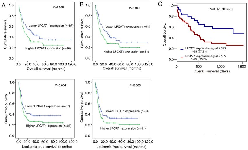
2 WANG et al: LPCAT FAMILY EXPRESSION IN AML
However, the potential roles and clinical implications of LPCAT1 expression that occur during the development of
LPCAT family members in AML remain poorly investigated. AML since PB contains blasts, and the collection of BM
Therefore, the aim of the present study was to investigate the samples from patients with AML in the medical laboratory of
prognostic role of the expression of LPCAT family members Jiangyin People's Hospital (Jiangyin, China) was challenging.
in AML, and to determine whether this has the potential to be PB samples were collected from the 20 controls and the
used as a new biomarker for the diagnosis and prognosis of 48 patients with AML at diagnosis, and from 15 of the patients
AML, and for the optimization of clinical decision‑making in with AML at complete remission (CR). Nucleated cells were
the treatment of the disease. obtained from PB using red blood cell lysis buffer (Beijing
Solarbio Science & Technology Co., Ltd.). Total RNA was
Materials and methods extracted from the PB nucleated cells using TRIzol® reagent
(Invitrogen; Thermo Fisher Scientific, Inc.). Reverse transcrip‑
Patients. The first cohort included in the present study tion was performed to synthesize cDNA from the RNA using
comprised 173 patients with AML for whom LPCAT family the PrimeScript™ RT reagent kit (Takara Bio, Inc.) according
(LPCAT1/2/3/4) expression data were available from The to the manufacturer's instructions.
Cancer Genome Atlas (TCGA) database (TCGA‑LAML,
NEJM 2013) (https://cancergenome.nih.gov/ and http://www. Quantitative PCR (qPCR) analysis. qPCR analysis was
cbioportal.org/) (15). The clinical and molecular characteris‑ conducted to detect LPCAT1 and GAPDH transcripts using
tics obtained for the cohort included age (median, 58 years; TB Green Premix Ex Taq™ II (Takara Bio, Inc.). The primers
age range, 18‑88 years), sex (81 male and 92 female), white used were as follows: LPCAT1 forward, 5'‑ACCTATTCC
blood cell (WBC) counts, peripheral blood (PB) blasts, bone GAGCCATTGACC‑3' and reverse, 5'‑CCTA ATCCAG CT
marrow (BM) blasts, French‑American‑British (FAB) subtype, TCTTGCGAAC‑3'; and GAPDH forward, 5'‑AATCCCATC
karyotype and the frequencies of known common genetic muta‑ ACCATCT TCCAG‑3' and reverse, 5'‑GAGCCCCAGCCT
tions, as well as gene expression levels. Due to the independent TCTCCAT‑3'. GAPDH served as the reference gene. qPCR
disease entity of patients with FAB‑M3, the non‑M3 AML conditions were as follows: 95˚C for 30 sec, followed by
cohort was used in the survival analysis. Following induction 40 cycles of 95˚C for 10 sec, 60˚C for 1 min, 72˚C for 1 min,
chemotherapy, 100 of the patients received only chemotherapy and 80˚C for 30 sec. The relative LPCAT1 transcript level was
as a consolidation treatment, whereas 73 patients underwent calculated using on the 2‑∆∆Cq method (20).
hematopoietic stem cell transplantation (HSCT) with/without
chemotherapy as a consolidation treatment. This cohort of Statistical analysis. The Mann‑Whitney U test or the
patients with AML was used for the identification of LPCAT Kruskal‑Wallis test followed by Dunn's post hoc test was used
family members whose expression was associated with for the comparison of continuous variables, and Pearson's χ2 or
prognosis, and for the analysis of the clinical implications of Fisher's exact test were used for the comparison of categorical
LPCAT1 expression. variables. The effect of LPCAT1 expression on leukemia‑free
A second cohort was also included, comprising 78 survival (LFS) and overall survival (OS) was analyzed using
patients (median age, 62 years, age range 18‑85 years) with the Kaplan‑Meier method with log‑rank test, and Cox regression
cytogenetically normal AML (CN‑AML) from a Gene analysis. Two‑tailed P‑values of 18 years were included. Patients with was associated with LFS, whereas LPCAT1/2 expression showed
antecedent hematological diseases or therapy‑associated a trend being associated with LFS (Table I). However, despite
AML were excluded. The study protocol was approved by the the well‑known prognostic factors (age, WBC and ELN risks)
Institutional Ethics Committee of Jiangyin People's Hospital, showing significant associations, only LPCAT1 expression was
and all the participants provided written informed consent. identified as an independent prognostic biomarker in AML by
This cohort of patients with AML was used to validate the Cox regression multivariate analysis, and exhibited a significant
changes of LPCAT1 expression in patients with AML. association with OS (Table I). Furthermore, Kaplan‑Meier
analysis also demonstrated that patients with higher expres‑
Sample preparation, RNA isolation and reverse transcription. sion levels of LPCAT1 exhibited significantly shorter OS time
PB samples were examined to identify the changes in compared with those with lower expression of LPCAT1 amongONCOLOGY LETTERS 21: 105, 2021 3
Table I. Cox regression analysis of variables for survival in patients with acute myeloid leukemia.
A, Overall survival
Univariate analysis Multivariate analysis
‑‑‑‑‑‑‑‑‑‑‑‑‑‑‑‑‑‑‑‑‑‑‑‑‑‑‑‑‑‑‑‑‑‑‑‑‑‑‑‑‑‑‑‑‑‑‑‑‑‑‑‑‑‑‑‑‑‑‑‑‑‑‑‑‑‑‑‑‑‑‑‑‑‑‑‑‑‑‑ ‑‑‑‑‑‑‑‑‑‑‑‑‑‑‑‑‑‑‑‑‑‑‑‑‑‑‑‑‑‑‑‑‑‑‑‑‑‑‑‑‑‑‑‑‑‑‑‑‑‑‑‑‑‑‑‑‑‑‑‑‑‑‑-‑‑‑‑‑‑‑‑‑‑‑‑‑‑
Variables HR (95% CI) P‑value HR (95% CI) P‑value
Age 1.040 (1.027‑1.054)4 WANG et al: LPCAT FAMILY EXPRESSION IN AML
Figure 1. Impact of LPCAT1 expression on the survival of patients with AML. Data on patients with AML are derived from TCGA database (TCGA‑LAML,
NEJM 2013) (15) and a Gene Expression Omnibus dataset (GSE12417) (16). Kaplan‑Meier survival curves of OS and LFS in patients with AML. OS and LFS
in (A) total AML and (B) non‑M3 AML from TCGA. (C) Survival analysis performed on GSE12417 using the online web tool GenomicScape [GenomicScape
ID, GS‑DT‑26; Metzeler, acute myeloid leukemia 1 (Affy U133 Plus 2)]. LPCAT1, lysophosphatidylcholine acyltransferase 1; AML, acute myeloid leukemia;
TCGA, The Cancer Genome Atlas; OS, overall survival; LFS, leukemia‑free survival.
or had not received HSCT after induction chemotherapy were
compared in the lower and higher LPCAT1 expression groups.
In the higher LPCAT1 expression group, the patients who
had undergone HSCT following induction therapy exhibited
significantly longer OS and LFS times compared with those
who only received chemotherapy after induction therapy in
both total‑AML and non‑M3‑AML cases (Fig. 4A‑D). In the
lower LPCAT1 expression group, no significant differences
were observed in OS and LFS between patients who had
undergone HSCT and those who had not among total‑AML
and only in LFS among non‑M3‑AML cases (Fig. 4E‑H).
These results suggest that patients with AML and higher
LPCAT1 expression may benefit from HSCT, and LPCAT1
expression may guide the decision of whether to select HSCT
Figure 2. LPCAT1 expression in AML. LPCAT1 transcript level in controls,
or chemotherapy as an appropriate treatment for patients with
patients with newly diagnosed AML and patients with AML who achieved AML after induction therapy.
CR, as detected by reverse transcription‑quantitative PCR. LPCAT1, lyso‑
phosphatidylcholine acyltransferase 1; AML, acute myeloid leukemia; Discussion
CR, complete remission.
Studies have demonstrated the biological role of LPCAT1 in
diverse human cancer types. For example, Morita et al (12)
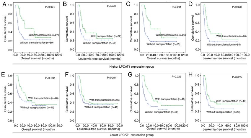
Moreover, LPCAT1 expression was further compared between reported that the overexpression of LPCAT1 promoted the
patients with and without NPM1, IDH1, CEBPA and TP53 proliferation, migration and invasion of hepatocellular carci‑
gene mutations, and the differences between the wild‑type and noma cells. In addition, in oral squamous cell carcinoma
mutant groups were observed to be statistically significant for cell lines, the knockdown of LPCAT1 resulted in decreased
all four genes (Fig. 3). cellular proliferation, invasiveness and migration, as well
as decreased intercellular platelet‑activating factor (PAF)
Patients with AML and higher LPCAT1 expression may concentration and PAF receptor expression (13). Furthermore,
benefit from HSCT. To investigate whether HSCT is able Du et al (10) revealed that LPCAT1‑knockdown in clear cell
to overcome the adverse outcomes associated with higher renal cell carcinoma inhibited cell proliferation, migration
expression of LPCAT1, the survival times of patients who had and invasion, and induced cell cycle arrest at the G0/G1 phase.ONCOLOGY LETTERS 21: 105, 2021 5
Table II. Association of LPCAT1 expression with clinicopathological characteristics in patients with acute myeloid leukemia.
LPCAT1 expression
‑‑‑‑‑‑‑‑‑‑‑‑‑‑‑‑‑‑‑‑‑‑‑‑‑‑‑‑‑‑‑‑‑‑‑‑‑‑‑‑‑‑‑‑‑‑‑‑‑‑‑‑‑‑‑‑‑‑‑‑‑‑‑‑‑‑‑‑‑‑‑‑‑‑‑‑‑‑‑‑‑‑‑‑‑‑‑‑‑‑‑‑‑‑‑
Parameters Low (n=87) High (n=86) P‑value
Sex, male/female 42/45 39/47 0.761
Age, median (range), years 57 (18‑82) 58.5 (22‑88) 0.311
Median WBC count (range), x109/l 18.7 (0.5‑297.4) 15.6 (0.4‑137.2) 0.808
Median PB blasts (range), % 50 (0‑98) 18 (0‑94) 0.001
Median BM blasts (range), % 72 (30‑99) 73 (30‑100) 0.449
FAB classification, n6 WANG et al: LPCAT FAMILY EXPRESSION IN AML Figure 3. Associations of LPCAT1 expression with gene mutations in AML. Data on patients with AML are derived from TCGA database (TCGA‑LAML, NEJM 2013) (15). LPCAT1 expression in AML patients with and without NPM1, IDH1, CEBPA and TP53 mutations. LPCAT1, lysophosphatidylcholine acyltransferase 1; AML, acute myeloid leukemia; NPM1, nucleophosmin 1; IDH1, isocitrate dehydrogenase 1; CEBPA, CCAAT/enhancer‑binding protein α; TCGA, The Cancer Genome Atlas; WT, wild‑type; mu, mutant. Figure 4. Effect of HSCT on the survival of patients with AML in groups with different LPCAT1 expression levels. Data on patients with AML are derived from TCGA database (TCGA‑LAML, NEJM 2013) (15). Kaplan‑Meier survival curves of (A) OS and (B) LFS among whole‑cohort AML in patients with higher LPCAT1 expression. Kaplan‑Meier survival curves of (C) OS and (D) LFS among patients with non‑M3 AML and higher LPCAT1 expression. Kaplan‑Meier survival curves of (E) OS and (F) LFS among whole‑cohort AML in patients with lower LPCAT1 expression. Kaplan‑Meier survival curves of (G) OS and (H) LFS among patients with non‑M3 AML and lower LPCAT1 expression. HSCT, hematopoietic stem cell transplantation; AML, acute myeloid leukemia; LPCAT1, lysophosphatidylcholine acyltransferase 1; OS, overall survival; LFS, leukemia‑free survival.
ONCOLOGY LETTERS 21: 105, 2021                                                      7

Moreover, short hairpin RNA‑mediated LPCAT1 silencing in              aberrant cytoplasmic delocalization of NPM1 mutants. NPM1
lung adenocarcinoma not only abrogated cell proliferation,            mutants have been demonstrated to maintain the leukemic
migration and invasion in vitro, but also arrested tumor growth       state through homeobox gene overexpression (21). The present
and brain metastases in vivo (11). Mechanically, LPCAT1               study identified a link between LPCAT1 and NPM1 muta‑
dysregulation has been shown to at least partially affect the         tions in patients with AML. However, the exact association
progression of lung adenocarcinoma through the PI3K/AKT               between LPCAT1 expression and NPM1 mutations remains
signaling pathway via the targeting of MYC transcription (11).        poorly defined. Further studies are required to determine the
However, studies investigating the direct role of LPCAT1 in           potential mechanism by which the overexpression of LPCAT1
AML are lacking. Therefore, further functional and clinical           contributes to the leukemogenesis caused by NPM1 mutations.
studies are required to investigate the potential role of LPCAT1          In conclusion, the findings of the present study indicate that
during leukemogenesis in AML.                                         LPCAT1 expression may serve as an independent prognostic
    The present study investigated the clinical implications          biomarker to guide the treatment choice between HSCT and
of the expression of LPCAT family members in AML, and                 chemotherapy in patients with AML.
demonstrated that LPCAT1 expression is significantly associ‑
ated with the survival of patients with AML. In addition, the         Acknowledgements
present study indicated that LPCAT1 expression may act as a
potential biomarker to guide the choice between HSCT and              Not applicable.
chemotherapy for the further treatment of patients with AML
after induction therapy. These results emphasize the potential        Funding
of LPCAT1 expression as a valuable indicator for the clinical
management of AML. Although this is, to the best of our               This study was supported by Scientific Research Projects from
knowledge, the first study to report the clinical significance of     the Wuxi Commission of Health and Family Planning (grant
LPCAT1 expression in AML, a number of studies have demon‑             no. MS201642).
strated the prognostic role of LPCAT1 expression in numerous
solid tumors. For example, Zhou et al (5) reported that LPCAT1        Availability of data and materials
expression was associated with the progression of prostate cancer
independently of patient ethnicity and age, prostate‑specific         TCGA database (TCGA‑LAML, NEJM 2013; https://cancerge‑
antigen level and the positivity of surgical resection margins,       nome.nih.gov/ and http://www.cbioportal.org/) and GEO
and suggested that it may be a novel biomarker for the diagnosis      (GSE12417) data are available online. The other datasets used
and prognosis of prostate cancer, as well as for studying its         and/or analyzed during the current study are available from
pathogenesis. Furthermore, Grupp et al (6) demonstrated that          the corresponding author on reasonable request.
high LPCAT1 expression independently predicted a high risk
of biochemical recurrence in prostate cancer. In breast cancer,       Authors' contributions
LPCAT1 expression has been shown to be significantly asso‑
ciated with tumor grade and TNM stage, as well as increased           JL conceived and designed the study. KW, ZW and YS analyzed
proliferative activity, negative estrogen receptor status, negative   the data. WT, XX and YC performed the experiments; KW
progesterone receptor status, positive human epidermal growth         and JL wrote the paper. All authors read and approved the final
factor receptor 2 status, and deletions of phosphatase and tensin     manuscript.
homolog and cyclin‑dependent kinase inhibitor 2A (7,8), with
multivariate analysis revealing that upregulated LPCAT1               Ethics approval and consent to participate
expression is an independent predictor of early tumor recur‑
rence in breast cancer (8). Uehara et al (9) reported that LPCAT1     All procedures performed in the study were approved by the
expression was positively associated with tumor differentiation       Ethics Committee of Jiangyin People's Hospital and complied
and negatively associated with the depth of tumor invasion,           with the 1964 Declaration of Helsinki and its later amend‑
lymph node metastasis and tumor stage in gastric cancer. In           ments or comparable ethical standards. Informed consent was
addition, Du et al (10) revealed that LPCAT1 expression was           obtained from all patients included in this study.
significantly associated with higher tumor grade, higher TNM
stage, larger tumor size and shorter OS time in clear cell renal      Patient consent for publication
cell carcinoma. Moreover, LPCAT1 expression was found to
negatively impact prognosis in lung adenocarcinoma (11). Thus,        Not applicable.
it may be inferred that LPCAT1 expression plays a key role in
AML and in the treatment response following HSCT. However,            References
the associations between LPCAT1 expression and AML biology
require further investigation.                                         1. Estey E and Döhner H: Acute myeloid leukaemia. Lancet 368:
    The NPM1 gene encodes a multifunctional protein that                  1894‑1907, 2006.
                                                                       2. Döhner H, Estey E, Grimwade D, Amadori S, Appelbaum FR,
shuttles between the nucleus and cytoplasm, and has prominent             Büchner T, Dombret H, Ebert BL, Fenaux P, Larson RA, et al:
nucleolar localization. NPM1 mutations are common in AML                  Diagnosis and management of AML in adults: 2017 ELN recom‑
and comprise a unique subtype in the 2016 WHO classification              mendations from an international expert panel. Blood 129:
                                                                          424‑447, 2017.
of hematopoietic neoplasms (19). NPM1 mutations represent              3. Santos CR and Schulze A: Lipid metabolism in cancer. FEBS
the most common genetic lesion in adult AML and cause the                 J 279: 2610‑2623, 2012.
8 WANG et al: LPCAT FAMILY EXPRESSION IN AML
4. Law SH, Chan ML, Marathe GK, Parveen F, Chen CH and 14. Mansilla F, da Costa KA, Wang S, Kruhøffer M, Lewin TM,
Ke LY: An updated review of lysophosphatidylcholine metabo‑ Orntoft TF, Coleman RA and Birkenkamp‑Demtröder K:
lism in human diseases. Int J Mol Sci 20: 1149, 2019. Lysophosphatidylcholine acyltransferase 1 (LPCAT1) overexpres‑
5. Zhou X, Lawrence TJ, He Z, Pound CR, Mao J and Bigler SA: sion in human colorectal cancer. J Mol Med (Berl) 87: 85‑97, 2009.
The expression level of lysophosphatidylcholine acyltransferase 1 15. Cancer Genome Atlas Research Network; Ley TJ, Miller C, Ding L,
(LPCAT1) correlates to the progression of prostate cancer. Exp Raphael BJ, Mungall AJ, Robertson A, Hoadley K, Triche TJ Jr,
Mol Pathol 92: 105‑110, 2012. Laird PW, et al: Genomic and epigenomic landscapes of adult de
6. Grupp K, Sanader S, Sirma H, Simon R, Koop C, Prien K, novo acute myeloid leukemia. N Engl J Med 368: 2059‑2074, 2013.
Hube‑Magg C, Salomon G, Graefen M, Heinzer H, et al: High 16. Metzeler KH, Hummel M, Bloomfield CD, Spiekermann K,
lysophosphatidylcholine acyltransferase 1 expression indepen‑ Braess J, Sauerland MC, Heinecke A, Radmacher M, Marcucci G,
dently predicts high risk for biochemical recurrence in prostate Whitman SP, et al: An 86‑probe‑set gene‑expression signature
cancers. Mol Oncol 7: 1001‑1011, 2013. predicts survival in cytogenetically normal acute myeloid
7. Lebok P, von Hassel A, Meiners J, Hube‑Magg C, Simon R, leukemia. Blood 112: 4193‑4201, 2008.
Höflmayer D, Hinsch A, Dum D, Fraune C, Göbel C, et al: 17. Kassambara A, Rème T, Jourdan M, Fest T, Hose D, Tarte K
Up‑regulation of lysophosphatidylcholine acyltransferase 1 and Klein B: GenomicScape: An easy‑to‑use web tool for gene
(LPCAT1) is linked to poor prognosis in breast cancer. Aging expression data analysis. Application to investigate the molecular
(Albany NY) 11: 7796‑7804, 2019. events in the differentiation of B cells into plasma cells. PLoS
8. Abdelzaher E and Mostafa MF: Lysophosphatidylcholine Comput Biol 11: e1004077, 2015.
acyltransferase 1 (LPCAT1) upregulation in breast carcinoma 18. Bennett JM, Catovsky D, Daniel MT, Flandrin G, Galton DA,
contributes to tumor progression and predicts early tumor recur‑ Gralnick HR and Sultan C: Proposed revised criteria for the
rence. Tumour Biol 36: 5473‑5483, 2015. classification of acute myeloid leukaemia. A report of the
9. Uehara T, Kikuchi H, Miyazaki S, Iino I, Setoguchi T, French‑American‑British Cooperative Group. Ann Intern
Hiramatsu Y, Ohta M, Kamiya K, Morita Y, Tanaka H, et al: Med 103: 620‑625, 1985.
Overexpression of lysophosphatidylcholine acyltransferase 1 19. Arber DA, Orazi A, Hasserjian R, Thiele J, Borowitz MJ,
and concomitant lipid alterations in gastric cancer. Ann Surg Le Beau MM, Bloomfield CD, Cazzola M and Vardiman JW: The
Oncol 23 (Suppl 2): S206‑S213, 2016. 2016 revision to the World Health Organization classification of
10. Du Y, Wang Q, Zhang X, Wang X, Qin C, Sheng Z, Yin H, myeloid neoplasms and acute leukemia. Blood 127: 2391‑2405,
Jiang C, Li J and Xu T: Lysophosphatidylcholine acyltrans‑ 2016.
ferase 1 upregulation and concomitant phospholipid alterations 20. Livak KJ and Schmittgen TD: Analysis of relative gene expres‑
in clear cell renal cell carcinoma. J Exp Clin Cancer Res 36: sion data using real‑time quantitative PCR and the 2(‑Delta Delta
66, 2017. C(T))method. Methods 25: 402‑408, 2001.
11. Wei C, Dong X, Lu H, Tong F, Chen L, Zhang R, Dong J, Hu Y, 21. Falini B, Brunetti L, Sportoletti P and Martelli MP:
Wu G and Dong X: LPCAT1 promotes brain metastasis of lung NPM1‑mutated acute myeloid leukemia: From bench to bedside.
adenocarcinoma by up‑regulating PI3K/AKT/MYC pathway. Blood 136: 1707‑1721, 2020.
J Exp Clin Cancer Res 38: 95, 2019.
12. Morita Y, Sakaguchi T, Ikegami K, Goto‑Inoue N, Hayasaka T, This work is licensed under a Creative Commons
Hang VT, Tanaka H, Harada T, Shibasaki Y, Suzuki A, et al: Attribution-NonCommercial-NoDerivatives 4.0
Lysophosphatidylcholine acyltransferase 1 altered phospholipid International (CC BY-NC-ND 4.0) License.
composition and regulated hepatoma progression. J Hepatol 59:
292‑299, 2013.
13. Shida‑Sakazume T, Endo‑Sakamoto Y, Unozawa M, Fukumoto C,
Shimada K, Kasamatsu A, Ogawara K, Yokoe H, Shiiba M,
Tanzawa H and Uzawa K. Lysophosphatidylcholine acyltrans‑
ferase1 overexpression promotes oral squamous cell carcinoma
progression via enhanced biosynthesis of platelet‑activating
factor. PLoS One 10: e0120143, 2015.You can also read