PET/CT in therapy control of infective native aortic aneurysms - Nature
←
→
Page content transcription
If your browser does not render page correctly, please read the page content below
www.nature.com/scientificreports
OPEN PET/CT in therapy control
of infective native aortic aneurysms
Lars Husmann 1*, Martin W. Huellner1, Nadia Eberhard2, Bruno Ledergerber2,
Marisa B. Kaelin2, Alexia Anagnostopoulos2, Ken Kudura1, Irene A. Burger1,
Carlos‑A. Mestres3, Zoran Rancic4 & Barbara Hasse2
Infective native aortic aneurysms (INAA) are aneurysms arising from infection of the aortic wall.
Treatment is demanding with 5-year survival rates between 53 and 55%. The aim of our study was
to evaluate the usefulness of 18F-fluorodeoxyglucose positron emission tomography/computed
tomography (PET/CT) in the long-term monitoring of patients with proven INAA. Fifty-three PET/
CT were performed in 15 patients with INAA in this single-center retrospective cohort study and
retrospective analysis of prospectively collected Vascular Graft Cohort Study (VASGRA) data. Median
metabolic activity (as measured by maximum standardized uptake value, SUVmax) of the aneurysms
at the initial PET/CT was high (6.8 (IQR 5.7–21.8)), and lower at the last PET/CT prior to the end of
antimicrobial therapy (3.9 (IQR 2.7–6.8); n = 11) as well as in the first PET/CT after the end of the
treatment (3.9 (IQR 3.0–4.4);n = 6). Compared to the course of C-reactive protein alone, PET/CT
provided different (> 20% difference in trend) or altering (opposed trend) information on the course
of disease in at least 14 comparisons (56%) in 11 patients (73%). The one-year and five-year freedom
from all-cause lethality was 92% (95% confidence interval 57%-99%). As compared to the course of
C-reactive protein, PET/CT provides different and occasionally altering information in therapy control
of INAA.
Surgical and medical treatment of infective native aortic aneurysms (INAA) is demanding and the imminent
rupture of the arterial vessel wall requires immediate surgical c are1,2. Open surgical treatment includes resection
of the aneurysm, extensive local debridement, and revascularization by in situ reconstruction or extra-anatomic
bypass3,4. In recent years, there is increasing evidence that endovascular aortic repair (EVAR) of INAA may be
an equivalent treatment o ption5,6, especially in cases where the anatomic location of the aneurysm precludes
open surgical repair. Endovascular treatment of INAA frequently leads to secondary vascular graft infections
(VGI). Treatment of secondary graft infections generally involves long-term antimicrobial therapy7,8 depend-
ing on the respective microorganisms, graft location, and involved graft material. Based on the heterogeneity
of these factors, there are still many uncertainties with regard to the type and length of antimicrobial t herapy7.
18
F-fluorodeoxyglucose positron emission tomography/computed tomography (PET/CT) can be used to assess
the treatment response in VGI. Its impact on patient management has been investigated in three preliminary
studies9–11, with promising results. To date, PET/CT has not been investigated in treatment response assessment
of INAA. Thus, the aim of our study was to evaluate the usefulness of PET/CT in the long-term monitoring of
patients with proven INAA.
Methods
Study design and definitions. Eligible participants included (a) prospectively acquired patients aged
18 years or older with proven INAA and open and/or endovascular surgery enrolled in the Vascular Graft
Cohort Study (VASGRA), or (b) retrospectively acquired patients with proven INAA, who were examined at
least twice with PET/CT between the years 2005 and 2018. The study was approved by the local ethics com-
mittee, namely the Kantonale Ethikkomission Zürich (protocol number 2018-01904), and we obtained written
informed consent from all participants who were either prospectively enrolled or examined between the years
2016 and 2018; for subjects scanned between the years 2005 and 2015, written informed consent was waived due
to retrospective inclusion by the local ethics committee, namely the Kantonale Ethikkomission Zürich (protocol
1
Department of Nuclear Medicine, University Hospital Zurich/University of Zurich, Raemistrasse 100,
8091 Zurich, Switzerland. 2Division of Infectious Diseases and Hospital Epidemiology, University Hospital
Zurich/University of Zurich, Zurich, Switzerland. 3Clinic for Cardiac Surgery, University Hospital Zurich/University
of Zurich, Zurich, Switzerland. 4Clinic for Vascular Surgery, University Hospital Zurich/University of Zurich, Zurich,
Switzerland. *email: lars.husmann@usz.ch
Scientific Reports | (2021) 11:5065 | https://doi.org/10.1038/s41598-021-84658-z 1
Vol.:(0123456789)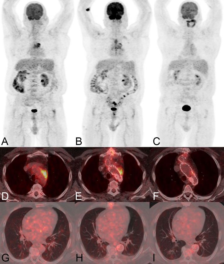
www.nature.com/scientificreports/
number 2018-01904). All procedures were performed in accordance with the 1964 Helsinki declaration and its
later amendments or comparable ethical standards.
Diagnosis of INAA was made in an overall appraisal of clinical presentation (pain, fever, sepsis), laboratory
(positive microbiological culture of aortic/aneurysmatic wall, presence of bacteria in thrombus or blood cul-
ture; elevation of inflammatory markers such as C-reactive protein and leucocytes) and imaging5. Diagnosis of
secondary VGI due to placement of stentgrafts relied on the MAGIC c riteria12.
In all prospectively enrolled patients, criteria for the termination of antimicrobial therapy were a combination
of the absence of clinical features of infection, normal CRP, and reduced metabolic activity in PET/CT. Cure of
INAA was defined as a combination of absence of clinical features, normal CRP, and reduced metabolic activ-
ity in PETCT at least three months after termination of antibiotic therapy (the latter only in the prospectively
enrolled patients).
For retrospective inclusion of patients, we performed a retrospective chart review in all patients with sus-
pected INAA, who were examined with PET/CT between the years 2005 and 2018, if the term “mycotic aneu-
rysm”, “infective aneurysm”, or “infected aneurysm” was mentioned in the written PET/CT report, and was found
to refer to an aneurysm of the thoracic, abdominal or pelvic arteries.
PET/CT examinations and patient follow‑up. The study design for all consecutively and prospectively
enrolled patients included consecutive PET/CT scans, at baseline, during follow-up on antimicrobial therapy,
and at the end of antimicrobial treatment. If feasible, a control PET/CT three months after the end of antimi-
crobial therapy was performed, to document continuous cure or possible signs for recurrence of infection. For
all retrospectively enrolled patients, PET/CT was performed depending on the clinical situation of the patient;
the reasons for patient’s referral are given in the results section. The exact time points of follow-up examinations
with regard to the baseline are demonstrated in Fig. 1. For all patients, baseline was defined as first PET/CT scan
at diagnosis of INAA.
We performed clinical follow-up of all patients by reviewing electronic patient charts. Patient data were
recorded at the time of imaging, and at the last recorded clinical visit (recorded until March 2020).
Recorded data at baseline and at follow-up included patient demographics, clinical information, laboratory
data (e.g. level of CRP and leucocyte count), results of microbiology, results from other diagnostic procedures,
and information about treatment.
PET/CT data acquisition and image analysis. Five different types of PET/CT scanners were used
within the study period between 2005 and 2018, i.e. a Discovery ST16, a Discovery VCT, two Discovery MI a
Discovery 690 and a Discovery 710 (all GE Healthcare, Waukesha, WI). Body weight, height, and blood glucose
level were measured prior to imaging (blood glucose levels < 12 mmol/l were accepted13). All PET/CT examina-
tions followed basic study protocols: patients fasted for at least four hours, body-weight adjusted intravenous
injection of FDG14, standardized uptake time of 60 min in supine position, non-enhanced CT scans for attenu-
ation correction, data acquisition with arms overhead whenever possible.
All PET/CT examinations were independently analysed by two double board certified radiologists and nuclear
medicine physicians, blinded to all clinical patient data. Readers determined the FDG uptake pattern in the
aneurysm or graft to be focal or d iffuse15. A consensus reading was performed if results differed. Furthermore,
both readers quantified the FDG-uptake in the aneurysm or in the vascular graft by measuring the maximum
standardized uptake value (SUVmax) in the aneurysm or graft as well as in the liver and mediastinal bloodpool
(the latter as background for reference). In case SUVmax measurements were not identical among both readers
concerning the measurements in the aneurysm or graft, a consensus reading was performed; in case of discordant
SUVmax values for the background measurements, the mean was calculated. To compensate for differences in the
sensitivity of the different PET/CT scanner generations between the years 2005 and 2018, and resulting differ-
ences in quantitative PET parameters (i.e. SUVmax), we calculated relatively scanner-independent FDG uptake
ratios (i.e. SUVratio was defined as uptake in the aneurysm of graft in relation to the mediastinal blood pool).
To determine differences between the course of C-reactive protein and SUVratio we calculated trends between
consecutive examinations, and defined an arbitrary cut-off of > 20% between differences in trends as relevant.
Furthermore, for all baseline and follow-up PET/CT examinations, readers determined whether a INAA or a
VGI was present or not, using a visual 4-point grading score. Score 1 (no signs for infected aortic aneurysm/VGI)
and score 2 (most likely a non-infected aneurysm/VGI) were considered negative for infected aortic aneurysm,
while score 3 (suspicion of infected aortic aneurysm/VGI) and score 4 (clear signs of infected aortic aneurysm/
VGI) were considered positive for infection.
Statistical analyses. Variables were expressed as median and interquartile range (25th, 75th percentiles)
or percentages. Kaplan–Meier estimates were used to describe survival at 1 and 5 years. We compared sizes and
visual grading scores of INAA from initial and last CT scans using nonparametric pairwise Wilcoxon signed-
rank test. Mixed-effects multilevel linear regression were applied to analyze the individual changes of SUVmax,
SUVratio and CRP. Statistical analysis were performed using commercially available software (Stata/SE, Version
15.1, StataCorp, College Station, Texas).
Results
Patient population. Seven out of 21 (33%) patients with confirmed INAA were prospectively enrolled,
while 14 (66%) patients were retrospectively included. Five out of 14 retrospective patients were excluded due to
lack of written consent and one out of the seven prospective patients was excluded due to lack of clinical follow-
up after the first PET/CT.
Scientific Reports | (2021) 11:5065 | https://doi.org/10.1038/s41598-021-84658-z 2
Vol:.(1234567890)
www.nature.com/scientificreports/

                                  Figure 1.  Courses over time of metabolic activity in PET/CT (SUVmax and SUVratio in the two graphs on
                                  the left) and CRP (graph on the right) in patients with proven infective native aortic aneurysms. Note: PET/CT
                                  provides additional or altering information (as defined in the methods section) on the course of disease in at
                                  least 14 comparisons (56%) in 11 patients (73%), as compared to CRP alone (i.e. patients: 02, 03, 04, 05, 06, 07,
                                  08, 09, 11, 14, 15). Abbreviations: PET: positron emission tomography; SUV: standardized uptake value; CRP:
                                  C-reactive protein.

                                     Thus, the final patient population consisted of 15 patients. All patients were eventually treated with open
                                  and/or endovascular repair (six prior to the first PET/CT, seven prior to the second, and two prior to the third
                                  PET/CT) (Table 1).
                                     At the time of the initial PET/CT examination, patients had a median age of 61 years (IQR 54–85), one patient
                                  (7%) was female, six (40%) were smokers or had a history of smoking, two (13%) patients were diabetic, and six
                                  (40%) had renal insufficiency. The diagnoses of INAA were confirmed by blood culture (n = 11; 73%), culture or
                                  PCR from tissue obtained during surgical revision (n = 2; 13%) and serology (n = 2, 13%) (Table 2).

                                  PET/CT examinations. We performed a total of 53 PET/CT scans in 15 patients (Figs. 1, 2, 3, Table 3) after
                                  intravenous injection of a median of 353 Megabecquerel of FDG (IQR 334—400). Initial imaging was per-
                                  formed for nine (60%) patients before and for six (40%) patients after vascular intervention, 11 (73%) received
                                  antimicrobial therapy at the time of initial imaging. The median visual 4-point grading score was 4 (11 × score 4,
                                  3 × score 3, 1 × score 2). Eventually, all patients underwent vascular intervention with graft placement, and were
                                  treated with antibiotics (Tables 1 and 2). All aneurysms (or grafts) had increased focal FDG uptake on the initial
                                  PET/CT examination (SUVmax 6.8 (IQR 5.7–21.8); SUVmax aneurysm/graft to blood pool background ratio
                                  2.7 (IQR 2.2–7.3)). FDG uptake in all aneurysms/grafts (100%) was higher than liver background (SUVmax 3.4
                                  (IQR 2.7–6.1) (Figs. 1, 2, 3, Table 3).
                                      The first follow-up PET/CT (112 (IQR 95–721) days after the first PET/CT) was performed in all patients, and
                                  13 (87%) patients received antimicrobial therapy at the time (in one patient antimicrobial therapy was started
                                  after the second PET/CT in another therapy was already terminated). The median visual 4-point grading score

Scientific Reports |   (2021) 11:5065 |                https://doi.org/10.1038/s41598-021-84658-z                                                     3

                                                                                                                                              Vol.:(0123456789)
www.nature.com/scientificreports/
Outcome
Days after end
Location of Days after initial of antimicrobial Signs for
Pat Age Vascular surgery Surgical technique aneurysm Reoperation operation treatment Status recurrence
01 62 Y-Graft Open Infrarenal aorta Second look 1938 1832 Cured No
02 58 Y-EAP Endovascular Infrarenal aorta n.a 1198 826 Cured No
T-EVAR debranch-
03 64 Endovascular Aortic arch n.a 1451 1346 Cured No
ing
T-EVAR debranch-
04 70 Endovascular Aortic arch n.a 287 n.a Ongoing* n.a
ing
Descending tho-
05 82 T-EVAR Endovascular n.a 758 643 Cured no
racic aorta
Resection of EVAR
Acute bridging Extraanatomic
06 48 EVAR bifurcation Hybrid Infrarenal aorta Y-graft 579 334 Cured No
prothesis Open abdomen
treatment
Resection of parts of
TEVAR
Supra-/juxta-renal the duodenum due
07 50 Renovisceral Hybrid 2585 n.a Ongoing Stable disease
aorta to fistula
debranching
VAC-on-vessel
Descending tho-
08 76 T-EVAR Endovascular n.a 1289 1183 Cured No
racic aorta
Resection of EVAR
Extraanatomic
Acute bridging
09 56 Hybrid Iliac artery Y-graft 2926 2508 Cured No
EVAR
Open abdomen
treatment
Debranching
A. mesenterica Prognostic laparos-
Descending tho-
10 71 superior and T. Hybrid copy due to peri- 1059 293 Cured No
racic aorta
coeliacus aortic abscess
EVAR
Descending
Viscerales debanch-
11 52 TA-EVAR Hybrid thoracic aorta/ 227 n.a Cured No†
ing
suprarenal
12 40 Y-EAP Endovascular Infrarenal aorta n.a 2798 n.a Cured No‡
Renoviscerales
Open abdomen
13 61 debranching Open Juxtarenal aorta 3163 3109 Cured No
treatment
Graft implantation
Open abdomen
14 61 Graft implantation Open Infrarenal aorta 1919 n.a Ongoing Stable disease
treatment
15 85 EVAR Endovascular Infrarenal aorta n.a 1931 804 Cured No
Table 1. Patient demographics, data on interventional procedures, and patient outcome data of the final study
population in patients with proven infective native aortic aneurysms. Pat: Patient number; na, not applicable.
*Patient died due to gastrointestinal bleeding, no relation to thoracic INAA. † Patient developed 1.5 years
later an infected pancreatic cyst. ‡ The active intravenous drug user died 8 years later due to aortic valve
endocarditis.
decreased to 3 (3 × score 4, 8 × score 3, 2 × score 2, 2 × score 1). The first follow-up showed an overall decrease in
SUVmax (5.5 (IQR 3.0–8.2) and SUVmax ratio (2.2 (IQR 1.6–2.9)); SUVmax decreased in 13 and increased in
two patients, SUVmax ratio decreased in 10 and increased in five patients (Figs. 1, 2, 3, Table 3).
In 11 patients, PET/CT was performed prior to the termination of the antimicrobial therapy. Compared to
background activity, PET/CT showed focally increased FDG uptake in nine patients (82%) and diffuse increased
FDG uptake in two patients (18%) with SUVmax 3.9 (IQR 2.7–6.8) and SUV ratio 1.7 (IQR 1.2–2.9). The FDG
uptake in the aneurysm/graft was higher than blood pool background in eight (73%) patients (Fig. 2) and higher
than liver background in seven (64%) patients. The median CRP was 12.0 mg/L (IQR 3–218). Of note, the median
time difference between the last PET/CT prior to the termination of antimicrobial therapy and the actual end of
antimicrobial therapy was a median 30 days (IQR 18–1059), and six examinations (55%) were considered to be
directly linked to the decision to end treatment.
A total of eight PET/CT were performed after termination of the antimicrobial therapy treatment in six
patients (SUVmax in all eight PET/CT: 4.2 (IQR 3.3–4.7); SUVmax in the first six PET/CT 3.9 (IQR 3.0–4.4).
Compared to background activity, six (75%) of these examinations showed increased and focal FDG uptake
(SUVmax 4.2 (IQR 3.3–4.7), SUV ratio 1.7 (IQR 1.5–4.1)), in two examinations (25%) the uptake was considered
diffuse; FDG uptake was higher than blood pool background (SUVmax 2.2 (IQR 2.0–2.9)) and liver background
(SUVmax 3.2 (IQR 2.7–3.6)) in all examinations (100%). The median visual 4-point grading score after termina-
tion of the antimicrobial therapy was 3 (0 × score 4, 5 × score 3, 2 × score 2, 1 × score 1).
The comparison of the last PET/CT prior to the termination of antimicrobial therapy to the first PET/CT after
termination of antimicrobial therapy (feasible in six patients) showed less focal FDG-uptake (from five to four)
Scientific Reports | (2021) 11:5065 | https://doi.org/10.1038/s41598-021-84658-z 4
Vol:.(1234567890)www.nature.com/scientificreports/
Antimicrobial treatment
Pat Microbiology* Intravenous Oral Strategy Duration (days) Complications
01 Escherichia coli Piperacillin/tacobactam Ciprofloxacin Prolonged 136 None
02 Streptococcus agalactiae Cetriaxone Amoxicillin/clavulanic acid Prolonged 382 None
03 Candida albicans Caspofungin Fluconazol Prolonged 192 None
04 Streptococcus agalactiae Penicillin Clindamycin Prolonged 289 Port infection
05 Streptococcus agalactiae Ceftriaxone Clindamycin Prolonged 220 None
Ciprofloxacin
06 Staphylococcus aureus Flucloxacillin Prolonged 261 None
Rifampicin
Streptococcus gallolyticus
Daptomycin Fluconazole
07 Candida albicans Lifelong Infinite None
Ertapenem Amoxicillin
Pediococcus acidilactici
Ceftriaxone
08 Streptococcus pneumoniae Moxifloxacin Prolonged 122 Vestibular toxicity due to gentamicin
Gentamicin
Bacteroides thetaiotaomicron/ Clostrid- Amoxicillin/clavulanic acid
09 Piperacillin/tacobactam Prolonged 458 None
ium perfringens Ciprofloxacin
Doxycycline
10 Coxiella burnetii na Prolonged 783 None
Plaquenil
Ceftriaxone
11 Streptococcus pneumoniae Clindamycin Prolonged 118 None
Gentamicin
Ciprofloxacin
12 Staphylococcus aureus Flucloxacillin Prolonged 176 None
Rifampicin
13 Salmonella typhi Ceftriaxon Ciprofloxacin Prolonged 99 None
(Doxycycline)
14 Coxiella burnetii na (Plaquenil) Lifelong Infinite Allergy to doxycyclin
Minocycline
15 Porphyromonas gingivalis Penicillin Clindamycin Prolonged 1139 None
Table 2. Data on microbiology and antimicrobial of the final study population in patients with proven
infective native aortic aneurysms. Pat: Patient number; na, not applicable. *Microbiology resulted from blood
culture (n = 11), culture or PCR from tissue obtained during surgical revision (n = 2) and serology (n = 2).
and decreasing FDG uptake in three patients for SUVmax and in two patients for SUVratio, while the uptake
increased in three patients for SUVmax and in four patients for SUVratio (SUVmax from 3.8 to 3.9 (IQR 2.5–4.4
and 3.0–4.4) and SUV ratio from 1.6 to 1.7 (IQR 0.9–1.9 and 1.5–4.1)). Notably, the two patients with a signifi-
cantly rising SUV ratio (from 0.7 to 2.0 and from 0.7 to 1.2; patient 9 and 13 in Table 3) had no signs of infection
at the last clinical follow-up, which was more than five and more than eight years after the last PET/CT and
without antimicrobial therapy. None of these patients had clinical signs of new VGI at the last clinical follow-up.
Sizes of INAA, measured on CT images, decreased significantly by 1.45 cm on average from the initial to the
last examination (p = 0.016, Table 3). In parallel, visual grading scores for INAA decreased by 1.2 on average
(p < 0.01, Table 3).
Discrepancies between trends of SUVratio and CRP. With 53 PET/CT performed in 15 patients we
were able to calculate 38 trends of SUVratio and 25 trends of CRP (immediate data of CRP missing for 13 PET/
CT).
The decrease or increase of SUVratio was paralleled by decreasing or increasing CRP values in 16 of 25 (64%)
trends, while it was opposed in nine (36%) examinations. Nineteen comparisons (76%) showed either opposed
trends (as mentioned above, n = 9) and/or differences between trends of CRP and SUVratio > 20%. For these
trends, PET/CT provided a conclusive explanation for the difference in five comparisons (26%) in three patients
(i.e. additional foci of infection with different response to therapy: patient 05, 11, and 14 in Table 3 and Fig. 2). No
apparent reason for high differences in trend could be determined for four comparisons (21%) in four patients
(i.e. three opposed trends between first and second PET/CT in patient 04, 07, and 11 (Table 3) and a high 46%
increase in CRP paralleled by a low 11% increase of SUVratio in patient 09 in Table 3). In five comparisons of
five patients, CRP values showed very good or complete responses to therapy at the time of the first follow-up
PET/CT, which were paralleled by less pronounced partial responses in SUVratio (patient 02, 03, 06, 08, and
15); further clinical follow-up could not determine which methods better described the actual clinical response
to therapy (as all other clinical parameters defining cure of INAA were normal/unchanged). The final five com-
parisons of differences between trends of CRP and SUVratio which were > 20%, were rated to be errors as the
values were very low and partly normal, i.e. between the fourth and fifth PET/CT of patient 02 (Table 3), CRP
values decreased by 80% from 2.1 to 1.3, which was paralleled by a 20% increase in SUV ratio from 1.65 to 1.84.
Hence, PET/CT provided additional or altering information on the course of disease in at least 14 compari-
sons (56%) in 11 patients (73%), as compared to CRP alone.
Patient outcome. Patients were clinically followed for a median of 1496 days (IQR 961–3187 days) after
their initial PET/CT examination and for a median of 1005 days (IQR 683–3109 days) after their last PET/CT
Scientific Reports | (2021) 11:5065 | https://doi.org/10.1038/s41598-021-84658-z 5
Vol.:(0123456789)www.nature.com/scientificreports/
Figure 2. PET/CT of an 82-year old male patient (patient 05 in Tables 1, 2 and 3) with a vascular graft
infection and spondylodiscitis due to Streptococcus agalactiae showed a new focal FDG uptake in the wall of
the thoracic aorta in September 2017. Both readers rated the thoracic finding as a infective native aneurysm
despite the fact that the vessel diameter was not pathologically widened. The first PET/CT follow-up in revealed
a progression of the aneurysm in size with stable increased FDG uptake; at the same time the FDG uptake of
the spondylodiscitis increased while it partially decreased in the vascular graft infection; C-reactive protein
and white blood cell count decreased. After subsequent thoracic endovascular repair with an Endurant II Stent
Graft Systems (MEDTRONIC), two further PET/CT follow-up, before and after termination of antimicrobial
therapy (223 days of therapy) showed faint residual FDG uptake in all sites of infection. At the last clinical
follow-up in January 2019 the patient was in good clinical condition with no sign of infection. Note: Panels A-D
show maximum intensity reconstructions of PET; Panels E-P show fused PET/CT images. Abbreviations: PET:
positron emission tomography; CT: computed tomography; FDG: 18F-fluorodeoxyglucose.
examination. The one-year and five-year freedom from all-cause mortality was 92% (95% confidence interval
[CI] 57–99%), for both. Overall mortality was 3.5% (0.9–13.8) per year. Two events occurred in 57.8 years of
Scientific Reports | (2021) 11:5065 | https://doi.org/10.1038/s41598-021-84658-z 6
Vol:.(1234567890)www.nature.com/scientificreports/
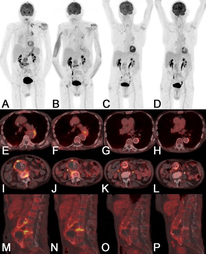
Figure 3. A 70-year old male patient (patient 04 in Tables 1, 2 and 3) presented with chest pain and signs for
infection (C-reactive protein 122 mg/L). The initial PET/CT examination showed strongly increased FDG-
uptake in the wall of an aortic arch aneurysm and no other infectious foci. Both PET/CT readers suspected a
infective native aneurysm; which was clinically confirmed (Streptococcus agalactiae in blood cultures). At first
PET/CT follow-up (ongoing antimicrobial therapy, after thoracic endovascular repair with a Conformable
GORE TAG Thoracic Endoprosthesis and debranching) a strong residual focal FDG-uptake was detected
adjacent to the graft, in line with a secondary vascular graft infection. The second PET/CT follow-up (ongoing
antimicrobial therapy), showed only very faintly increased FDG-uptake adjacent to the graft. However, a new
pneumonia in the right lower lobe was incidentally detected. The patient died 60 days after the last PET/CT
due to a gastrointestinal bleeding not related to the thoracic infective native aortic aneurysm. Note: Panels A-C
show maximum intensity reconstructions of PET; Panels D-I show fused PET/CT images. Abbreviations: PET:
positron emission tomography; CT: computed tomography; FDG: 18F-fluorodeoxyglucose.
follow-up time; i.e. two patients (13%) died because of reasons considered unlikely to be related to INAA; one
died due to gastrointestinal bleeding (location of the INAA was in the aortic arch), the other due to aortic valve
endocarditis (after more than six years after successful treatment of the INAA).
The two patients (13%) on continuous antibiotic therapy, showed higher metabolic activity in the INAA
(SUVmax 7.9 and 4.7, SUVratio 2.4 and 1.6) than the mean metabolic activity of those patients that were scanned
prior to the termination of antimicrobial treatment (as mentioned above: SUVmax 3.9 (IQR 2.7–6.8) and SUV
Scientific Reports | (2021) 11:5065 | https://doi.org/10.1038/s41598-021-84658-z 7
Vol.:(0123456789)www.nature.com/scientificreports/
Initial morphologic Morphologic signs
PET/CT signs in CT at last CT
Size INAA in cm
visual imaging Size INAA in cm
Pat 1 2 3 4 5 6 score visual imaging score
SUVmax 5.4 4.4 4.3 4.2 4.7 *Stranding, fluid, †
Stranding
CRP (mg/L) 199 n.a n.a 6 7 gas
01
Days to start of AB −3 − 112 − 230 − 328 − 740 7.1 3.4
Days to end of AB 133 24 − 94 − 192 − 604 4 3
SUVmax 9.3 6.9 5.3 4.1 4.4 †
Stranding, fluid, CE *Lymph
CRP (mg/L) 7 4 3 2 1
02
Days to start of AB − 79 − 192 − 284 − 380 − 475 6.7 4.3
Days to end of AB 303 190 98 2 − 93 4 3
†
SUVmax 7.8 6.4 Stranding, CE, †
None
CRP (mg/L) 27 3 lymph
03
Days to start of AB − 45 − 175 2.7 Not measurable
Days to end of AB 147 17 4 3
SUVmax 10 8.2 3.9 †
Stranding, CE *None
CRP (mg/L) 122 35 n.a
04
Days to start of AB 6 − 64 − 230 4.6 Not measurable
Days to end of AB n.a n.a n.a 4 3
†
SUVmax 6.2 5.7 3.8 3.5 Stranding, CE, †
Stranding, lymph
CRP (mg/L) 95 72 15 19 lymph
05
Days to start of AB − 21 − 68 − 214 − 303 4.4 4.4
Days to end of AB 199 152 6 − 83 4 3
†
SUVmax 11 6.8 Stranding, CE, *Stranding, fluid, CE,
CRP (mg/L) 145 15 lymph gas, lymph
06
Days to start of AB −3 − 148 6.6 4.9
Days to end of AB 258 113 4 3
SUVmax 6.1 7.9 6.8 7.0 8.3 7.9 *Stranding, fluid,
*Stranding, fluid, gas
CRP (mg/L) 17 4 2 2 1 n.a gas
07
Days to start of AB − 818 − 1011 − 1170 − 1337 − 1515 − 1702 3.3 3.6
Days to end of AB n.a n.a n.a n.a n.a n.a 3 4
SUVmax 7.9 3.3 †
Stranding, fluid, CE *Stranding
CRP (mg/L) 69 1
08
Days to start of AB −6 − 101 5.5 3.1
Days to end of AB 116 21 4 3
SUVmax 4.0 2.6 3.9 2.0 4.4
*Stranding, lymph *Stranding
CRP (mg/L) 11 16 n.a n.a 3
09
Days to start of AB − 71 − 176 − 325 − 421 − 520 2.7 2.7
Days to end of AB 387 282 133 37 − 62 3 2
SUVmax 22 5.7 †
Stranding, fluid, CE *Stranding
CRP (mg/L) 31 n.a
10
Days to start of AB − 11 − 721 6.6 4.0
Days to end of AB 772 62 4 3
SUVmax 4.5 3.9 2.8 †
Stranding, fluid, CE *Stranding
CRP (mg/L) 100 218 129
11
Days to start of AB 40 0 − 153 6.0 6.4
Days to end of AB 158 118 na 3 1
SUVmax 6.8 2.4 2.6 2.0 †
*Stranding, fluid Lymph
CRP (mg/L) 146 n.a n.a n.a
12
Days to start of AB −4 − 98 − 464 − 2602 5.8 Not measurable
Days to end of AB n.a n.a n.a n.a 4 1
†
SUVmax 1.1 2.3 Stranding, fluid,
*Stranding
CRP (mg/L) 23 n.a CE, gas
13
Days to start of AB − 20 − 163 4.1 3.3
Days to end of AB 79 − 64 2 2
SUVmax 6.6 5.5 5.8 4.1 5.9 4.7
*Stranding, fluid *Stranding, fluid
CRP (mg/L) 10 15 4 3 3 4
14
Days to start of AB 24 − 253 − 618 − 933 − 1027 − 1646 8.3 4.2
Days to end of AB n.a n.a n.a n.a n.a n.a 4 2
SUVmax 7.2 2.1 *Stranding, fluid,
Lymph
CRP (mg/L) 126 9 lymph
15
Days to start of AB 6 − 96 4.5 4.1
Days to end of AB 1145 1043 4 1
Table 3. Patient demographics, follow-up PET/CT and CT findings of the final study population in patients
with proven infective native aortic aneurysms. Pat: Patient number; AB: antimicrobial treatment; n.a.: not
applicable; stranding: fat stranding; fluid: fluid collection; CE: contrast enhancement, gas: gas formation;
lymph.: lymph adenopathy. *Non-enhanced CT;† contrast-enhanced CT.
Scientific Reports | (2021) 11:5065 | https://doi.org/10.1038/s41598-021-84658-z 8
Vol:.(1234567890)www.nature.com/scientificreports/
ratio 1.7 (IQR 1.2–2.9)). The conditions of these two patients were determined to be stable at the last clinical
visit 261, and 729 days after the last performed PET/CT.
One patient (7%) showed recurrent signs of infection at the last clinical visit, which were attributed to an
infected pancreatic cyst. All other patients (n = 10, 67%) did not show any signs of infection at the last clinical
visit and all were without antimicrobial therapy (Table 1).
Among patients alive without antimicrobial therapy, yearly changes of SUVmax, SUVratio and CRP were
-3.5 (95% CI -5.5 to -1.5, p < 0.01), -1.0 (-1.7 to -0.3, p < 0.01) and -58 (-96 to -20, p < 0.01), respectively. Among
patients still on antimicrobial therapy or who died, yearly changes of SUVmax, SUVratio and CRP were -0.39
(95% CI -0.81 to 0.03, p = 0.07), -0.20 (-0.33 to -0.08, p < 0.01) and -4.6 (-14.2 to 5.1, p = 0.35), respectively.
Discussion
To the best of out knowledge, this is the first study investigating the role of PET/CT in therapy control of INAA.
Our study results show: (i) PET/CT adds additional information in therapy control of INAA. (ii) Metabolic
activity in the aneurysms remains slightly elevated after the end of antimicrobial therapy and therefore should
not be mistaken for persistent infection.
All patients of the present study were treated with open and/or endovascular repair, and as this treatment in
INAA frequently leads to secondary graft infections, we may compare the present study results to the findings of
three previous publications9–11 on therapy control with PET/CT in VGI. In line with these s tudies9–11, we found
that in addition to laboratory and clinical information, consecutive PET/CT examinations were a valuable source
of information for treatment monitoring, often displaying additional and sometimes even opposed results to
clinical and laboratory parameters.
Previous studies have documented a correlation between the course of CRP and SUVmax in therapy control
of patients with VGI10. In the present study, only a minority of comparisons (i.e. 24%) of CRP and the metabolic
activity in the aneurysm showed a similar course in therapy control of patients with INAA. The majority of cases
displayed large discrepancies between trends of CRP and metabolic activity or even opposed courses. The latter,
may partly be explained by additional foci of infection with different response to therapy, which was in line with
results of a previous study, showing correlation between CRP and SUVmax only in a subpopulation of patients
with VGI without additional infectious f oci9.
However, we could identify another group of comparisons with no apparent reason for the large discrepan-
cies between trends of CRP and metabolic activity, which may indicate, that CRP and metabolic activity in the
aneurysm are two unrelated identities, which may have to be evaluated independently, when treating patients
with INAA. Because of such discrepancies and the overall severity of INAA in general, we believe that in all
patients with INAA, decisions for further therapy should always be based also on other parameters such as
clinical aspects (general health status, fever, clinical signs of INAA and/or graft infection), and other laboratory
parameters (CRP, ESR, leucocytes).
Furthermore, we documented in line with previous reports16–18, that the metabolic activity in INAA is initially
high or very high, and comparable to the initial presentation of VGI in PET/CT9–11,15,19–25. Under antimicrobial
therapy the metabolic activity generally d ecreases9–11. However, the metabolic activity remains above background
level before and even after the end of medical treatment; despite the fact that we did not observe any cases of
recurrent infection. High rates of residual metabolic activity were also observed in patient populations with VGI
at the end of medical treatment. Husmann et al.10 observed a complete metabolic response to treatment in only
33% of patients with VGI, while we could not observe a complete response in any of our patients. The reason
for this difference remains unclear, and may possibly be due to differences in the course of two similar but yet
different diseases, or it may be due to bias as our study cohort was considerably smaller in number. Notably,
residual metabolic activity (i.e. SUVmax values slightly above background level) in the aneurysms after the end
of medical treatment should not be mistaken for persistence of infection in INAA, but may possibly represent a
sterile ongoing inflammatory reaction, as none of our patients showed signs of recurrence during the follow-up
of this series and an excellent patient outcome was observed.
Two patients died due to reasons not considered unrelated to INAA. The one-year and five-year freedom from
all-cause mortality was 93%, and was much higher than in previously published studies, which found five-year
survival rates between 53%26 and 55%7. Whether disease monitoring with PET/CT contributed to the excellent
clinical outcome in the present study still remains unanswered. However, we could not find any other obvious
systematic difference, which may account for the improved clinical outcome.
Limitations of the study. The present study population is heterogeneous, with varying numbers of fol-
low-up PET/CT examinations, as well as prospectively and retrospectively included patients, which precludes
standardized PET/CT intervals. Furthermore, due to the lack of comparable data in the literature, we defined an
arbitrary cut-off of > 20% between differences in trends of SUVratio and CRP as relevant.
Despite these limitations, the results of the present study appear consequential and should be confirmed in
further studies.
Conclusion
As compared to the course of C-reactive protein, PET/CT provides different and occasionally altering information
in therapy control of INAA. Of note, metabolic activity in the aneurysms remains slightly elevated even after the
end of antimicrobial therapy and should not be mistaken for persistent infection.
Received: 27 October 2020; Accepted: 19 February 2021
Scientific Reports | (2021) 11:5065 | https://doi.org/10.1038/s41598-021-84658-z 9
Vol.:(0123456789)www.nature.com/scientificreports/
References
1. Wilson, W. R. et al. Vascular graft infections, mycotic aneurysms, and endovascular infections: a scientific statement from the
American Heart Association. Circulation 134, e412–e460 (2016).
2. Sorelius, K., Budtz-Lilly, J., Mani, K. & Wanhainen, A. Systematic review of the management of mycotic aortic aneurysms. Eur. J.
Vasc. Endovasc. Surg. 58, 426–435 (2019).
3. Muller, B. T. et al. Mycotic aneurysms of the thoracic and abdominal aorta and iliac arteries: experience with anatomic and extra-
anatomic repair in 33 cases. J. Vasc. Surg. 33, 106–113 (2001).
4. Johansen, K. & Devin, J. Mycotic aortic aneurysms: a reappraisal. Arch. Surg. 118, 583–588 (1983).
5. Sorelius, K. et al. Nationwide study of the treatment of mycotic abdominal aortic aneurysms comparing open and endovascular
repair. Circulation 134, 1822–1832 (2016).
6. Ding, N. et al. CT texture analysis predicts abdominal aortic aneurysm post-endovascular aortic aneurysm repair progression.
Sci. Rep. 10, 12268 (2020).
7. Sorelius, K. et al. Endovascular treatment of mycotic aortic aneurysms: a European multicenter study. Circulation 130, 2136–2142
(2014).
8. Wanhainen, A. et al. Editor’s choice—European Society for Vascular Surgery (ESVS) 2019 clinical practice guidelines on the
management of abdominal aorto-iliac artery aneurysms. Eur. J. Vasc. Endovasc. Surg. 57, 8–93 (2019).
9. Husmann, L. et al. (1)(8)F-FDG PET/CT for therapy control in vascular graft infections: a first feasibility study. J. Nucl. Med. 56,
1024–1029 (2015).
10. Husmann, L. et al. The role of FDG PET/CT in therapy control of aortic graft infection. Eur. J. Nucl. Med. Mol. Imaging. 45,
1987–1997 (2018).
11. Machelart, I. et al. Graft infection after a Bentall procedure: a case series and systematic review of the literature. Diagn. Microbiol.
Infect. Dis. 88, 158–162 (2017).
12. Lyons, O. T. et al. Diagnosis of aortic graft infection: a case definition by the management of aortic graft infection collaboration
(MAGIC). Eur. J. Vasc. Endovasc. Surg. 52, 758–763 (2016).
13. Eskian, M. et al. Effect of blood glucose level on standardized uptake value (SUV) in (18)F- FDG PET-scan: a systematic review
and meta-analysis of 20,807 individual SUV measurements. Eur. J. Nucl. Med. Mol. Imaging. 46, 224–237 (2019).
14. Morand, G. B. et al. Maximum standardized uptake value (SUVmax) of primary tumor predicts occult neck metastasis in oral
cancer. Sci. Rep. 8, 11817 (2018).
15. Sah, B. R. et al. Diagnostic performance of F-FDG-PET/CT in vascular graft infections. Eur. J. Vasc. Endovasc. Surg. 49(4), 455–464
(2015).
16. Husmann, L. et al. Diagnostic accuracy of PET/CT and contrast enhanced CT in patients with suspected infected aortic aneurysms.
Eur. J. Vasc. Endovasc. Surg. 59, 972–981 (2020).
17. Ben Shimol, J. et al. The utility of PET/CT in large vessel vasculitis. Sci. Rep. 10, 17709 (2020).
18. Hannsberger, D., Heinola, I., di Summa, P. G. & Sorelius, K. The value of 18F-FDG-PET-CT in the management of infective native
aortic aneurysms. Vascular. https://doi.org/10.1177/1708538120987971 (2021).
19. Husmann, L. & Hasse, B. PET-CT in vascular graft infections. Zentralbl Chir. 142, 502–505 (2017).
20. Keidar, Z., Engel, A., Hoffman, A., Israel, O. & Nitecki, S. Prosthetic vascular graft infection: the role of 18F-FDG PET/CT. J. Nucl.
Med. 48, 1230–1236 (2007).
21. Keidar, Z. & Nitecki, S. FDG-PET in prosthetic graft infections. Semin. Nucl. Med. 43, 396–402 (2013).
22. Hasse, B. et al. Vascular graft infections. Swiss. Med. Wkly. 143, w13754 (2013).
23. Tokuda, Y. et al. Detection of thoracic aortic prosthetic graft infection with 18F-fluorodeoxyglucose positron emission tomography/
computed tomography. Eur J. Cardiothorac. Surg. 43, 1183–1187 (2013).
24. Fukuchi, K. et al. Detection of aortic graft infection by fluorodeoxyglucose positron emission tomography: comparison with
computed tomographic findings. J. Vasc. Surg. 42, 919–925 (2005).
25. Husmann, L. et al. Comparing diagnostic accuracy of (18)F-FDG-PET/CT, contrast enhanced CT and combined imaging in
patients with suspected vascular graft infections. Eur. J. Nucl. Med. Mol. Imaging. 46, 1359–1368 (2019).
26. Luo, C. M. et al. Long-term outcome of endovascular treatment for mycotic aortic aneurysm. Eur. J. Vasc. Endovasc. Surg. 54,
464–471 (2017).
Acknowledgements
We are grateful to our patients for their participation in the study. We thank C. Mueller/ S. Bajrami, study nurses
and Ch. Laich /C. Voegtli for administrative assistance. The members of the VASGRA Cohort Study are (in
alphabetical order): A. Anagnostopoulos, B. Hasse (PI), N. Eberhard, M. Hoffmann, L. Husmann, R. Kopp, B.
Ledergerber, Z. Rancic, C.A. Mestres, R. Zbinden, A. Zinkernagel.
Author contributions
B.H. and L.H. designed the study. L.H., M.H., B.L. and B.H. analysed the data. L.H. wrote the first draft, and L.H.
and B.H. wrote the final version of the manuscript. The remaining investigators (A.A., N.E., M.K., K.K., I.A.B.,
C.A.M., Z.R.) contributed to data collection and interpretation of the data, reviewed drafts of the manuscript,
and approved the final manuscript.
Funding
This study was financed within the framework of the Vascular Graft Cohort Study (VASGRA), supported by the
Swiss National Science Foundation (SNF) grant 320030_184918/1. This work was also supported by the Clinical
Research Priority Program of the University of Zurich for the CRPP Precision medicine for bacterial infections.
M.H. is a recipient of investigator initiated study grants by GE Healthcare. The funders had no role in study
design, data collection and analysis, decision to publish, or preparation of the manuscript.
Competing interests
The authors declare no competing interests.
Additional information
Correspondence and requests for materials should be addressed to L.H.
Reprints and permissions information is available at www.nature.com/reprints.
Scientific Reports | (2021) 11:5065 | https://doi.org/10.1038/s41598-021-84658-z 10
Vol:.(1234567890)www.nature.com/scientificreports/
Publisher’s note Springer Nature remains neutral with regard to jurisdictional claims in published maps and
institutional affiliations.
Open Access This article is licensed under a Creative Commons Attribution 4.0 International
License, which permits use, sharing, adaptation, distribution and reproduction in any medium or
format, as long as you give appropriate credit to the original author(s) and the source, provide a link to the
Creative Commons licence, and indicate if changes were made. The images or other third party material in this
article are included in the article’s Creative Commons licence, unless indicated otherwise in a credit line to the
material. If material is not included in the article’s Creative Commons licence and your intended use is not
permitted by statutory regulation or exceeds the permitted use, you will need to obtain permission directly from
the copyright holder. To view a copy of this licence, visit http://creativecommons.org/licenses/by/4.0/.
© The Author(s) 2021
Scientific Reports | (2021) 11:5065 | https://doi.org/10.1038/s41598-021-84658-z 11
Vol.:(0123456789)You can also read