Transbronchial cryobiopsy for diagnosing parenchymal lung diseases: real-life experience from a tertiary referral center
←
→
Page content transcription
If your browser does not render page correctly, please read the page content below
Original article: Clinical research

SARCOIDOSIS VASCULITIS AND DIFFUSE LUNG DISEASES 2021; 38 (1); e2021004         DOI: 10.36141/svdld.v38i1.11029           © Mattioli 1885

Transbronchial cryobiopsy for diagnosing parenchymal lung
diseases: real-life experience from a tertiary referral center
Demet Turan1, Efsun Gonca Uğur Chousein1, Aysu Sinem Koç 2, Mustafa Çörtük 1, Zeynep Yıldırım1,
Bariş Demirkol 1, Mehmet Akif Özgül 1, Halit Çınarka1, Neslihan Akalın3, Aytül Hande Yardımcı4,
Erdoğan Çetinkaya1
1
  University of Health Sciences Turkey, Yedikule Chest Disease and Thoracic Surgery Education and Research Hospital, Department of Chest
Diseases, Istanbul, Turkey; 2Ministry of Health, Dr. Yaşar Eryilmaz Dogubayazit State Hospital, Clinic of Chest Diseases, Agri, Turkey;
3
  University of Health Sciences Turkey, Yedikule Chest Diseases and Thoracic Surgery Education and Research Hospital, Department of
Pathology, Istanbul, Turkey; 4University of Health Sciences Turkey, Basaksehir Cam and Sakura City Hospital, Department of Radiology,
Istanbul, Turkey

Abstract. Background and Objectives: Transbronchial cryobiopsy (cryo-TBB) is increasingly being used in
the diagnosis of diffuse parenchymal lung diseases (DPLD). Varying diagnostic success and complication rates
have been reported. Herein we report our experience with cryo-TBB, focusing on diagnostic yield, factors affect-
ing diagnosis, and safety. Methods: This retrospective study was conducted in a tertiary referral chest diseases
hospital. Data regarding the patients, procedures, complication rates, diagnostic yield, and the final diagnosis
made by a multidisciplinary committee at all diagnosis stages were evaluated. Results: We recruited 147 patients
with suspected DPLD. The definitive diagnosis was made pathologically in 98 of 147 patients (66.6%) and us-
ing a multidisciplinary approach in 109 of 147 (74.1%) cases. The number of samples had a significant effect
on diagnostic success. Histopathologic diagnostic yield and diagnostic yield with a multidisciplinary committee
after a single biopsy were 50%, and histopathological diagnostic yield and diagnostic yield with multidiscipli-
nary committee increased to 71.4% and 85.7%, respectively, with a second biopsy (p = 0.034). The incidence
of mild-to-moderate hemorrhage was 31.9%; no severe hemorrhage occurred. Pneumothorax rate was 15.6%,
and the mortality rate was 0.68%. Conclusions: Cryo-TBB has sufficient diagnostic yield in the context of a
multidisciplinary diagnosis with acceptable complication rates. Performing at least 2 biopsies and from at least
2 segments increases diagnostic success.

Key words: Transbronchial cryobiopsy, bronchoscopy, parenchymal lung diseases, diagnostic yield

Received: 26 November 2020                                             Introduction
Accepted after revision: 23 February 2021

Correspondence: Demet Turan                                                 Diffuse parenchymal lung diseases (DPLDs) are
Department of Chest Diseases, University of Health Sciences            a heterogeneous group of lung parenchymal disorders
Turkey, Yedikule Chest Disease and Thoracic Surgery Education          with varying treatment options and prognoses. Given
and Research Hospital
Belgrat Kapi Yolu Street No:1, Zeytinburnu                             this heterogeneity, arriving at a differential diagnosis
Istanbul, 34020, Turkey                                                is critical (1, 2). However, arriving at a differential
Tel: 0090 505 638-43-06                                                diagnosis could be challenging given the similar and
E-mail: drdemetturan@gmail.com
2                                                    SARCOIDOSIS VASCULITIS AND DIFFUSE LUNG DISEASES 2021; 38 (1); e2021004

overlapping features between diseases. In most cases,           Methods
further invasive procedures are required for accurate
diagnosis following appropriate radiologic and phys-                 This retrospective study was held in Yedikule
iological evaluations (3). Surgical lung biopsy (SLB)           Chest Diseases and Thoracic Surgery Hospital inter-
continues to be the gold standard in identifying the            ventional pulmonology unit between January 2014
possible causes of DPLDs and their histopathologic              and December 2019. . The total number of patients
patterns despite the associated mortality and mor-              eligible for cryobiopsy was 155. Among those,
bidity (4-6). The 60-day mortality rate after SLB               4 patients developed desaturation immediately after
has been reported to range from 2% to 4%, signify-              the intubation with a rigid bronchoscope, the cryo
ing the need for less-invasive diagnostic procedures            probe could not be moved distally in 3 patients,
(5, 7). Among these minimally invasive methods,                 and 1 patient developed arrhytmia. Exclud-
conventional transbronchial forceps biopsy offers a             ing these 8 cases, the study included data from
less-invasive approach; however, its diagnostic yield is        147 patients. The diagnostic steps are detailed in the
low owing to small biopsy samples and crush artefacts           scheme (Figure 1) Inclusion criteria were DPLD
(8, 9). Transbronchial cryobiopsy (cryo-TBB), which             patients who could not be differentially diagnosed
seems to be safe procedure with lower complication              with clinical, laboratory, immunological and high
and mortality rates compared than SLB in DPLD                   resolution computed tomography (HRCT) data
diagnosis (4). Comparing the results from published             and were indicated for surgical lung biopsy and
cryo-TBB case series is challenging given the differ-           evaluated with cryo-TBB. All patients had pulmo-
ences in technical details of the procedure such as             nary function tests (PFTs), complete blood count,
the use of bronchial blockers, non-use of fluoroscopy,          HRCT, and echocardiography. The procedure was
and performing the procedure through an intubation              not applied to patients who had bleeding tendency
tube. These different procedural approaches can also            (INR> 1.5, Platelet count  40 mmHg,
tion rates (10).                                                uncontrolled cardiac arrhythmia, unstable angina,
     This study aimed to report our experience with             severe hypoxemia (pO2
SARCOIDOSIS VASCULITIS AND DIFFUSE LUNG DISEASES 2021; 38 (1); e2021004 3 (FVC)
4                                                       SARCOIDOSIS VASCULITIS AND DIFFUSE LUNG DISEASES 2021; 38 (1); e2021004

cryobiopsy in the exclusion criteria were included                    comparing the groups. Categorical values were
in the bleeding assessment. Chest x-ray control was                   analyzed using the Fisher exact test. The Mann–
planned 2 hours after the procedure to check for an                   Whitney U test and paired samples t tests were used
iatrogenic pneumothorax.                                              to compare continuous variables. A p value < 0.05
     Clinical information, radiological features, and                 was considered statistically significant.
biopsy results were then evaluated by a multidisci-
plinary committee including clinicians, radiologists,                 Results
and pathologists. This study was approved by the
ethics committee of our institution.                                       147 patients were enrolled to the study. Among
                                                                      those, 4 patients developed desaturation immediately
Statistical Analysis                                                  after the intubation with a rigid tracheoscopy, the
                                                                      cryo probe could not be moved distally in 3 patients,
     Statistical analysis was performed using IBM                     and 1 patient developed arrhytmia. Excluding these
SPSS Windows 22.0. Continuous variables were                          8 cases, the study included data from 147 patients.
presented as means ± standard deviations and medians                       The average age of the patients analyzed in the
(min-max) and categorical variables as numbers and                    study was 56.4 ± 13 and 82 of them were female
percentages. Parametric test assumptions (normality                   (55.8%) and 65 were male (44.2%). The baseline
and homogeneity of variances) were checked before                     characteristics of the patients are provided in Table 1.

                     Table 1. Patients’ characteristics at baseline and procedural details
                      Patient number (n)                                                     147

                      Mean age (years±SD)                                                56.4±13

                      Gender n (%) Female                                                82 (55.8)

                                        Male                                             65 (44.2)

                      Smoking (Pack-years) (n=95, mean±SD)                              12.5±16.8

                      CCI (n)      0-2                                                        90

                                   >3                                                         57

                      FVC % predicted (Mean±SD)                                         82.4±18.5

                      DLCO% predicted (Mean)                                           59.01±18.1

                                          Right lung (n,%)                              134 (91.2)

                                          Left lung (n,%)                                13 (8.8)
                      Biopsy
                                          One segment                                         73
                      location
                                          Two different segments                              69

                                          Three different segments                            5

                      Mean biopsy number (n±SD)                                              3.3±1

                      Freezing duration (seconds, min-max)                           4.8±1.2 s (4-10)

                      Small axis diameter (mm±SD)                                        2.8±0.6

                      Large axis diameter (mm±SD)                                        6.9±2.3

                     CCI Charlson Comorbidity index, FVC: Forced vital capacity, DLCO: Carbon monoxide
                     diffusion capacity, cTBB: Transbronchial lung cryobiopsy, SD: standard deviation
SARCOIDOSIS VASCULITIS AND DIFFUSE LUNG DISEASES 2021; 38 (1); e2021004 5
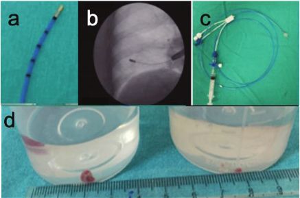
The number of biopsies taken from the right biopsy materials were 2.8 ± 0.6 mm, 6.9 ± 2.3 mm,
and the left lung were 134 (91.2%) and 13 (8.8%), respectively (Figure 2d).
respectively. Biopsies were taken from one segment Histopathological diagnosis was provided in
in 73 (49.6%) patients, from two different segments 98/147 cases (66.6%). The most common diagno-
in 69 (46.9%) patients, and from three different sis was nonspecific interstitial pneumonia (NSIP)
segments in 5 (3.4%) patients. Cryo-TBB was (n=46, 46.9%). Fourteen cases were diagnosed with
performed on different segments of the same lobe granulomatous inflammation (n=14, 14.3%), 12 were
in 61 cases and two different lobes and different malignancy (n=12, 12.2%), 6 with usual interstitial
segments in 15 cases . The mean biopsy number per pneumonia (UIP) (n=6, 6.1%), 6 with organizing
patient was 3.3 ± 1 (range 1-6). Mean freezing time pneumonia (OP) (n=6, 6.1%), and 6 with hypersensi-
during the procedure was 4.8 ± 1.2 seconds (range: tivity pneumonitis (HP) (n=6, 6.1%). The pathological
4-10). The shortest and longest diameters of the diagnoses and diagnostic yield are showed in Table 2.
Table 2. Histopathological and multidisciplinary diagnoses and diagnostic yield
Diagnosis n (%)
Non-Specific Interstitial Pneumonia 46 (46.9)
HISTOPATHOLOGICAL DIAGNOSIS
Hypersensitivity Pneumonia 6 (6.1)
Usual Interstitial Pneumonia 6 (6.1)
Organizing Pneumonia 6 (6.1)
Granulomatous Inflamation 14 (14.3)
Malignancy 12 (12.2)
Adenocarcinoma 10
Breast cancer metastas 1
Lymphoma 1
Othersa
8 (8.2)
DIAGNOSIS
Non-diagnostic 49
Diagnosis n (%)
Non-Specific Interstitial Pneumonia 38 (34.8)
MULTIDISCIPLANARY COUNCIL
Hypersensitivity Pneumonitis 20 (18.3)
Idiopathic pulmonary fibrosis 4 (3.7)
Sarcoid 12 (11)
DIAGNOSIS
Organizing Pneumonia 7 (6.4)
Malignancy 12 (12.2)
Adenocarsinoma 10
Breast cancer metastas 1
Lymphoma 1
Othersb
15 (13.8)
Non-diagnostic 38
Othersa: Alveolar microlithiasis (1), Non-spesific inflammation (1), Mosaic pattern (1), Histiocytosis X (1), Folicular bronchio-
litis (1), Chronic eosinophilic pneumonia (1), Acute Lung Injury (1), Unclassifiable interstitial lung disease (1)
Othersb: Rheumatoid lung disease (1), Tuberculosis (1), Drug induced lung disease (1), Alveolar microlithiasis (1), Nonresol-
ving pneumonia (1), Mosaic attenuation (1), Histiocytosis X (1), Follicular bronchiolitis (1), Acute interstitial pneumonia (1),
Unclassifiable interstitial lung disease (1)
SD: standard deviation6 SARCOIDOSIS VASCULITIS AND DIFFUSE LUNG DISEASES 2021; 38 (1); e2021004
A final diagnosis was made in 109 (74.1%) Complications occurred in 77 (49.6%) of
of 147 cases evaluated by the multidisciplinary 155 patients (patients who were excluded from cryo-
committee. The most common diagnosis was NSIP TBB and patients who included in the study). 69
in 38 patients. Other diagnoses are shown in Table 2. (46.9%) patients who developed complications due
SLB was performed in 26 (17.6%) out of to the cryo-TBB procedure included in the study.
38 patients who could not be diagnosed after Hemorrhage developed in 47 (31.9%) patients;
multidisciplinary evaluation, 21 patients were grade 1 in 28 (19%) patients, grade 2 in 19 (12.9%)
diagnosed while pathological diagnosis could not be patients. Grade 3 hemorrhage was not observed in
made in 5 patients (11 UIP, 5 HP, 2 Adenocarcinoma, any patient. Pneumothorax was developed in 23
1 NSIP, 1 Emphysema, 1 Anthracosis). The remaining (15.6%) patients after the procedure. Of those with
12 patients were patients who were not suitable for pneumothorax, 14 (60.9%) had a tube thoracostomy.
SLB due to their general condition or who did not Respiratory failure occurred in 3 patients (2%), 2 of
accept a further examination. these patients were discharged with a short-term
Overall histopathological yield and diagnostic noninvasive mechanical ventilation application in
yield with multidisciplinary approach were 66,6% the pulmonology wards, 1 patient was admitted to an
and 74,1%, respectively. Histopathological diagnos- intensive care and administered invasive mechanical
tic yield and diagnostic yield with multidisciplinary ventilation and died on the 15th day (Table 4).
approach after a single biopsy was 50%, and histo- When the factors affecting the development of
pathological diagnostic yield and diagnostic yield pneumothorax and hemorrhage were evaluated, there
with multidisciplinary approach increased to 71.4% was no significant difference in pneumothorax rates
and 85.7% by a second biopsy (p = 0.034). No fur- between patients who underwent 1 and 2 biopsies and
ther increase in diagnostic yield was observed when those who had more than 3 biopsies (Table 3). While
more than two samples were taken (Table 3). pneumothorax was detected in 13.7% of patients who
The histopathological diagnostic yield was underwent single-segment biopsy, and in 17.6% of
61.9% when multiple biopsies were taken from a patients who underwent 2 or more segment biopsies,
single segment, and 69.8% with multidisciplinary but it was not statistically significant. There was no
approach, while the histopathological diagnostic significant difference between pneumothorax rates
yield was 72.9% when multiple biopsies were taken in patients who underwent a biopsy from the same
from different segments of the same lobe and 81.1% and different lobes. The pneumothorax rate was 6.3%
with multidisciplinary approach. There is no statisti- when a biopsy was taken from the upper lobes, and
cally significant difference in diagnostic yield when 18.3% when a biopsy was taken from the lower lobes.
biopsy is taken from single or different lobes. No statistically significant difference was found in
Table 3. Diagnostic yield according to the number of samples
3 biopsy
Overall (diagnostic yield)
diagnostic yield (%) 1-2 biopsy (diagnostic yield) (n=31) (n=116) p value*
20 (64.51%)
78 (67.2%) 0.995
Pathological diagnosis 66.6 1 biopsy (n=10) 2 biopsy (n=21) p=0.244*
5 (50%) 15 (71.4%)
23 (74.1%)
Diagnosis with
Multidisciplinary 74.1 1 biopsy (n=10) 2 biopsy (n=21) 86 (74.1%) 0.775
comittee p=0.034*
5 (50%) 18 (85.7%)
*p value of the comparison between 1 and 2 biopsies
**p value of the comparison between 1-2 and ≥3 biopsies.SARCOIDOSIS VASCULITIS AND DIFFUSE LUNG DISEASES 2021; 38 (1); e2021004 7
Table 4. Safety outcomes and length of hospital stay (n=147) histopathologic diagnostic yield with cryo-TBB
Complications n (%) was 66.6%. When these histopathologic diagnoses
Pneumothorax 23/147 (total) were combined with a multidisciplinary approach,
Oxygen therapy 9 (39.1) the diagnostic yield was 74.1%. The most frequently
Tube Thoracostomy 14 (60.9) made histopathologic diagnoses and histopathologic
Hemorrhage Grade 1: 28/147 (19) diagnosis rates differ among the published reports by
Grade 2: 19/147 (12.9)
Grade 3: 0/147
centers where the procedures were performed. Sev-
eral studies have reported diagnostic yields between
Respiratory failure 3/147 (2.0)
44% and 91% (13, 15-17). This wide distribution in
Mortality within 30 days 1/147 (0.7) histopathologic diagnosis rates could be attributed
to the different equipment used at the centers, dif-
Length of Hospital stay fering experience, and the differing perspectives of
Discharged same day 114 (77.5)
Discharged next day pathologists.
1-3 days 33/147 (22.4) The higher diagnostic yield with cryo-TBB over
>4 days conventional TBB has been attributed to the larger
Hospital stay (mean days±SD) 15/33 (45.5)
biopsy size (18-20). The sample size may affect the
18/33 (54.5) diagnostic yield. Although the optimal sample size
4.3±2.8 for cryo-TBB materials remains unestablished in the
literature, some pathologists contend that samples of
5 mm diameter (equivalent to the size of the full area
terms of hemorrhage rates in biopsies taken from the seen with a 4X microscope objective lens) are suf-
same or different lobes and biopsies taken from the ficient (21). In our study, the shortest mean diameter
upper and lower lobes. was 2.8 ± 0.6 mm and the longest was 6.9 ± 2.3 mm.
Wälscher et al reported the average diameter of the
Discussion biopsy sample in their study to be 5 mm (22). How-
ever, our diagnostic success rate is similar to that
Cryo-TBB is used for the diagnosis of DPLD in the study by Wälscher et al (22). In the study by
as an alternative to SLB at many centers. Our study Ravaglia (10) that included 699 patients, the shortest
presents real-world data from patients who under- diameter was 4.6 ± 1.2 mm, and the longest diameter
went cryo-TBB at our tertiary referral hospital with was 6.3 ± 1.9 mm; the histopathologic diagnostic
experience in interventional bronchoscopy. We yield was 87.8%. In comparison, the short diameter
found a sufficient histopathologic diagnostic yield in our study was smaller. We suppose that the dif-
of 66.6%, and when these histopathologic diagno- ferences in diagnostic yield could be because of the
ses were combined with clinical and radiographic differences in the biopsy diameter. Considering the
information using a multidisciplinary approach, the effect of the heterogeneity of the disease and the dis-
diagnostic yield reached 74.1% for specific diagnoses tribution of parenchymal pathology on the diagnosis,
in most cases. In addition, cryo-TBB was associated usually more than 1 biopsy samples (mean number
with a lower pneumothorax rate and controllable of biopsies per patient, 3.3 ± 1) were collected in
hemorrhagic complications. our study. The optimal number of biopsies for cryo-
Reports from various centers indicate the diag- TBB remains undetermined in the literature. Similar
nostic yield in DPLD with cryo-TBB to range from to those in the 2 studies by Ravaglia, the diagnostic
50% to 100% and the complication rates (e.g., pneu- yield in our study significantly increased in patients
mothorax) to range from 1% and 30% (4, 11, 12). with 2 biopsies instead of 1 (10, 23). No significant
These varying results are probably because of the lack difference was observed in the diagnostic yield with
of standardization in patient selection and cryo-TBB ≥2 biopsies.
techniques. A 2019 review assessed the literature on The “Transbronchial Cryobiopsy for the Diag-
cryo-TBB and furnished an evidence-based expert nosis of Interstitial Lung Diseases: CHEST Guide-
panel report (14). However, the cryo-TBB technique line and Expert Panel Report” published in 2019
remains unstandardized. In our series of 147 cases, recommends collecting biopsy samples from at least8 SARCOIDOSIS VASCULITIS AND DIFFUSE LUNG DISEASES 2021; 38 (1); e2021004
2 different regions (14). We performed cryo-TBB on strategy (≥1 regions, lower or upper lobes) in our
different lobes in patients with radiographic inter- study.
lobar heterogeneity. In patients with diffuse radio- While 77.5% of our patients were discharged on
graphic patterns in both the upper and lower lobes, the same day, the average hospital stay among our
cryo-TBB was generally performed on different seg- hospitalized patients was 4.3 ± 2.8 days. In the study
ments of the same lobe. In our study, we found that by Ravaglia et al (4), the average hospital staying
collecting 2 biopsy samples from different parts of among patients who underwent SLB was 6.1 days.
the same lobe or from different lobes significantly In terms of hospital stay, cryo-TBB could be consid-
increased the diagnostic yield. In line with our obser- ered as a cost-effective method.
vation, Ravaglia, in 2 distinct studies, reported that Studies have reported mortality rates associ-
collecting biopsy samples from 2 different locations ated with cryo-TBB to range between 0% and 4.1%
significantly increased diagnostic yield (8, 23). (4, 24, 33, 34). The mortality rate among our patients
Pneumothorax is one of the most common com- was 0.7%: 3 patients needed non-invasive mechani-
plications reported to be associated with cryo-TBB. cal ventilation after the procedure, and 2 of these
However, the incidence of pneumothorax varies sig- patients recovered in a short time (1 exitus). How-
nificantly across publications, ranging from 1% to ever, 1 patient was taken to the intensive care unit
30% (4, 8, 11, 18-20, 24-26). Deep sedation and jet and invasive mechanical ventilation was needed on
ventilation have been reported to increase the inci- the 5th day; the patient subsequently died because
dence of pneumothorax (4). In fact, in the study by of diffuse alveolar damage and respiratory failure.
Alvarez et al, the incidence of pneumothorax on per- In a retrospective study by Ravaglia et al, patients
forming cryo-TBB with local anesthesia and under who underwent cryo-TBB and SLB were compared
conscious sedation was 4.7% (12). In our study, the (0.3% vs 2.7%), and the mortality rate in the SLB
incidence of pneumothorax was 15.6%, and all pro- group was higher than that in our study (4). In the
cedures were performed under deep sedation by intu- study by Hutchinson that included 12,000 patients
bation with a rigid bronchoscope. The incidence of who were examined using SLB, mortality rate was
pneumothorax could also be affected by the sample reported to be 1.7% among elective cases, respec-
size and technical. In fact, in the study by Ravaglia et tively. This result is higher than the mortality rates in
al. (10), the incidence of pneumothorax was higher patients who underwent cryo-TBB in our study (35).
in case of biopsy from >1 and/or upper lobes. In our Our study has some limitations. First, it is a
study, while the incidence of pneumothorax due to retrospective study. Second, we could not compare
cryo-TBB was unaffected by the number of biopsies, results with FOB or SBL without general anesthe-
we observed that it increased in cases of biopsies per- sia and jet ventilation. Furthermore, this study did
formed on the lower lobes and on multiple regions. not include a control group, and cryo-TBB was not
Hemorrhage is another common complication compared with surgical lung biopsy in the same
associated with cryo-TBB, with reported incidence population.
between 2.5% and 87% (8, 12, 18-20, 22, 24, 25, 27,
28). In our study, in accordance with the literature, Conclusion
grade 1 and grade 2 hemorrhages occurred in 31.9%
of the patients. Hemorrhages were easily controlled Our results show that cryo-TBB has sufficient
with adrenaline and cold saline in addition to the use diagnostic success in most DPLD cases. The diag-
of the Fogarty balloon placed in the bronchus in each nostic yield increases with at least 2 biopsies and
patient. Six patients needed absorbable hemostat use. biopsies from at least 2 segments. Cryo-TBB has a
None of the patients had a life-threatening bleeding higher diagnostic success, controllable complication
requiring transfusion, intensive care unit follow-up, and lower mortality rates; it can hence be considered
or surgical intervention. Although hemorrhage has as a first step in the diagnostic strategy for DPLD
been reported to be more common in biopsies on the in patients requiring invasive diagnosis with a multi-
lower lobes (10), the incidence of bleeding was not disciplinary committee at a center with expertise in
associated with the number of samples or sampling these procedure.SARCOIDOSIS VASCULITIS AND DIFFUSE LUNG DISEASES 2021; 38 (1); e2021004 9
Statement of Ethics: This research comply with the guidelines for 11. Iftikhar IH, Alghothani L, Sardi A, Berkowits D, Musani AI.
human studies and was conducted ethically in accordance with the Transbronchial lung cryobiopsy and video-assisted thoracoscopic
World Medical Association Declaration of Helsinki. All authors lung biopsy in the diagnosis of diffuse parenchymal lung disease:
state that subjects (or their parents or guardians) have given their a meta-analysis of diagnostic test accuracy. Ann Am Thorac Soc.
written informed consent and that the study protocol was approved 2017;14:1197–211.
by the Yedikule Chest Disease and Thoracic Surgery Education 12. Bango-Alvarez A, Ariza-Prota M, Torres-Rivas H, et al. Transbron-
and Research Hospital ethical committee on human research. chial cryobiopsy in interstitial lung disease: experience in 106 cases
– how to do it. ERJ Open Res. 2017;3:00148–2016
13. Hetzel J, Maldonado F, Ravaglia C, et al. Transbronchial Cryobi-
opsies for the diagnosis of diffuse parenchymal lung diseases: ex-
pert statement from the Cryobiopsy working group on safety and
Conflict of Interest: The authors have no conflicts of interest to utility and a call for standardization of the procedure. Respiration.
declare. 2018;95:188–200.
14. Maldonado F, Danoff SK, Wells AU et al. Transbronchial Cryo-
biopsy for the Diagnosis of Interstitial Lung Diseases: CHEST
Guideline and Expert Panel Report CHEST (2019), doi: https://doi.
org/10.1016/j.chest.2019.10.048.
Authors’ Contributions: All authors participated in the study de- 15. DiBardino DM, Haas AR, Lanfranco AR, Litzky LA, Sterman D,
sign and/or or implementation, analysis, and interpretation of the Bessich JL. High complication rate after introduction of transbron-
data. All authors had full access to the data, participated in manu- chial cryobiopsy into clinical practice at an Academic Medical Center.
script development, and gave final approval before submission. Ann Thorac Soc. 2017;14:851–7.
16. Lentz RJ, Taylor TM, Kropski JA, et al. Utility of flexible broncho-
scopic cryobiopsy for diagnosis of diffuse parenchymal lung diseases.
J Bronchology Interv Pulmonol. 2018;25:88–96.
References 17. Ussavarungsi K, Kern RM, Roden AC, Ryu JH, Edeli ES. Transbron-
chial cryobiopsy in diffuse parenchymal lung disease: retrospective
1. Mikolasch TA, Garthwaite HS, Porter JC. Up- date in diagnosis and analysis of 74 cases. Chest. 2017;151: 400–408
management of intersti- tial lung disease. Clin Med (Lond). 2016 18. Babiak A, Hetzel J, Krishna G et al. Transbronchial cryobiopsy: a new
Dec; 16 Suppl 6:s71–8. tool for lung biopsies. Respiration. 2009; 78(2):203–8
2. Poletti V, Ravaglia C, Gurioli C et al. Invasive diagnostic tech- 19. Yarmus L, Akulian J, Gilbert C, et al. Cryoprobe transbronchial lung
niques in idiopathic interstitial pneumonias. Respirology. 2016 biopsy in patients after lung transplanta- tion: a pilot safety study.
Jan;21(1):44–50. Chest. 2013 Mar; 143(3):621–6.
3. Nead MA, Morris DG. InterstitialLungDisease: A Clinical Over 20. Kropski JA, Pritchett JM, Mason WR et al. Bronchoscopic
view and General Approach. In: Fishman AP, Elias JA, Fishman JA, cryobiopsy for the diagnosis of diffuse parenchymal lung disease.
Grippi MA, Senior RM, Pack AI. Fishman’s Pulmonary Diseases PLoS One. 2013 Nov;8(11):e78674.
and Disorders. 4th edition McGrawHill 2008; 1105-24. 21. Colby TV, Tomassetti S, Cavazza A, Dubini A, Poletti V. Transbron-
4. Ravaglia C, Bonifazi M, Wells AU et al. Safety and Diagnostic Yield chial cryobiopsy in diffuse lung disease: update for the pathologist.
of Transbronchial Lung Cryobiopsy in Diffuse Parenchymal Lung Arch Pathol Lab Med. 2017;141:891–900.
Diseases: A Comparative Study versus Video-Assisted Thoracoscopic 22. Wälscher J, Groß B, Eberhardt R et al. Transbronchial Cryobiopsies
Lung Biopsy and a Systematic Review of the Literature. Respiration. for Diagnosing Interstitial Lung Disease: Real-Life Experience from
2016;91(3):215-27. doi: 10.1159/000444089. a Tertiary Referral Center for Interstitial Lung Disease. Respiration
5. Travis WD, Costabel U, Hansell DM, et al. An official American 2018 DOI: 10.1159/000493428
Thoracic Society/European Respiratory Society statement: update 23. Ravaglia C, Wells AU, Tomassetti S, et al. Transbronchial lung cryo-
of the international multidisciplinary classification of the idiopath- biopsy in diffuse parenchymal lung disease: comparison between bi-
ic interstitial pneumonias. Am J Respir Crit Care Med 2013; 188: opsy from 1 segment and biopsy from 2 segments - diagnostic yield
733–748. and complications. Respiration. 2017;93:285–92
6. Raghu G, Collard HR, Egan JJ, et al. An official ATS/ERS/JRS/ 24. Hagmeyer L, Theegarten D, Wohlschlager J, et al. The role of trans-
ALAT statement: idiopathic pulmonary fibrosis: evidence-based bronchial cryobiopsy and surgical lung biopsy in the diagnostic al-
guidelines for diagnosis and management. Am J Respir Crit Care gorithm of interstitial lung disease. Clin Respir J. 2016;10:589–95.
Med 2011; 183: 788–824. 25. Sharp C, McCabe M, Adamali H, Medford AR. Use of transbron-
7. Kreider ME, Hansen-Flaschen J, Ahmad NN et al. Complications of chial cryobiopsy in the diagnosis of interstitial lung disease-a system-
video-assisted thoraco- scopic lung biopsy in patients with interstitial atic review and cost analysis. QJM. 2017;110:207–14.
lung disease. Ann Thorac Surg. 2007 Mar; 83(3):1140–4. 26. Gershman E, Fruchter O, Benjamin F, et al. Safety of
8. Pajares V, Puzo C, Castillo D, et al. Diagnostic yield of transbronchial cryo-transbronchial biopsy in diffuse lung diseases: analysis of three
cryobiopsy in interstitial lung disease: a randomized trial. Respirology. hundred cases. Respiration. 2015;90:40–6.
2014; 19:900–6 27. Fruchter O, Fridel L, Rosengarten D, Raviv Y, Rosanov V,
9. Tomasetti S, Cavazza A, Colby TV et al. Transbronchial biopsy is Kramer MR. Transbronchial cryo-biopsy in lung transplantation
useful in predicting UIP pattern. Raspir Res.2012 Oct 29;13:96. doi: patients: First report. Respiralogy (2013)18, 669-673. Doi:10.1111/
10.1186/1465-9921-13-96 resp.12037.
10. Ravaglia C, Wells AU, Tomassetti S et al..Diagnostic yield and risk/ 28. Griff S, Ammenwerth W, Schonfeld N, et al. Morphometrical analy-
benefit analysis of trans-bronchial lung cryobiopsy in diffuse par- sis of transbronchial cryobiopsies. Diagn Pathol. 2011;6:53.
enchymal lung diseases: a large cohort of 699 patients. BMC Pulm 29. Casoni GL, Tomassetti S, Cavazza A, et al. Transbronchial lung
Med. 2019; 19: 16. doi: 10.1186/s12890-019-0780-3 cryobiopsy in the diagnosis of fibrotic interstitial lung diseases. PLoS
One. 2014;9:e86716.10 SARCOIDOSIS VASCULITIS AND DIFFUSE LUNG DISEASES 2021; 38 (1); e2021004
30. Echevarria-Uraga JJ, Perez-Izquierdo J, Garcia-Garai N, et al. Use- 34. Sriprasart T, Aragaki A, Baughman R, et al. A Single US Center Ex-
fulness of an angioplasty balloon as selective bronchial blockade de- perience of Transbronchial Lung Cryobiopsy for Diagnosing Intersti-
vice after transbronchial cryobiopsy. Respirology. 2016;21:1094–9. tial Lung Disease With a 2-Scope Technique. Journal of bronchology
31. Linhas R, Marcoa R, Oliveira A, Almedida J, Neves S, Campaninha & interventional pulmonology. 2017;24(2):131-135
S. Transbronchial lung cryobiopsy: associated complications. Rev Port 35. Hutchinson JP, Fogarty AW, McKeever TM, Hubbard R. In-hos-
Pneumol. 2017;23:331–7. pital mortality after surgical lung biopsy for interstitial lung dis-
32. Tomic R, Cortes-Puentes GA, Murugan P, Joo Kim H, Amin K, ease in the United States. 2000–2011. Am J Respir Crit Care Med.
Dincer HA. Acute exacerbation of interstitial lung disease after cryo- 2016;193:1161–7
biopsy. J Bronchology Interv Pulmonol. 2017;24:319–22
33. Cascante JA, Cebollero P, Herrero S, et al. Transbronchial Cryobiopsy
in Interstitial Lung Disease: Are We on the Right Path? Journal of
bronchology & interventional pulmonology. 2016;23(3):204-209You can also read