A Study on Anatomical Variations of Hyoid-thyroid Complexes of Adult Post-mortem Specimens from Sri Lanka
←
→
Page content transcription
If your browser does not render page correctly, please read the page content below
International Journal of Medical Science and Health Research

                                                                              Vol. 5, No. 03; 2021

                                                                                 ISSN: 2581-3366

A Study on Anatomical Variations of Hyoid-thyroid Complexes of Adult Post-
                   mortem Specimens from Sri Lanka
                              Hulathduwa S. R1, Colombage S.M2
1.
   Senior Lecturer, Department of Forensic Medicine, Faculty of Medical Sciences, University of
                          Sri Jayawardenepure, Nugegoda, Sri Lanka.
 2.
    Former Consultant Judicial Medical Officer, Colombo South Teaching Hospital, Kalubowila,
                                           Sri Lanka.

 doi: 10.51505/ijmshr.2021.5303               URL: http://dx.doi.org/10.51505/ijmshr.2021.5303

Abstract
Studies done on anatomical variations of laryngeal skeleton are limited throughout the world.
Some are based on numerically constricted sample-sizes. This is the pioneer research conducted
in Sri Lanka. An awareness of the anatomical variations of this important area is crucial for the
accurate interpretation of postmortem findings specially when pressure on the neck is suspected.
The study was conducted to identify the common anatomical variations of the hyoid-thyroid
complexes, their correlation with the age and sex, using 241 adult postmortem laryngeal
skeletons. Ethical clearance was obtained from the ethics review committee of the Colombo
South Teaching Hospital. After obtaining the written informed consent from the legal claiments
of the body. Hyoid and larynx were removed during routine autopsy, examined afresh after
manual de-fleshing, preserved in formalin and carefully dissected and subjected to X-ray.
Findings were manually tabulated for each case. Some crucial findings significantly diverge
from comparative studies done elsewhere. Fusion of the greater horn of hyoid was very variable
with no significant co-relation with the advancement of age. In 13% of cases lesser horns were
totally absent. In 80% of cases, lesser horns were symmetrical. In a minority of cases lesser
horns were unusual in anatomy. Significant variations of mobility of lesser horns, presence or
absence of projections on the body of the hyoid, shape of the hyoid, angle between the thyroid
laminae, presence of triticeous cartilages, their site and number, shortening of superior cornua of
thyroid, direction, length and angulation of the superior cornua, angle between thyroid laminae
etc. had been found. The presence of triticeous cartilages in 63% of cases in Sri Lanka is a
notable finding which is prone to be mistaken for a thyroid superior cornual fracture.

Keywords: laryngeal skeleton, triticeous cartilages, superior cornua (of thyroid), greater and
lesser horns (of hyoid)

Introduction
Evolution has given certain animals the special ability of generating sounds. Vocalization is
maximum among humans compared to all other animals including birds and primates. The
soundbox of the human body is the larynx which is located in the anterior compartment of the
neck, suspended from the hyoid bone spanning between third to sixth cervical vertebrae. It is
continuous inferiorly with the trachea and opens superiorly into the laryngeal part of the

www.ijmshr.com                                                                             Page 15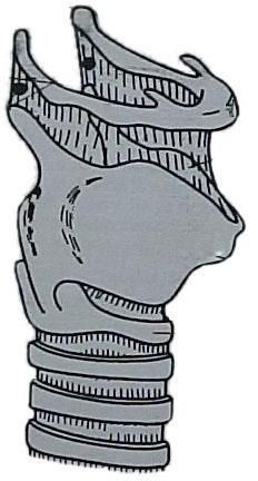
International Journal of Medical Science and Health Research
Vol. 5, No. 03; 2021
ISSN: 2581-3366
pharynx. The larynx is formed by a cartilaginous skeleton which is held together by ligaments,
membranes and muscles. (1) The normal adult hyoid-larynx complex is described as combination
of hyoid apparatus (i.e. styloid processes, stylohyoid ligaments and lesser horns of the hyoid),
body and greater horns of the hyoid bone, thyroid, cricoid and arytenoid cartilages and their
ligaments proper. The thyroid cartilage encompasses its superior and inferior cornua. Normal
length of the styloid process is generally described as 20–30 mm, though attention had not been
paid to the anatomy of the styloid process in our study. (2) Larynx consists of only one bone
which is the hyoid. It is a ‘U’ shaped structure located in the anterior neck. It lies at the base of
the mandible (approximately at the level of C3), where it acts as a site of attachment for the
anterior neck muscles. The hyoid bone is unique in the fact that it does not articulate with any
other bone as it is suspended in place by the muscles and ligaments attached to it. As far as the
structure is concerned, the hyoid is composed of a body, two greater and two lesser horns. The
body is the central part of the hyoid bone. It has an anterior convex surface and a concave
posterior surface. Greater horn projects from each end of the body in a posterior, superior and
lateral direction. It acts as a site of attachment for numerous neck muscles. Lesser horn arises
from the superior aspect of the hyoid bone near the origin of the greater horn and it projects
supero-posteriorly (towards the styloid process of the temporal bone). The stylohyoid ligament is
attached to the apex of the lesser horn. (3)
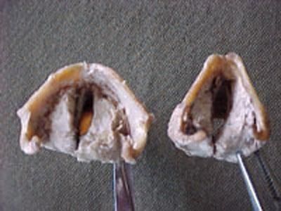
Hayoid greater horns
Hayoid body
Triticeous cartilages
Hayoid lesser horns
Thyroid superior cornua
Thyrohyoid membrane
Thyroid Laminae
Cricoid cartilage
Tracheal
Figure 1: anatomy of the larynx
The thyroid cartilage part of which forms the Adam's apple, is the largest and the uppermost of
the nine cartilages comprising the larynx or the voice box. It houses the vocal folds better known
as the vocal cords. The thyroid cartilage is composed of two plates termed thyroid laminae that
join in the front midline at an angle of 90 to 120 degrees. The visible protrusion it creates on the
www.ijmshr.com Page 16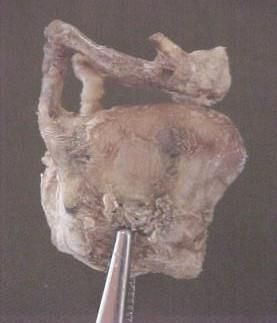
International Journal of Medical Science and Health Research

                                                                               Vol. 5, No. 03; 2021

                                                                                  ISSN: 2581-3366

front of the neck is generally more prominent in men because of the sharper angle. The thyroid
cartilage typically grows larger during the teenage years especially in boys and is considered as a
secondary sexual characteristic in males which goes hand in hand with deepening of their voice.
Thyroid cartilage plays a significant role in the production of the human voice providing
protection and support for the vocal folds. The muscles of the larynx act on the skeletal
structures including the thyroid cartilage to enhance the vibration of the vocal folds which is
necessary for precise vocalization. (4)
Anatomical variations of the hyoid-larynx complex occur in 4–30% of the general population.
Anomalies and variations of this complex are of great clinical importance for radiological
examination and interpretation as well as in operative surgery of the neck region. Very few
comprehensive studies had been conducted in the western world on this topic. This is the pioneer
research done in Sri Lanka. Practical difficulties encounterd in collecting an adequate sample
may be the reason for scarcity of research on this important area. An understanding and an
awareness of the anomalies and anatomical variations of this region is equally significant for the
forensic pathologist who conducts an autopsy and attempts to interpret naked eye findings of a
death with suspected fatal pressure or penetrating injuries over neck area such as manual
strangulation, ligature strangulation, hanging, firearm injuries and stabs. (2)

Anomalies of the hyoid bone involving differential development of the greater and lesser horns
could result in the asymmetrical development of greater horn or agenesis of the lesser horn. (5)
Atypical morphological variations including differences in the shape of the hyoid bone, osseous
extensions possibly resulting from Eagle Syndrome (ossification of the stylohyoid ligament) or
from unknown etiology have also been reported.

                                   Figure 2: human hyoid bone

Minor variations in hyoid-larynx comples consist of age-related fusion of the body with the
greater and/or lesser horns by ankyloses of the joints etc. (6) (7) Several anomalies and anatomical
variations are in record with relation to the thyroid cartilage as well. Thyroid cartilage
calcification, cystic changes in the thyroid cartilage, agenesis of the thyroid cornuae, presence of
triticeaous cartilages, ectopic superior thyroid cornuae, terminal segmentation of the thyroid
cornuae and buckled thyroid cartilage are some of them. (8) (2)
Morphological variations of the hyoid bone are closely related to the gender, race, body
proportions/built and the age. (9) European hyoids are broader and shorter than African ones.
Studies have found that the distal ends of the greater horns are significantly longer in women
than in men. Male hyoids are generally larger than female ones. (7) Inward curving and fattening

www.ijmshr.com                                                                              Page 17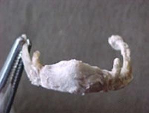
International Journal of Medical Science and Health Research
Vol. 5, No. 03; 2021
ISSN: 2581-3366
of the greater horns are typical for male hyoids. Male hyoids are more susceptible to age-related
modifications. Males show a higher degree of thyroid ossification ultimately leading to the
completely ossified os thyroideum which is more prone to fractures of the superior cornua due to
the absence of elasticity. Finally, muscle attachment sites also show some individual variations.
Certain minor variations occur so often that they cannot be considered as anatomical variations.
(2)
The synchondrosis between the greater horn and the body of the hyoid presents as a vertical
radiolucent line in neck radiographs which may be mistaken for a fracture. Since it is a normal
feature in many hyoid bones, especially in the younger age groups, it is important to identify and
recognize this condition as a normal variant. (10) The presence of triticeous cartilages at the end
of the superior cornua of the thyroid may also be mistaken for a fracture by the novice
pathologist.
Objectives
The objectives of the research was to find out the anatomical variations of the hyoid larynx
complexes in Sri Lankan population and compare the findings with the research done
elsewhere.
Methodology
The study had been conducted based at the Office of the Judicial Medical Officer, Colombo
South Teaching Hospital, Kalubowila, Sri Lanka. Data collection was done over a nine-month
period. Two hundred and forty-one (n=241) adult hyoid larynx complexes were collected during
routine autopsy after obtaining written informed consent from the legal claiment of the dead
body. Children below 18 years of age, those who died due to perforating and sharp trauma to
neck region and cases where consent had not been given by the relatives were excluded. Both
males and females were included. Collection and analysis of results was done at three stages.
The specimen was examined afresh after dissection from the neck. Then they were defleshed,
preserved for two weeks in 10% formol saline, de-fleshed again and examined. The clean
skeletal samples were subject to X-ray and X-rays examined by the researchers as well as by the
consultant radiologist of Kalubowila hospital. The results were recorded manually at each of the
three stages and compared. Final results were obtained after carefully considering the results at
each level specially in the rare event where there happens to be a slight discrepancy among the
findings at each of the three stages. Analysis was done using SPSS 25.
Results
The following variations were identified in relation to the hyoid bone.
The degree of fusion of the greater horn with the body of the hyoid bone could be of three
types as complete fusion, partial fusion and non-fusion. (Table 1)
Complete fusion Partial fusion Nonfusion
Figure 3: The degree of fusion of the greater horn with the body of the hyoid bone
www.ijmshr.com Page 18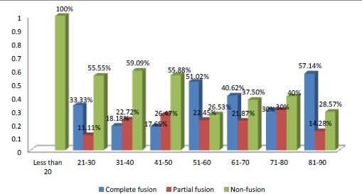
International Journal of Medical Science and Health Research
Vol. 5, No. 03; 2021
ISSN: 2581-3366
Degree of fusion Left % Right %
Complete fusion 33 35
Partial fusion 14 30
Non-fusion 53 35
Table 1: The degree of fusion of the greater horn with the body of the hyoid
In this study several variations were identified in relation to the lesser horn. Some
specimens did not contain a lesser horn on one side. Only 89% had lesser horn on the
right side and the presence of the lesser horn on the left side was 85%. (Table 2)
Lesser horn present Lesser horn absent
Figure 4: Presence or absence of lesser horn
Presence or absence of Left % Right %
lesser horn
Present 85.3 89.3
Absent 14.7 10.7
Table 2: The presence or absence of lesser horn
www.ijmshr.com Page 19International Journal of Medical Science and Health Research
Vol. 5, No. 03; 2021
ISSN: 2581-3366
There were variations in the site of attachment of the lesser horn. Some were attached to
the body of the hyoid while the others were attached to the greater horn. (Table 3)
Attached to the body of the hyoid Attached to the greater horn of hyoid
Figure 5: Site of lesser horn attachment
Site of attachment (lesser horn) Left% Right %
Body of hyoid 50.57 58.42
Greater horn of hyoid 49.43 41.58
Table 3: The site of attachment of lesser horn
www.ijmshr.com Page 20International Journal of Medical Science and Health Research
Vol. 5, No. 03; 2021
ISSN: 2581-3366
The symmetry of the lesser horns on either side was analyzed (out of the percentage of
hyoid bones where both lesser horns were present). Out of such specimens 78.% were
symmetrical and only 12% were found to be asymmetrical.
Symmetrical Asymmetrical
Figure 6: Symmetry of the lesser horns
symmetry
symmetrycal asymmetrycal
12%
88%
In this research, variations in the mobility of the lesser horns were studied. (as a
percentage of all hyoid bones collected for the study and not as a percentage of hyoid
bones where both lesser horns were present). 66.67% of left sided lesser horns were
mobile whereas on the right side it was 58.82%. Out of all specimens 18.63% of the left
sided lesser horns were immobile and on the right side the percentage of immobility was
28.43%. (Table 4)
Mobility of lesser horns Left% Right %
Mobile 66.67 58.82
Immobile 18.63 28.43
Table 4: The mobility of the lesser horn
www.ijmshr.com Page 21International Journal of Medical Science and Health Research
Vol. 5, No. 03; 2021
ISSN: 2581-3366
66.67%
70.00% 58.82%
60.00%
50.00%
40.00% 28.43%
30.00% 18.63%
20.00%
10.00%
0.00%
Mobile Immobile
Left Right
The presence of ‘exostosis-like projections’ on the superior surface of the body of the
hyoid bone was studied. Only 25.50% of all specimens had this variation. All the other
specimens (74.5%) had a smooth superior surface of the hyoid.
exostosis-like projections’ present smooth superior surface (no projections)
Figure 7: presence or absence of exostosis on superior surface
Presence of exostosis like projections on the
superior surface
26%
74%
smooth projections present
www.ijmshr.com Page 22International Journal of Medical Science and Health Research
Vol. 5, No. 03; 2021
ISSN: 2581-3366
The shape of the hyoid bone is mainly determined by the relative angulation of the two
greater horns. Three variations could be found based on the direction of the greater horn
as diverging, converging and parallel. (Table 5)
Diverging Converging Parallel
Figure 8: Variations in the direction of the greater horn/shape of the hyoid
Shape of the hyoid % of the variation
Diverging 71
Converging 18.62
Parallel 9.81
Table 5: Variations in the direction of the greater horn/shape of the hyoid
Direction of the greater horn
10%
19%
71%
Diverging Converging Parallel
The following variations were identified in relation to the thyroid cartilage
The angle of the thyroid cartilage is formed by the midline fusion of the two thyroid
laminae. This angle could be acute or obtuse. 24.51% of the specimens had an acute
angle which is less than 90 degrees and 75.49% had a wide angle which is more than 90
degrees.
www.ijmshr.com Page 23International Journal of Medical Science and Health Research
Vol. 5, No. 03; 2021
ISSN: 2581-3366
Acute angle between two
Wide angle between two laminae
laminae (900)
Figure 09: Variations in angle between two laminae
Triticeous cartilages are usually bilateral ovoid structures found as a non-essential
component of the laryngeal skeleton located centrally in the lateral thyrohyoid ligament
at the level of third and fourth cervical vertebrae (C3-C4). (11). Out of all specimens, 63%
had triticeous cartilages and 37% did not. (Table 6)
Triticeous cartilages % of the variation
Present 63
Absent 37
Table 6: Triticeous cartilages
www.ijmshr.com Page 24International Journal of Medical Science and Health Research
Vol. 5, No. 03; 2021
ISSN: 2581-3366
Among the specimens which contained triticeous cartilages, 62% had them bilaterally,
16% on the left side alone and 22% on the right side only. (Table 7)
Side of the attachment % of the variation
bilaterally 62
Right side only 22
Left side only 16
Table 7: Site of Triticeous cartilages attached
In this study we also noticed certain variations in the superior cornu of the thyroid
cartilage. Out of them; some specimens did not have a superior cornu of the thyroid on
both sides. Some contained a shortened superior cornu while in some specimens there
was no change in the length of the superior cornua. (Table 8)
Absence of the superior cornua Shortened superior cornua No change in the length of
superior cornua
Figure 10: Type of the variation of the superior cornua
www.ijmshr.com Page 25International Journal of Medical Science and Health Research
Vol. 5, No. 03; 2021
ISSN: 2581-3366
Type of the variation of the % variation on left side % variation on right side
superior cornua
Absence of the superior 1.82 1.78
cornua
Shortened superior cornua 54.5 60.70
No change in the length of 43.63 37.5
superior cornua
Table 8: Variations of the superior cornua in the presence of triticeous cartilagesn
Anterior posterior direction (orientation) of the superior cornua in relation to thyroid
laminae also showed some variations such as bending forward, being straight and
bending backward. (Table 9)
Bent forward Straight Bent backward
Figure 11: Direction of the superior cornua
Direction of the superior % variation on left side % variation on right side
cornua
Bent forward 7.8 5.9
Straight 21.6 20.6
Bent backward 69.6 71.6
Table 9: Variations of the direction of the superior horn
www.ijmshr.com Page 26International Journal of Medical Science and Health Research
Vol. 5, No. 03; 2021
ISSN: 2581-3366
There were significant variations in the length of the superior cornua. The measurements
are taken in mili meteres. (Table 10).
Length (mm) Left % Right %
0-5 5.8 5.8
6-10 9.8 8.8
11-15 48 50
16-20 29.4 27.4
21-25 7.8 8.8
Table 10: Length of the superior cornua
50%
48%
50.00%
45.00%
40.00%
35.00% 29.40%
27.40%
30.00%
25.00%
20.00%
15.00% 9.80%8.80%
7.80% 8.80%
10.00% 5.80% 5.80%
5.00%
0.00%
0-5mm 6-10mm 11-15mm 16-20mm 21-25mm
Left Right
The correlation of these variations with related to the age and the sex are also analyzed in
this study. The table below shows the variations in the fusion of greater horns of the
hyoid in relation to the advancement of age. This shows that there is no relationship
www.ijmshr.com Page 27International Journal of Medical Science and Health Research
Vol. 5, No. 03; 2021
ISSN: 2581-3366
between the degree of fusion of the greats horns with the body of the hyoid and the
advancement of age. (Table 11)
Age group (years) Complete fusion Partial fusion Non-fusion
1. Less than 20 100%
2. 21-30 33.33% 11.11% 55.55%
3. 31-40 18.18% 22.72% 59.09%
4. 41-50 17.65% 26.47% 55.88%
5. 51-60 51.02% 22.45% 26.53%
6. 61-70 40.62% 21.87% 37.5%
7. 71-80 30% 30% 40%
8. 81-90 57.14% 14.28% 28.57%
Table 11: Variations in the fusion of the greater horns with advancement of age.
When considering the correlation of the angle between the two thyroid laminae with the
gender, it was evident that, 80.5% of males had an acute angle and the remaining 19.5%
of the males had wide angles. In females, only 60% had acute angles and the remaining
40% had wide angles. This is one reason why thyroid cartilage becomes prominent
during puberty in males. (Table 12)
Gender Wide angle Acute angle
Male 19.5% 80.5%
Female 40% 60%
Table 12: Angle between two laminae of the thyroid cartilage in males and females
The presence of triticeous cartilages was commoner among males than among females.
(Table 13)
www.ijmshr.com Page 28International Journal of Medical Science and Health Research
Vol. 5, No. 03; 2021
ISSN: 2581-3366
Triticeous cartilages and Presence Absence
gender
Male 67.53% 32.47%
Female 52% 48%
Table 13. Tritieceous cartilages and gender
Some other variations found in this study were bilateral elongation of lesser horns, unilateral
elongation of lesser horn, unilateral absence of superior cornua, superior cornua connected to the
thyroid laminae with a fibrous band (non-fusion) and hyoid bone fused with thyroid superior
cornua etc. These were numerically insignificant for statistical analysis but some extreme
examples are elaborated with photographs below.
Bilateral elongation of lesser Unilateral elongation of Unilateral absence of
horns lesser horn superior cornua.
Superior cornua connected Hyoid bone fused with thyroid
to the thyroid laminae with a superior cornua.
fibrous band (non fusion)
Figure 12: Other variations
www.ijmshr.com Page 29International Journal of Medical Science and Health Research
Vol. 5, No. 03; 2021
ISSN: 2581-3366
Discussion
In this study several anatomical variations were identified in relation to the hyoid bone and the
thyroid cartilage. Some such variations were correlated with the age and the sex. The study
showed variations in the fusion of the greater horn of the hyoid with its body. The right side
showed fusion more frequently than left side and this finding had been previously observerd
similarly by other researchers. (2) The variations of the fusion are considered by some authors to
be important in forensic investigations when determining the age and the gender of the victims.
(12)
In contrary to that, in our study a significant correlation between the fusion of the greater
horn of the hyoid to its body and the age could not be established. The fusion of the greater horn
was very variable and non-fusion was fairly common even among the elderly age groups. In the
61-70 year age group, non-fusion was 37.5%. Within the 71-80 year age group and 81-90 year
age group, the percentage of non-fusions were 40% and 28.57% respectively.
Most of the specimens of the hyoid bone found in the current study had bilateral lesser horns.
Absence of a lesser horn on one side is relatively an uncommon finding according to our study.
These results tally well with other comparative studies. (2) Since the hyoid bone is a midline
structure, its symmetry has an anatomical significance. According to the findings of the current
research, the lesser horns of the hyoid bone were symmetrical in 78.43% of the cases. This re-
confirms the finding of some previous studies. (13) (14)
The direction of the greater horns of the hyoid principally determines the shape of the hyoid
bone. As stated above, three main variations of the shape were identified in our study according
to the direction of greater horn as parallel (9.8%), convergent (18.6%) and divergent (71%). A
similar study done in Greece during 1989 had found variations of the shape of hyoid in relation
to the direction of the greater horns with comparatively similar findings. (15)
The findings of our study indicate a great variation in the presence of triticeous cartilages in
relation to the superior cornua of the thyroid cartilage. We found the overall prevalence of the
triticeous cartilages in our study population to be 63%. This finding is supported by one
previous study which observed that the prevalence of triticeous cartilages is around 65% in
Japanese populations. (16) Contrasting results have also been reported in two Western studies. In
one of the above studies, among 86 dissected cadavers, a prevalence rate of 33% was noted. In
the other study which was limited to 40 cadavers, the prevalence of triticeous cartilages was
estimated as 30%. (17) (14). The difference in the findings may reflect that the prevalence of
triticeous cartilages may have an ethnic impact. In South East and Far East Asian populations the
prevelance tend to be higher than in the Western populations. Therefore, a multi-centred study
covering population sub-types across the globe to investigate the ethnic variations in the
prevalence of triticeous cartilages would be an interesting area for further research.
Anatomical variations of the hyoid-thyroid complexes are important in forensic pathological
interpretations. There are some anatomical variations in this important region of the neck which
might be misinterpreted as injuries. (18) (2) The presence of triticeous cartilages, for example,
could be mistaken for a fracture of the superior cornua of the thyroid cartilage. The presence of
accessary bones, fusion or ankylosis of the greater horn with the body of the hyoid, fusion of the
www.ijmshr.com Page 30International Journal of Medical Science and Health Research
Vol. 5, No. 03; 2021
ISSN: 2581-3366
superior cornua of the thyroid with the body of the hyoid bone, unilateral or bilateral absence of
greater horns of hyoid or superior cornua of thyroid, unilateral or bilateral elongation of the
lesser horns, absence of greater horns of the hyoid and superior cornua of the thyroid and
presence of fibrous bands among those structures etc. are some such noteworthy variations.
Therefore, when conducting an autopsy of a person died due to fatal pressure on the neck as in
hanging, manual strangulation or when other pathological conditions are expected in and around
the neck such as carcinomas or perforating injuries (including firearm injuries) an understanding
of the standard anatomy of this region as well as the possible variations (as mentioned above)
would prove to be of immense assistance for accurate interpretation of gross anatomical and
radiological findings. (6)
In a retrospective study done in Netherland using 284 postmortem examination findings of
deaths due to different forms of pressure on the neck, it was found that only 20% had fractures of
the hyoid bone and fractures of the thyroid cartilage were twice as common as in the hyoid,
amounting to 40%. (18). In our study, out of 241 cases there were only 11 cases with fatal
pressure on the neck (10 due to suicidal hanging and 01 due to homicidal ligature strangulation)
and none had laryngeal skeletal injuries. This is in keeping with the gnereal forensic
understanding that hanging leads to minimal internal injuries. The major difference between our
study and the one conducted in 2020 lies in the study population: our one is based on all adult
postmortems out of which only 11 being deaths due to fatal pressure on the neck while the 2020
study is based purely on deaths due to fatal pressure on the neck. Such studies are more
informative for the study and interpretation of laryngeal trauma in deaths due to neck violence
while our study is more informative in the understanding of anatomy and its variations in the
region. A study conducted by Graham in 2016 showed that a substantial pressure is needed for
the causation of damage to hyoid or thyroid and that the death could occur due to other
mechanisms well before the laryngeal skeleton is damaged. This study also showed that hyoid
bone fractures are less common than thyroid cartilage fractures. These findings are in keeping
with our results as well as the results of the 2020 study based on 284 fatal cases. (19).
Conclusion
The study was conducted using 241 adult cadavers subjected to medico-legal postmortem
examination in the JMO’s office at Colombo South Teaching Hospital, Kalubowila, Sri lanka.
Several anatomical variations were identified in the hyoid thyroid complexes. There correlations
to the age and sex were analyzed. According to the findings there was no significant correlation
between the fusion of the greater horn with the body of the hyoid and advancement of age. In
13% of the cases, lesser horns were absent at least on one side. In 80% of the cases, lesser horns
were symmetrical. An acute angle between two thyroid laminae was commoner among males.
Triticeous cartilages were present in 63% of the population and this was more in males than in
females. Sri Lankan average for the length of the superior cornua of the thyroid for males was
16.63 mm and for females it was 13.28mm.
www.ijmshr.com Page 31International Journal of Medical Science and Health Research
Vol. 5, No. 03; 2021
ISSN: 2581-3366
Recommendations
A more extensive multi-centre study should be carried out to find out the correlation between
above mentioned anatomical variations and their effects on post-mortem findings in cases of
fatal pressure on the neck. Ethnic and geographic variations of the hyoid-larynx complex could
be appreciated in the light of more widespread research.
References
1. Barnes, Sam. TeachMe Anatomy. [Online].; 2000 [cited 2021 march 10. Available from:
https://teachmeanatomy.info/neck/viscera/larynx/organ/#:~:text=The%20larynx%20is%2
0located%20in,laryngeal%20part%20of%20the%20pharyn
2. Bernadette S. de Bakker, Henri M. de Bakker, Vidija Soerdjbalie-Maikoe & Frederik G.
Dikkers. Variants of the hyoid-larynx complex, with implications for forensic science and
consequence for the diagnosis of Eagle's syndrome. nature. 2019;(15950): p. Scientific
Reports.
3. Jones, Oliver. TeachMe Anatomy. [Online].; 2021 [cited 2021 march 10. Available from:
HYPERLINK "https://teachmeanatomy.info/neck/bones/hyoid-bone/"
https://teachmeanatomy.info/neck/bones/hyoid-bone/.
4. www.healthline.com. [Online].; 2015 [cited 2021 march 09. Available from: HYPERLINK
"https://www.healthline.com/human-body-maps/thyroid-cartilage" \l "1"
https://www.healthline.com/human-body-maps/thyroid-cartilage#1 .
5. Minoru Hirano, MD, Keiichiro Yukizane, MD, Shigejiro Kurita, MD, Seishi Hibi, PhD.
Asymmetry of the Laryngeal Framework: A Morphologic Study of Cadaver Larynges.
sage. 1989; 98(02): p. 135-140.
6. Pinto, Deborrah C. The Laryngohyoid Complex in Medicolegal Death Investigations. PMC.
2016; p. PMC6474559.
7. M.S.Pollaen, D.H.Ubelaker. Forensic significance of the polymorphism of hyoid bone shape.
PMC. 1997; p. PMID: 9304837.
8. Dr.Grace Carpenter, Tom spencer. radiopedia. [Online]. [cited 2021 march 10. Available
from: HYPERLINK "https://radiopaedia.org/articles/thyroid-cartilage-1"
https://radiopaedia.org/articles/thyroid-cartilage-1.
9. K.M.Miller, P.L.Walker, R.L.O.Halloran. Age and sex-related variation in hyoid bone
morphology. PUBMED. 1998; 43(PMID: 9846390): p. 1138-1143.
10. D.S.Macdonald, Jankowski. The synchondrosis between the greater horn and the body of the
hyoid bone: a radiological assessment. PUBMED. 1990; 4(PMID: 2097227): p. 171-2.
11. Mansur Ahamad BDS, Richard Madden DDS, Loscar Perez DMD. Triticeous cartilage:
Prevalence on panoramic radiographs and diagnostic criteria. Oral Surgery, Oral
Medicine, Oral Pathology, Oral Radiology, and Endodontology. 2005; 992(2): p. 225-
230.
www.ijmshr.com Page 32International Journal of Medical Science and Health Research
Vol. 5, No. 03; 2021
ISSN: 2581-3366
12. AlJulaih, Ghadeer H.; Menezes, Ritesh G. Anatomy, Head and Neck, Hyoid Bone: StatPearls
Publishing; 2020.
13. Bhavna Kalyan, Rajan Kumar Singla, Ravi Kant Sharma. Symmetry and Isometry of Human
Adult Hyoid Bone: A Cadaveric Study. CHRISMED Journal of Health and Research.
2018; 5(2).
14. Nunzio Di Nunno, Salvatore Lombardo, Fulvio Costantinides, and Cosimo Di Nunno.
Anomalies and Alterations of the Hyoid-Larynx Complex in Forensic Radiographic
Studies. The American Journal of Forensic Medicine and Pathology. 2004; 25.
15. N. PAPADOPOULOS, G. LYKAKI-ANASTOPOULOU. The shape and size of the human
hyoid bone and a proposal. journal of anatomy. 1989; 163: p. 249-260.
16. Hiroshi Watanabe, Katsuyoshi Kurihara, Tatsuya Murai. A Morphometrical Study of
Laryngeal Cartilages. SAGE Journals. 1982; 22(04): p. 255-260.
17. Iain Wilson, corresponding author J. Stevens, J. Gnananandan, A. Nabeebaccus, A. Sandison,
and A. Hunter. Triticeal cartilage: the forgotten cartilage. Surgical and Radiologic
Anatomy. 2017; 39(10): p. 1135–1141.
18. Henri M. de Bakker, Moritz V. Warmbrunn, Peggy van den Biggelaar, Vidija
Soerdjbalie-Maikoe & Bernadette S. de Bakker. Correction to: Fracture patterns of the
hyoid-larynx complex after fatal trauma on the neck: retrospective radiological
postmortem analysis of 284 cases. International Journal of Legal Medicine. 2021: p.
1105–1113.
19. Michael A Graham, Marianna Sandomirsky, J Scott Denton. medscape. [Online].; 2016
[cited 2021 march 18. Available from: HYPERLINK
"https://emedicine.medscape.com/article/1988699-overview" \l "a3"
https://emedicine.medscape.com/article/1988699-overview#a3 .
20. A.G.Jurik. Ossification and Calcification of the Laryngeal Skeleton. sage journal. 1984;
25(1): p. 17-22.
21. Barnes, Sam. The Larynx.: TeachMe Anatomy; 2020.
22. Bernadette S. de Bakker, Henri M. de Bakker, Vidija Soerdjbalie-Maikoe & Frederik G.
Dikkers. Variants of the hyoid-larynx complex, with implications for forensic science and
consequence for the diagnosis of Eagle's syndrome. nature. 2019;(15950): p. Sci Rep 9.
23. G Friedrich 1, J Kainz, G H Schneider. Die Impression der Schildknorpelplatte--Eine
Differentialdiagnose zur Taschenfaltenhyperplasie [Impression of the thyroid cartilage
lamina--differential diagnosis in hyperplasia of the ventricular fold]. Laryngol Rhinol
Otol (Stuttg).. 1988 may: p. 232-9.
24. Iu L Lopatin, M A Furman. The significance of developmental anomalies of the thyroid
cartilage for the practice of forensic medical expertise. Sud Med Ekspert. 1992; 35(3): p.
10.
www.ijmshr.com Page 33International Journal of Medical Science and Health Research
Vol. 5, No. 03; 2021
ISSN: 2581-3366
25. J Koebke, K S Saternus. Morphology of the adult human hyoid bone. pubmed. 1979; 84(1):
p. 7-18.
26. Michael L. Bagnoli, Stanly G. Leban, Frank A Williams. Isolated fracture of the hyoid bone:
Report of a case. journal of oral and maxillofacial surgery. 1988; 46(4): p. 326-328.
27. P A Hudgins 1, J Siegel, I Jacobs, C R Abramowsky. The normal pediatric larynx on CT and
MR. pubmed. 1997; 18(2): p. 239-45.
www.ijmshr.com Page 34You can also read