Hodgkin and Reed-Sternberg cells in bone marrow aspirations of a patient with advanced classical Hodgkin lymphoma - HCTM
←
→
Page content transcription
If your browser does not render page correctly, please read the page content below
CASE STUDY

     Hodgkin and Reed-Sternberg cells in bone
       marrow aspirations of a patient with
      advanced classical Hodgkin lymphoma
                         Omayma Saad Eldeen Bakheet1, Nurasyikin Yusof1,
                            'HSDUWPHQWVRI3DWKRORJ\DQG0HGLFLQH
                                     8QLYHUVLWL.HEDQJVDDQ
                         Malaysia Medical center, Kuala Lumpur, Malaysia

       ABSTRACT
       Classical Hodgkin lymphoma (CHL) is a unique type of lymphoma because of the extraordinary and unexplained
       scarcity of its neoplastic Hodgkin Reed-Sternberg (HRS) cells that derived from clonal germinal center B cells with
       rearranged immunoglobulin genes bearing crippling mutations. The occurrence of these cells in the bone marrow
       aspirations are considered rare. Their presence is an expression of widely disseminated disease and it indicates
       poor prognosis. We report a case of a 24-year-old female with relapsed Hodgkin lymphoma, after eleven years
       in remission with the standard chemotherapy regime. She was initially diagnosed during childhood with CHL stage
       IIIB. On this current presentation, she was noted to have cervical lymphadenopathy during her antenatal check-up.
       The lymph nodes biopsy confirmed relapse of the disease; however, there was no evidence of bone marrow
       infiltration. She was given various chemotherapy regimes in which she was refractory to. At this point, repeated
       bone marrow aspiration interestingly revealed the presence of HRS cells. The immunophenotyping analysis by
       flow cytometry revealed a small population of cells expressing CD20, CD15, and CD30 that further supported the
       presence of HRS cells in the bone marrow aspirate. The bone marrow biopsy confirmed infiltration of scattered
       Reed-Sternberg cells and mononuclear Hodgkin cells in a reactive background. In conclusion, we highlight the
       presence of the rare HRS cells in this patient with advanced relapsed Hodgkin lymphoma.
       Keywords: Hodgkin Reed-Sternberg cells, classical Hodgkin lymphoma, bone marrow aspirates, flow cytometry.

       N Z J Med Lab Sci 2015; 69:

INTRODUCTION
Hodgkin Lymphoma (HL) is a group of lymphomas of mainly                from the same clonal B cells population. The rearranged
nodal origin with similar clinical and histological features (1). HL   immunoglobulin (Ig) genes of the tumor cells harbor a high load
encompasses 95% of classical Hodgkin lymphomas (CHL) and               of somatic hypermutations in the variable region of the Ig heavy
the rest are of       nodular lymphocyte predominant Hodgkin           chain genes usually without signs of ongoing mutations. These
lymphoma (NLPHL). These two entities differ in their aetiology,
                                                                       clonal rearrangements are usually detectable only in the DNA of
clinical features, histopathological features, immunophenotype
                                                                       isolated single HRS cells and not in whole tissue DNA (2).
and molecular genetic features. NLPHL is an uncommon
monoclonal B cell neoplasm characterized by large neoplastic
cells known as popcorn or lymphocyte predominant cells (LP             Hodgkin lymphoma (HL) is a curable disease in adults. With
cells) in an inflammatory background. NLPHL is considered to           modern treatment strategies majority of the HL patients with
be of germinal centre B-cell origin. It is managed differently         various anatomical stages and histological subtypes can be
from classical HL. The prognosis is good; however, late                cured (4). Improvement in its diagnosis and staging, treatment
relapses and transformation to high grade non-Hodgkin                  of the disease, as well as its supportive management, has
lymphoma can occur (1,2).
                                                                       contributed in the outcome of the disease (5). Nevertheless,
                                                                       relapse or progression of the disease after the initial treatment
Classical Hodgkin lymphoma (CHL) is a monoclonal lymphoid
neoplasm representing less than 1% of all de novo neoplasms            still occurs in 20-30% of the patients. The pathogenesis and
occurring each year worldwide (2). It is unique among                  mechanism of treatment failure are still not well understood
lymphomas due to the distinctive malignant multinucleated              partly due to the difficulties in studying the molecular phenotype
Hodgkin Reed-Sternberg (HRS) cells as well as the                      of the rare malignant HRS cells (3).
mononuclear Hodgkin cells (2,3). Generally, there are four
subtypes of CHL; nodular sclerosis, mixed cellularity,
                                                                       We report a case of advanced CHL with presence of Reed-
lymphocyte-rich, and lymphocyte-depleted. These subtypes
                                                                       Sternberg cells in the bone marrow where the diagnosis of CHL
may differ in the clinicopathological aspects, however, the
immunophenotype of the tumor cells remain the same. The                stage IIIB was made during childhood period. She was treated
distinctive Reed-Sternberg and Hodgkin cells are nearly always         successively with chemotherapy. However, the disease
CD30 positive. Most of the CHL cases are also CD15 positive            relapsed and these cells were demonstrated in her bone
whilst CD20 is positive with varied intensity in 30-40% of cases.      marrow samples only after the disease had disseminated and
Hodgkin cells and Reed-Sternberg (HRS) cells are derived               became refractory to chemotherapy.

                                                        NZ J Med Lab Science 2015
                                                                   24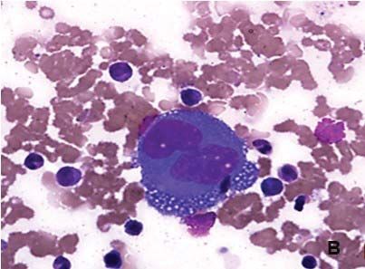
CASE REPORT underwent three cycles of plasmapheresis. A CT EUDLQ scan
A 24-year-old Chinese female was first diagnosed with classical was done and demonstrated left frontal infarct with
Hodgkin lymphoma at the age of 11 years. She was treated haemorrhagic transformation as well as marked perilesional
with the standard ABVD regime (Adriamycin, bleomycin, oedema and mass effect. However, lymphatic tumoral
vinblastine, dacarbazine) and achieved remission for 11 years infiltration could not be excluded. Eventually, she developed
until she presented to us again with a mass in the neck during multiorgan failure and succumbed to her condition.
pregnancy. One month post-partum, a biopsy from the cervical
lymph node was performed to reveal relapsed CHL, mixed
cellularity subtype. The computed tomography (CT) scan
demonstrated extensive cervical, mediastinal, axillary,
abdominal, and inguinal lymphadenopathy. At this point of time,
bone marrow examination showed no evidence of
lymphomatous infiltration. The patient was treated with five
cycles of BEACOPP (bleomycin, etoposide, doxorubicin,
cyclophosphamide, vincristine, procarbazine, prednisone)
chemotherapy. Mid-cycle assessment with PET scan 5 months
later showed evidence of active lymphoma in the chest node
and bone involvement. In view of these findings, the
chemotherapy regimen was changed to ICE (ifosfamide,
carboplatin, etoposide) and the patient was planned for
autologous stem cell transplantation (ASCT). Following
completing two cycles of ICE, stem cells harvesting was
attempted twice but unfortunately failed. In view of disease
progression, as evidenced by CT scan findings that
demonstrated worsening of the mediastinal, intra-abdominal
lymphadenopathy with lung, spleen, liver and urinary bladder
infiltration, chemotherapy regimen was escalated to modified
IGEV (ifosfamide, gemcitabine, vinorelbine).
Half way through this chemotherapy regime, her full blood count
exhibited mild anemia, mild leukocytosis, and severe
thrombocytopenia (haemoglobin: 111.0 g/L; MCV: 87.6 fl; MCH:
30.1 pg; white cells count: 10.1x 10⁹/L; platelet count: 4 x 10⁹/
L). The full blood picture showed occasional nucleated red cells
and a few atypical lymphocytes but no abnormal lymphoid cells.
There was no leukoerythroblastic picture noted. However, the
bone marrow aspirates demonstrated a normocellular marrow
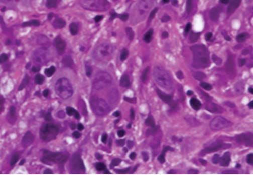
but there was an occasional presence of very large cells. These
cells exhibited binuclearity with inclusion-like nucleoli and
abundant cytoplasm resembling the typical Reed-Sternberg Figure 1. Bone marrow aspirates (A & B) showing Reed-
(RS) cells (Figure 1 A & B). There was also the presence of Sternberg cells- very large cells that exhibit paired nuclei
mononuclear Hodgkin cells, which are large cells of a similar with inclusion-like nucleoli and abundant cytoplasm. MGG
appearance but with a single nucleus. Interestingly, the x600.
immunophenotyping analysis demonstrated a small abnormal
population (2.39%) gated at the CD19 positive area which DISCUSSION
showed positivity for CD45 (dim), CD20 (dim), CD15, and Neoplastic cells, either the classical Reed-Sternberg (RS) cells
CD30. This small population consisted of large cells with or the Hodgkin cells characteristically represent only a minority
complex characteristics as evidenced from the forward and side of the cellular infiltrate with an incidence ranging from 0.1- 10%
scatter gating (Figure 2). The bone marrow biopsy showed
(2). Nevertheless, the confirmation of HL requires morphologic
infiltration by scattered Reed-Sternberg cells and mononuclear
Hodgkin cells in reactive background consisting of small diagnosis of the neoplastic cells with the appropriate cellular
lymphocytes, epithelioid histiocytes, and occasional neutrophils background along with the result of immunophenotyping as
and eosinophils (Figure 3). The immunohistochemistry staining cells resembling Reed-Sternberg cells can be found in cases of
results showed these cells to be CD30+ (strong and diffuse); B and T lymphomas, melanomas, sarcoma, and in some
CD15 and PAX5 were positive in a few of these cells. However, reactive conditions such as infectious mononucleosis, which are
they were negative for CD20. The reticulin stain exhibited common in populations across the globe (6).
coarse fibre network. These findings were concluded as bone
marrow infiltration by classical Hodgkin lymphoma. In 1998, Küppers et al. performed molecular studies in a single
cell of HRS cells in HL (7). Their study showed that HRS cells in
Her condition took a downfall when she developed sepsis CHL, as well as NLPHL, originate from the germinal center (GC)
secondary to the infected chemoport site, thus chemotherapy B cells in most cases, if not all. HRS cells in NLPHL represents
was delayed. Furthermore, her serum alkaline phosphatase and transformed antigen-selected GC B cells with evidence of
bilirubin levels were increasing in trend. Urgent ultrasound ongoing immunoglobulin (Ig) V gene mutation. Whereas HRS
revealed hepatosplenomegaly and multiple ill-defined cells in CHL appear to often or always derive from GC B cells
that have lost the capacity to express a functional antigen
hypoechoic lesions within the spleen, which may represent
receptor. Piccaluga et al. (6) also mentioned that HRS cells are
multiple abscesses or lymphomatous infiltration. Later, she
sustained by an autocrine and/or paracrine production of several
developed acute renal failure, which resulted in alteration in her cytokines, including IL-5, IL-8, IL-9, CCL- 5, and CCL-28. The
level of consciousness and a few episodes of seizures. Owing release of these molecules is also accountable for most of the
to the clinical features coupled with the full blood picture symptoms observed in patients with HL, in addition to the ability
findings of microangiopathic haemolytic anaemia (MAHA), she of the neoplastic cells to escape from growth controls and
was treated for thrombotic thrombocytopenic purpura (TTP) and immunosurveillance.
NZ J Med Lab Science 2015
25
by the bone marrow aspirate needle, and also in HL the bone
                                                                    marrow tends to have fibrous tissue, making their aspiration
                                                                    more difficult (10). Fibrosis is a common finding in Hodgkin’s
                                                                    lesions in the bone marrow and is not limited to nodular
                                                                    sclerosis or lymphocyte depletion variants (9). RS cells are
                                                                    found mainly in bone marrow of patients with generalized
                                                                    advanced stages of HL (10). Thus, the presence of RS cells in
                                                                    bone marrow is an expression of widely disseminating disease
                                                                    (11). Therefore, features of diffuse fibrosis associated with
                                                                    polyploid RS cell variants or large abnormal mononuclear cells
                                                                    with huge nucleoli are sufficient evidence for the determination
                                                                    of marrow involvement on random biopsy (11).

                                                                    Morphologic findings and immunohistochemical stains are vital
                                                                    in the diagnosis of CHL (12). In the majority of cases, flow
                                                                    cytometry analysis is of little or no value to the detection of HRS
                                                                    and the diagnosis of CHL, as neoplastic cells are rarely seen in
                                                                    the cytological preparations (1), and it usefulness is limited to
                                                                    tissue samples such as lymph nodes (12). Recently, Fromm
                                                                    and Wood (13) described a method of identifying HRS cells in
                                                                    lymph nodes by flow cytometry using a single-tube (6-colour
                                                                    assay) with high sensitivity and specificity. Also, they proposed
                                                                    that   this    method       might     obviate     the   need     for
                                                                    immunohistochemistry in many cases. Furthermore flow
                                                                    cytometry offered several potential benefits in the diagnosis of
                                                                    CHL in which it is more sensitive in equivocal or possibly
                                                                    negative cases by morphology. It has a rapid turnaround time
                                                                    with significant cost effectiveness compared to the elaborate
                                                                    immunohistochemical panels. In our present case we have
                                                                    concluded that there was a presence of HRS cells in our
                                                                    analysis using a 4-colour assay flow cytometry in the aspirate
                                                                    samples.

                                                                    According to the WHO classification (2), Hodgkin and Reed-
                                                                    Sternberg cells are almost always positive for CD30 and positive
                                                                    for CD15 in 75-85% of cases. Both CD30 and CD15 are typically
Figure 2. Flow cytometry immunophenotyping analysis                 present in a membrane pattern increasing in the GoIgi area. For
showing the small (2.39%) abnormal populations (highlight-          B-cell markers, HRS cells are positive for CD20 on a minority of
red). These cells expressed CD45 (dim), CD19, CD20, CD15,           the neoplastic cells with varied intensity in 30-40% of cases,
and CD30.                                                           CD79a is less often expressed. The B-cell nature of HRS cells
                                                                    can be demonstrable in approximately 95% of cases by their
                                                                    expression of the B-cell specific activator protein PAX5 and it is
                                                                    usually weaker than that of reactive B cells. Even though HRS
                                                                    cells are usually negative for CD45 (2), the total absence of
                                                                    CD45 is unlikely (13).

                                                                    CD20 is important in the regulation of human B-cell growth and
                                                                    differentiation. It is present on most mature normal and
                                                                    neoplastic B lymphocytes (14). The prognostic significance of
                                                                    CD20 expression in CHL is controversial and a matter of
                                                                    ongoing debate. A review of the literature showed that the
                                                                    expression of CD20 was not associated with different clinical
                                                                    and laboratory features among equivalently treated patients
                                                                    (14) and has no prognostic significance for the failure-free
                                                                    survival and overall survival in CHL patients (14,15). One study
                                                                    on 248 CHL patients found that failure-free and overall survival
                                                                    were reduced considerably in CD20-positive patients as
Figure 3. Bone marrow biopsy section showing focally                compared with CD20-negative patients (16). Whereas in
hypercellular marrow space displaying infiltration by scattered     another study of 119 CHL patients showed a significantly higher
Hodgkin cells and Reed-Sternberg cells (arrow). H&E x 600.          frequency of disease relapses in the CD20-negative group and
                                                                    a better failure-free and overall survival in the CD20-positive
Bone marrow involvement is rare in patients with HL. Its            group (17).
incidence varies between 4% and 14% in the series reported
during the past 20 years (8). As the bone marrow lacks              Previously, bone marrow biopsy (BMB) was the recommended
lymphatics, infiltration of the bone marrow by Hodgkin's            approach for staging in newly diagnosed patients with HL.
lymphoma indicates vascular dissemination of the disease            Recently, positron emission tomography/computed tomography
(stage IV). The incidence of bone marrow involvement in             (PET/CT) is required for staging and response assessment in
Hodgkin's lymphoma varies with the histologic subtype: 10% in       lymphoma according to the recommendations whenever it is
classical Hodgkin's mixed cellularity, approximately 1% in          available (18-20). Studies have shown that conventional
lymphocyte predominant and lymphocyte rich CHL, and 3% in           staging can detect bone marrow involvement in only
nodular sclerosis subtype (9). The reason for this rarity can be    5–8% of patients, whereas PET/CT staging can detect bone
due to the scattered focal lesions which may not be aspirated       marrow involvement in up to 18% of the cases (20).
                                                      NZ J Med Lab Science 2015
                                                                 26
0RUHRYHU+/LV>)@IOXRURGHR[\JOXFRVHDYLGDOPRVWDOOWKXV 7. Küppers R, Hansmann ML, Rajewsky K. Clonality and
3(7&7XVLQJ)'*LVPRUHDFFXUDWHWKDQ&7IRUVWDJLQJLQ+/ germinal centre B-cell derivation of Hodgkin/Reed-Sternberg
DQG 1+/ ZLWK LQFUHDVHG VHQVLWLYLW\ SDUWLFXODUO\ IRU H[WUDQRGDO cells in Hodgkin's disease. Ann Oncol 1998; 9 Suppl 5: S17-
20.
GLVHDVH
8. Vassilakopoulos TP, Angelopoulou MK, Constantinou N,
Furthermore, PET/CT leads to change in stage in 10% to 30% Karmiris T, Repoussis P, et al. Development and validationof
of patients, more often upstaging, although alteration in a clinical prediction rule for bone marrow involvement in
patients with Hodgkin lymphoma. Blood 2005; 105: 1875-
management occurs in fewer patients, with no demonstrated
impact on overall outcome (18). El-Galaly et al. (12), in a cohort 1880.
study, investigated whether BMB can add useful information to 9. Kar R, Dutta S, Tyagi S. Clinically unsuspected Hodgkin's
FDG-PET/CT staging in patients with HL. They concluded that lymphoma diagnosed primarily from bone marrow trephine
PET/CT can accurately detect marrow involvement therefore biopsy: report of six cases. Indian J Pathol Microbiol 2008;
51: 186-189.
the evaluation by BMB is often unnecessary. Likewise, Adams
et al. made the same observation for the appropriate use of 10. Bouroncle BA. Sternberg-Reed Cells in the peripheral blood
of patients with Hodgkin's disease. Blood 1966; 27: 544-
FDG-PET/CT to replace BMB in newly diagnosed HL (22). They
also mentioned that the major advantages of FDG-PET/CT over 556.
BMB are the fact that it is non invasive and able to visualize the 11. Lukes RJ. Criteria for involvement of lymph node, bone
entire bone marrow therefore eliminating any sampling errors. marrow, spleen, and liver in Hodgkin's disease. Cancer Res
1971; 31: 1755-1767.
Nevertheless, biopsy is recommended to confirm residual
12.Roshal M, Wood BL, Fromm JR. Flow cytometric detection
disease and to exclude false-positive uptake with FDG before
of the classical Hodgkin lymphoma: clinical and research
starting second-line therapy (20). applications. Adv Hematol 2011; 2011:387034.
We reported a case of refractory classical Hodgkin lymphoma in 13. Fromm JR, Thomas A, Wood BL. A Six-Color Flow
which Hodgkin Reed-Sternberg (HRS) cells were only Cytometry Assay for Immunophenotyping classical Hodgkin
demonstrated in bone marrow aspirations and trephine biopsy Lymphoma in Lymph Nodes. Am J Clin Pathol 2014;
sections only when the disease disseminated. The patient’s 141:388-396.
bone marrow aspirations revealed the typical morphologic 14.Rassidakis GZ, Medeiros LJ, Viviani S, Bonfante V, Nadali
characteristics of Reed-Sternberg cells. Whereas, the trephine GP, Vassilakopoulos TP, et al. CD20 expression in Hodgkin
and Reed-Sternberg cells of classical Hodgkin’s disease:
biopsy sections showed infiltration by scattered Reed- associations with presenting features and clinical outcome.J
Sternberg cells and mononuclear Hodgkin cells in reactive Clin Oncol 2002; 20: 1278-1287.
background. Immunophenotyping analysis by flow cytometry 15.Fu XH, Wang SS, Huang Y, Xiao J, Zhai LZ, Huang QC, et
and immunohistochemistry confirmed the presence of HRS al. [Prognostic significance of CD20 expression in Hodgkin
cells. and Reed-Sternberg cells of classical Hodgkin’s lymphoma].
Ai Zheng 2008; 27: 1197-1203. [Article in Chinese].
AUTHOR INFORMATION 16. Portlock CS, Donnelly GB, Qin J, Straus D, Yahalom J,
Omayma Saad Eldeen Bakheet, MBBS DPath, Zelenetz A, et al. Adverse prognostic significance of CD20
Haematopathologist1 positive Reed-Sternberg cells in classical Hodgkin's disease.
Br J Haematol 2004; 125: 701-708.
Nurasykin Yusof, MBChB FRCPA, Consultant Haematologist1
17.Tzankov A, Krugmann J, Fend F, Fischhofer M, Greil R,
Siti Fadilah Abdul Wahid, MD MMed PhD FRCPE, Senior
Dirnhofer S. Prognostic significance of CD20 expression in
Consultant Haematologist & Head of Cell Therapy Center2 classical cases Hodgkin lymphoma: a clinicopathological
Suria Abdul Aziz, MBBS MPath, Lecturer/ study of 119 cases. Clin Cancer Res 2003; 9: 1381-1386.
Specialist Haematology1 18. Cheson BD, Fisher RI, Barrington SF, Cavalli F, Schwartz
Departments of 1Pathology and 2Medicine, Universiti LH, Zucca E, et al. Recommendations for initial evaluation,
Kebangsaan Malaysia Medical Fenter, Kuala Lumpur, Malaysia staging, and response assessment of Hodgkin and non-
Hodgkin lymphoma: the Lugano Classification. J Clin Oncol
Author for correspondence: Dr O Bakheet, Department of 2014; 32: 3059-3067.
Pathology, Universiti Kebangsaan Malaysia Medical center, 19. Barrington SF, Mikhaeel NG, Kostakoglu L, Meignan M,
Jalan Yaakob Latiff, Bandar Tun Razak, 56000, Cheras, Kuala Hutchings M, Müeller SP, et al. The role of imaging in the
staging and response assessment of lymphoma: consensus
Lumpur, Malaysia. Email: ombakheet@gmail.com of the International Conference on Malignant Lymphomas
Imaging Working Group. J Clin Oncol 2014; 32: 3048-3058.
REFERENCES 20. Follows GA, Ardeshna KM, Barrington SF, Culligan DJ,
1. Bain BJ, Clark DM, Wilkins BS. Bone Marrow Pathology. Hoskin PJ, Linch D, et al. Guidelines for the first line
4thEdition. Wiley-Blackwell, UK. 2009. management of classical Hodgkin lymphoma. Br J Haematol
2. Swerdlow SH, Campo E, Harris NL, Jaffe ES, Pileri SA,Stein 2014; 166: 34–49.
H, et al. WHO Classification of Tumours of the 21.El-Galaly TC, d’Amore F, Mylam KJ, de Nully Brown P,
Haematopoietic and Lymphoid Tissue. 4th Edition. IARC, Bögsted M, Bukh A, et al. Routine bone marrow biopsy has
Lyon, France. 2008. little or no therapeutic consequence for positron emission
3. Steidl C, Diepstra A, Lee T, Chan FC, Farinha P, Tan K, etal. tomography/computed tomography-staged treatment-naive
Gene expression profiling of microdissected Hodgkin Reed- patients with Hodgkin lymphoma. J Clin Oncol 2012; 30:
Sternberg cells correlates with treatment outcome inclassical 4508–4514.
Hodgkin lymphoma. Blood 2012; 120: 3530-3540. 22.Adams HJ, Kwee TC, de Keizer B, Fijnheer R, de Klerk JM,
Littooij AS, et al. Systematic review and meta-analysis onthe
4. Diehl V, Stein H, Hummel M, Zollinger R, Connors JM. diagnostic performance of FDG-PET/CT in detecting bone
Hodgkin's lymphoma: biology and treatment strategies for marrow involvement in newly diagnosed Hodgkin lymphoma:
primary, refractory, and relapsed disease. Hematology Am is bone marrow biopsy still necessary? Ann Oncol 2014; 25:
Soc Hematol Educ Program 2003; 225-247. 921–927.
5. Klimm B, Schnell R, Diehl V, Engert A. Current treatmentand
immunotherapy of Hodgkin’s lymphoma. Haematologica Copyright: © 2015 The authors. This is an open-access article
2005; 90: 1680-1692. distributed under the terms of the Creative Commons Attribution
6. Piccaluga PP, Agostinelli C, Gazzola A, Tripodo C, Bacci F, License, which permits unrestricted use, distribution, and
Sabattini E, et al. Pathobiology of Hodgkin lymphoma. Adv reproduction in any medium, provided the original authors and
Hematol 2011; 2011:920898. source are credited.
NZ J Med Lab Science 2015
27You can also read