UNDERLYING CONDITIONS IN A SHAR-PEI DOG WITH KIDNEY DISEASE - CASE STUDY
←
→
Page content transcription
If your browser does not render page correctly, please read the page content below
6                                                                                       Rev Rom Med Vet (2020) 30 | 2 : 6-10
                                                                                        ISSN: 1220-3173; E-ISSN: 2457-7618

                       UNDERLYING CONDITIONS IN A SHAR-PEI DOG
                          WITH KIDNEY DISEASE - CASE STUDY
                           AFECŢIUNI ASOCIATE CU BOALA RENALĂ
                              A SHAR-PEIULUI - STUDIU DE CAZ
                                                                           Roxana Mariana IGNĂTESCU (ŢÎMPĂU)1),
                                                                           Ana-Maria GOANȚĂ1), Carmen IONIȚĂ1),*),
                                                                          Ana-Maria BĂDULESCU1), D.S. SORESCU1),
                                                                                                      L. IONIȚĂ1)

     Renal disease in a Shar-Pei dog is a condition that            Boala renală a Shar-Peiului este o manifestare cli-
should make one consider Shar-Pei Fever, but diseases          nică care ar trebui întotdeauna asociată cu febra Shar-
such as nephrotic syndrome, renal amyloidosis, glo-            Peiului, fiind necesară includerea mai multor boli pe lis-
merulonephritis, diabetic ketoacidosis, also Lyme di-          ta diagnosticului diferențial, cum ar fi: sindromul nefro-
sease (in endemic areas) should also be on the list of         tic, amiloidoza renală, glomerulonefrita, cetoacidoza
differential diagnoses, due to their lack of specific clini-   diabetică, boala Lyme (în zonele endemice) datorită si-
cal findings. The aim of this paper is to bring into atten-    militudinii constatărilor clinice și paraclinice. Scopul
tion the underlying conditions of the Shar-Pei breed's         acestei lucrări este de evidenția condițiile preexistente
renal disease in order to provide a more relevant prog-        asociate cu boala renală în cazul Shar-Peiului pentru a
nosis. With the presentation of this clinical case, we try     oferi un prognostic vital mai relevant. Odată cu prezen-
to broaden the range of differential diagnoses in kid-         tarea acestui caz clinic, ne dorim să extindem gama di-
ney disorders and to help the clinician to establish the       agnosticelor diferențiale în tulburările renale și să aju-
specific therapeutic protocol in each case. A seven            tăm clinicianul să stabilească protocolul terapeutic spe-
years old female Shar-Pei was presented with clinical          cific pentru fiecare afecțiune în parte. O femelă Shar-
signs of intermittent weakness, acute generalized oe-          Pei de șapte ani a fost prezentată cu semne clinice de
dema, decreased appetite, weight loss, and lethargy.           slăbiciune intermitentă, edeme generalizate, inapeten-
It was diagnosed with an acute renal injury one day            ță, scădere în greutate și letargie. Acesta a fost diag-
before the presentation. Physical examination re-              nosticată cu insuficiență renală acută cu o zi înainte și a
vealed normothermia, tachycardia and tachypnea, ar-            primit recomandare de hemodializă. Examinarea fizică
terial hypertension, moderate dehydration, mild ulce-          a evidențiat normotermie, tahicardie și tahipnee, hi-
ration of the oral cavity, and pale mucous membranes.          pertensiune arterială, grad moderat de deshidratare,
Also, significant oedema of the head and limbs was             ulcerație ușoară a cavității orale și mucoase palide. De
observed. Further diagnostic investigations revealed           asemenea, a fost observat edem pregnant la nivelul
hyperglobulinaemia, hypoalbuminaemia, mild hypo-               membrelor și capului. Investigațiile suplimentare au
glycaemia, as well as elevated serum activities of li-         evidențiat hiperglobulinemie, hipoalbuminemie, hipo-
pase, amylase, creatinine, blood urea nitrogen, cal-           glicemie ușoară, valori crescute ale lipazei, amilazei,
cium, and phosphorus. Blood gases and electrolytes             creatininei, ureei, calciului și fosforului, precum și mo-
were significantly modified. Urinalysis revealed protei-       dificări semnificative ale ionogramei, iar analizele de
nuria. Without a morphopathologic diagnosis, suppor-           urină au evidențiat proteinurie. În absența unui diag-
tive and symptomatic treatment was initiated.                  nostic histopatologic de certitudine, a fost inițiat un tra-
              Keywords: Shar-Pei fever, amyloidosis,           tament de susținere și simptomatic.
              glomerulonephritis, nephrotic syndrome                      Cuvinte cheie: febră Shar-Pei, amilodoză,
                                                                                  glomerulonefrită, sindrom nefrotic

   A 7-year-old, neutered Shar-Pei female was pre-             for last two weeks. The dog was born and raised in
sented to the Clinic of the Faculty of Veterinary Medi-        Bucharest, Romania and it lived with another clinically
cine Bucharest for acute general swelling, lameness,           healthy neutered Shar-Pei male. One day before the
decreased appetite, progressive weight loss, lethargy,         presentation, the dog had been evaluated in a private
                                                               clinic and it was diagnosed with acute renal injury.
1) University of Agronomic Sciences and Veterinary Medicine,       During the physical examination, the rectal tempe-
   Faculty of Veterinary Medicine, Bucharest, Romania          rature was 38.7 °C, pulse rate was 167 bpm, respi-
*) Corresponding author: ionitacarmen63@yahoo.com              ratory rate was 52 rpm and systolic blood pressure
Rev Rom Med Vet (2020) 30 | 2 7
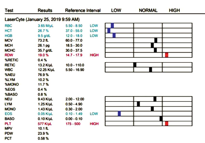
was 190 mm Hg. The dog was estimated to be 8% 26.7%, reference range (RR) 37-55% and hemoglo-
dehydrated based on moderate loss of skin turgor, dry bin 9.5 g/dL, RR: 12-18g/dL, red blood cells 3.65M/
mucous membranes, weak rapid pulses, enophthal- µL, RR: 5.5-8.5M/µL, mild thrombocytosis (PLT 577K/
mia. There were mild ulcerations of the oral cavity. µL, RR: 175-500), eosinopenia (Eos 0.05 K/µL, RI:
Thoracic and pelvic limbs were swollen with pitting oe- 0.10-1.49).
dema and also the face and the neck were significantly Findings on the chemistry panel were: significantly
oedematous. No subcutaneous nodules or other kinds increased kidney parameters: Creatinine 28.2 mg/dL,
of abnormalities were noted. RR: 0.5-1.8 mg/dL, Blood Urea Nitrogen (BUN) 199
ELISA SNAP 4Dx and Urine Protein Creatinine ratio mg/dL, RR: 7-27 mg/dL and pancreatic enzymes:
(IDEXX Laboratories, Westbank, Maine, USA) had Amylase: 1972 U/L, RR: 500-1500 U/L, Lipase 4684 U/
been performed at the private veterinary clinic and the L, RR: 200-1800 U/L, moderate hypoalbuminemia (Al-
result was negative for Borrelia burgdorferi, Ehrlichia bumin 2.0 g/dL, RR, 74–143 g/dL), hyperglobulinemia
canis, Anaplasma phagocytophilum, and Dirofilaria (Globulins 4.9 g/dL, RR: 2.5-4.5 g/dL), hypercalcemia
immitis antigen. Urine Protein/Creatinine (UP/C) ratio (Calcium 24.5 mg/dL, RR: 9-12 mg/dL), hyperphos-
was 0.8. According to the recommendations from the phatemia (Phos 23 mg/dL, RI: 2.5-6.8). The hepatobi-
2004 ACVIM Forum Consensus Statement (Small liary parameters: Alanine Aminotransferase (ALT), Ga-
Animal): Assessment and Management of Proteinuria mma-Glutamyl Transferase (GGT), Total Bilirubin (T
in Dogs and Cats, a severe azotemic dog with UP/C ≥ BIL), Alkaline Phosphatase (ALKP) were in normal ran-
0.5 and inactive urine sediment is considered to be ges. Chemistry results for the analyzer run were mul-
significantly proteinuric (6). tiplied by the dilution factor for a dilution of 1 in 3 total.
Venous blood gases and electrolytes performed at
INVESTIGATIONS IN THE FIRST DAY the same time revealed normochloraemic metabolic
acidosis: pH 7.23, RR: 7.31-7.42, HCO3 12 mmol/L,
All the bloodwork was carried out at the laboratory RR: 20-29, PCO2 31 mmHg, RR: 32-49, AnGap 35
of the clinic of the Faculty of Veterinary Medicine. mmol/L, tCO2 12.9 mmol/L, RR: 21-31, Na 153 mmol
Complete blood count performed at presentation /L, RR: 144-160, K 5.4 mmol/L, RR: 3.5-5.8, Cl 112
revealed mild non-regenerative anemia (hematocrit mmol/L, RR: 109-122.
Fig. 1. Complete blood count, Shar-Pei breed, neutered female, 7-year-old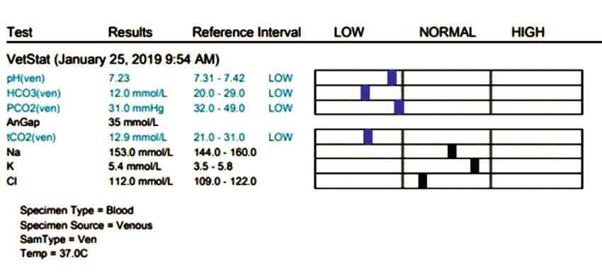
8                                                                                       Rev Rom Med Vet (2020) 30 | 2

         Fig. 2. Venous blood gases and electrolytes, Shar-Pei breed, neutered female, 7-years-old

   A direct blood smear was also performed and re-         sing: crystalloids (polyionic solution) and levo amino
vealed the presence of microfilaria.                       acids completed with sodium bicarbonate (bicarbo-
   Ultrasound examination revealed normal shaped           nate deficiency (meq) = body weight (kg) x 0.3 x (de-
kidneys of normal size according to the animal's           sired bicarbonate – patient bicarbonate) (13). Also,
weight and size and with little distinction between        beta erythropoietin was taken into account to sti-
cortical and medullary tissue; the hyperechoic cortex      mulate erythropoiesis (100 UI/kg each other day,
suggesting renal parenchymal disease.                      three days per week), which was associated with
                                                           cyanocobalamin (50 µg/kg IM once per week), iron
           DIFFERENTIAL DIAGNOSIS                          supplements, vitamin C and folic acid (14).
                                                               The next day, blood work reevaluation showed an
    Differential diagnoses, in this case, were nume-       insignificant decrease in creatinine (26.7 mg/dL), BUN
rous, and the clinical signs were not diagnostic for any   197 mg/dL, phosphorus 18.1 mg/dL and calcium 24.2
specific disease. The mild normochromic, normocytic        mg/dL. Blood gas analysis revealed a slight improve-
and non-regenerative anaemia can be due to acute           ment of the metabolic acidosis: pH 7.26, HCO3 15.6
haemorrhage or haemolysis and it can also be asso-         mmol/L, PCO2 38 mmHg, AnGap 33 mmol/L, tCO2 16.8
ciated with inflammatory disease (not in this case),       mmol/L. Electrolytes were insignificantly modified: Na
bone marrow disorders, maturation abnormalities, or        157 mmol/L, K 5.5 mmol/L, Cl 114 mmol/L.
erythropoietin deficiency due to renal disease.                Patient clinic status was almost the same, except
    Severe hypertension with acute impairment of one       for the urine output, which became normal (2 ml/kg
or more organ systems (especially central nervous sys-     per day). Systolic arterial pressure was 20 mmHg.
tem, cardiovascular, renal) may determine irreversible     Treatment was completed with Prednisolone (1 mg/kg
ischemic kidneys, which causes renal vasoconstriction,     BID), Spironolactone (2 mg/kg BID) and Amlodipine
increased aldosterone secretion and salt retention,        (0.25 mg/kg daily).
which elevate blood pressure even further (12).                After 24 hours the patient status was reevaluated:
    Hypoalbuminemia may occur with decreased pro-          general oedema was still significant, systolic arterial
duction of albumin secondary to liver disease or infla-    pressure was 133/77 mm Hg, no clinical improvement
mmation, but in this case, it is probably the conse-       of the animal status was noted.
quence of a glomerulonephropathy which leads to                Acid-base balance was restored, but significant
protein loss via the kidneys (proteinuria). Also, serum    elevated values of Creatine (25.9 mg/dL), Blood Urea
concentrations of albumin were significantly lower in      Nitrogen (196 mg/dL), Calcium (23.7 mg/dL) and
hypocobalaminemic Shar-Pei (median: 2.5 g/dl) than         Phosphorus (17.3 mg/dL) were observed. Mild hyper-
in normocobalaminemic Shar-Pei (median: 2.9 g/dl; P        natremia was still detected: 167 mmol/L. The therape-
< 0.0001) (4, 11).                                         utic protocol remained the same.
                                                               The persistently elevated levels of the kidney para-
            THERAPEUTIC APPROACH                           meters (Creatinine > 25 mg/dL, Blood Urea Nitrogen
                                                           > 190 mg/dL) even after 48 hours of continuous rate
   In the first day of hospitalization, treatment inclu-   infusion and the ultrasonographic renal structure su-
ded correcting the fluid and electrolyte imbalance u-      ggested significant renal damage.
Rev Rom Med Vet (2020) 30 | 2 9
Table 1
Evolution of renal parameters during hospitalization,
Shar-Pei breed, neutered female, 7-year-old
Due to increased vascular fragility, the peripheral associated with inflammatory disease. Amyloid A pro-
veins were not an option any longer. We recommended tein is formed by the polymerization of the amino acid
central venous catheterisation, but anaesthesia was terminal portion of serum amyloid A protein. It was
considered to be a high risk for the patient. The risk- established that affected Shar-Pei have increased va-
benefit assessment for the patient was concluded with lues of the serum interleukin-6 (a cytokine involved in
the suggestion of euthanasia, considering that in this the serum amyloid A protein synthesis and initiates
situation haemodialysis would be more risky than the acute phase response of the inflammation), cha-
helpful. racterized by hepatic production of acute-phase
proteins, fever, and neutrophil mobilization (10). If in
the liver will be present the amyloidosis then high le-
vels of alkaline phosphatase (>102 IU/L), alanine
transaminase (>100 IU/L), aspartate transaminase
(>100 IU/L), and total bilirubin (>0.4 mg/dl) may be
present, findings which were not present in this case.
The nephropathies that arise with the involvement
of glomeruli in the processes that initiate renal injury
are defined as the primary glomerulopathies, and the
glomerular changes occur after renal tubular or whole
nephron damage are defined as the secondary glome-
rulopathies (8). The two major categories of glomeru-
lopathies in dogs are renal amyloidosis and glomerulo-
nephritis. A definitive diagnosis is realised by using a
renal biopsy. This allows differentiation between amy-
loidosis and glomerulonephritis. A variety of morpho-
Fig. 3. Severe oedema, Shar-Pei breed, logic patterns may be seen with glomerular pathology
neutered female, 7-year-old (e.g., membranous, and membranoproliferative), and
the differentiation is of prognostic or therapeutic im-
OUTCOME AND FOLLOW-UP portance (1).
Nephrotic syndrome occurs as a result of glomeru-
The owners chose at-home euthanasia to be per- lar disease and is characterised by proteinuria, hypo-
formed by their previous veterinarian. On the next day, albuminaemia, hyperlipidaemia, and oedema (2).
they announced the death of the animal. Unfortunately, In the case presented above, hypoalbuminemia
they declined necropsy of the animal and subsequent may have contributed to general oedema. However, as
morphopathologic examination of kidneys. the development of oedema is dependent not only on
the oncotic pressure of albumin but also on vascular
DISCUSSION endothelial integrity and hydrostatic pressure (7), it
might explain general oedema. Many Shar-Peis have a
According to the results of the investigations per- history of recurrent fever and swelling of the tibio-tar-
formed both previously and immediately after the arri- sal joints (commonly called Shar-Pei fever or Shar-Pei
val in our clinic, the origin of the lesion was considered swollen hock syndrome) before renal amyloidosis de-
to be renal. Based on patient signalmen [familial pre- velops, even if in this case the owner said that this is
disposition, clinical renal disease and ultrasonographic the first time when the dog was ill. Proteinuria with
renal parenchymal involvement (5)], the case was di- renal amyloidosis can be massive and may lead to the
agnosed as familial renal amyloidosis. The amyloidosis development of the nephrotic syndrome. This syn-
reported in Shar-Pei is believed to be a reactive one drome in Chinese Shar-Pei is believed to be an auto-
because the primary protein involved, amyloid A, is somal recessive inherited trait (10).10 Rev Rom Med Vet (2020) 30 | 2
Conditions that are known to be associated with natu- 3. Fakhouri F., Frémeaux-Bacchi V., Noël L.H., Cook H.T.,
rally occurring glomerulonephritis in the dog include pyo- Pickering M.C., (2010), C3 glomerulopathy: a new cla-
metra, neoplasia, systemic lupus erythematosus, dirofila- ssification. Nature reviews. Nephrology, 6(8):494-499
riasis, Lyme disease, canine adenovirus I infection, pan- 4. Grützner N., Heilmann R.M., Cranford S.M.,
creatitis, and diabetes mellitus (8, 15). In the case presen- Holzenburg A., Suchodolski J.S., Steiner J.M., (2015),
ted above, only microfilaria discovered accidentally could Inflammatory, immunological, and intestinal disease
be considered an underlying cause of glomerulonephritis. biomarkers in Chinese Shar-Pei dogs with marked hy-
Due to the complex clinical findings, supportive the- pocobalaminemia, Journal of Veterinary Diagnostic In-
rapy should be provided as indicated to maintain hydra- vestigation, 27(1):31-40
tion and to resolve the metabolic acidosis. According to 5. Lee S.G., Moon H.S., Han J.H., Yoon B.I., Hyun C.,
the International Renal Interest Society, immunosuppre- (2007), Familial renal amyloidosis in a Shar Pei dog.
ssive treatment of dogs with proteinuric kidney diseases Korean J Vet Res, 47(2):255-257
even if in the absence of a histopathologic diagnosis is re- 6. Lees G.E., Brown S.A., Elliott J., Grauer G.E., Vaden
commended (3, 8, 9). Also, additional antihypertensive S.L., American College of Veterinary Internal Medicine,
agents may be needed if hypertension (systolic blood pre- (2005), Assessment and management of proteinuria in
ssure >160 mm Hg) persists after initiation of angioten- dogs and cats: 2004 ACVIM Forum Consensus State-
sin-converting enzyme inhibitor therapy: Amlodipine: 0.1 ment (small animal). J Vet Intern Med, 19(3):377-385.
mg/kg PO twice daily. The dose can be titrated upward 7. Littman M.P., (2011), Protein-losing nephropathy in
weekly (to 0.2-0.4 mg/kg PO twice daily) if indicated; small animals, Vet Clin North Am Small Anim Pract,
blood pressure should be monitored weekly until normo- 41:31-36
tensive, and then once every three to six months. 8. Littman M.P., Daminet S., Grauer G.F., Lees G.E., van
Dongen A.M., (2013), Consensus recommendations
CONCLUSIONS for the diagnostic investigation of dogs with suspected
glomerular disease, Journal of Veterinary Internal Me-
Dramatic renal disease in a Shar-Pei dog should in- dicine, 27(1):19-26
clude more than Acute/Cronic Kidney Disease in its diffe- 9. Pressler B., Vaden S., Gerber B., Langston C., Polzin
rential diagnosis, so one should consider all relevant cli- D., (2013), Consensus guidelines for immunosuppre-
nical findings: familial Shar-Pei amyloidosis as a result of ssive treatment of dogs with glomerular disease ab-
Shar-Pei Fever: the history of recurrent fever and swelling sent a pathologic diagnosis, Journal of Veterinary In-
of the tibiotarsal joints, massive proteinuria, general ternal Medicine, 27(1):55-59
signs of renal injury; glomerulonephritis: proteinuria, 10. Rivas A.L., Tintle L., Meyers-Wallen V., Scarlett J.M.,
polyuria/polydipsia, hematuria, oedema; nephrotic syn- van Tassell C.P., Quimby F.W., (1993), Inheritance of
drome: simultaneous proteinuria, hypoalbuminaemia, renal l amyloidosis in Chinese Shar-Pei dogs, J Hered,
hyperlipidaemia and oedema. The Shar-Pei's renal di- 84:438-442
sease may be associated with underlying conditions such 11. Rozanski E., (2010), Using blood gases in practice
as glomerulonephritis, amyloidosis, nephrotic syndrome (Proceedings).Available at: https://www.dvm360.
in order to offer a correct and fair prognosis, although a com/view/using-blood-gases-practice-proceedings,
certain diagnosis of each of these conditions is difficult (Accessed: 01.04.2019)
due to similarities in the laboratory and clinical signs and 12. Sangle N., (2018), Malignant hypertension and
absence of histopathological diagnosis. It is necessary to accelerated nephrosclerosis, Available at: https://
use basic examination methods for each case of renal di- www.pathologyoutlines.com/topic/kidneymalignanthy
sease that should include: abdominal ultrasonography, per.html [Accessed: 01.05.2019]
cardiologic evaluation, blood pressure measurement, 13. Viţălaru B.A., 2016, Pet medicine emergencies [in Ro-
blood and urine testing and also, complex polymerase manian], Ed. Printech, Bucharest, Romania
chain reaction (PCR) analyses. Ideally, the investigations 14. Viţălaru B.A., Ştefănescu A., (2017), Treatment proto-
should be concluded with a renal biopsy, histopathological cols of anemia in dogs with chronic kidney disease by
and immunologic examination to obtain the final diagno- stimulating erythropoiesis using erythropoietin beta,
sis, which is still difficult to obtain because of patient sta- Lucrări Ştiinţifice –USAMV, 50(2):302-305
tus, risks, costs, and owners' reluctance. 15. Vladimirescu A., Dumitrescu G.,Ionescu L., Necsulescu
M., Moraru V., Popescu D., Bicheru S., Danes D.,
REFERENCES Baraitareanu S., Ciulacu-Purcarea V., Nicolescu G.,
(2016), Real-Time PCR studies regarding the borrelia
1. Carr A.P., (2008), Protein losing kidney disease: What burgdorferi, francisella tularensis, tick borne encepha-
to do (Proceedings), Available at: https://www.dvm litis virus (TBEv) and Crimeean Congo hemorrhagic fe-
360.com/view/protein-losing-kidney-disease-what- ver virus (CCHFv) occurrence in the Romanian ticks.
do-proceedings, [Accessed: 01.05.2019] International Journal of Infectious Diseases, 45(Suppl
2. Carter A.J., Van Heerden J., (1994), Aortic thrombosis 1):193-194.
in a dog with glomerulonephritis. Journal of the South
African Veterinary Association, 65(4):189-192You can also read