A novel SPTB gene mutation in neonatal hereditary spherocytosis: A case report
←
→
Page content transcription
If your browser does not render page correctly, please read the page content below
EXPERIMENTAL AND THERAPEUTIC MEDICINE 20: 3253-3259, 2020

                           A novel SPTB gene mutation in neonatal
                           hereditary spherocytosis: A case report
    YANG LIU1*, JIE ZHENG2*, LI SONG1, YULIAN FANG3,4, CHAO SUN1, NA LI1, GELI LIU5 and JIANBO SHU3,4

       1
        Department of Neonatalogy, Tianjin Children's Hospital, The Pediatric Clinical College in Tianjin Medical University,
      Tianjin 300074; 2Graduate College of Tianjin Medical University, Tianjin 300070; 3Tianjin Key Laboratory of Prevention
          and Treatment of Birth Defects; 4Tianjin Pediatric Research Institute, Tianjin Children's Hospital, Tianjin 300134;
                5
                  Department of Pediatrics, Tianjin Medical University General Hospital, Tianjin 300052, P.R. China

                                      Received November 10, 2019; Accepted June 19, 2020

                                                   DOI: 10.3892/etm.2020.9062

Abstract. The aim of the present study was to enhance the           Introduction
understanding of the diagnosis and treatment of neonatal
hereditary spherocytosis (HS). Gene sequencing and analysis         Hereditary spherocytosis (HS), which is also known as
was performed for the crucial splicing signals on the exons         Minkowski‑Chauffard disease, is a heterogeneous disease
and introns of the 302 known pathogenic genes [including            and a type of non‑immune hemolytic anemia that is identified
ANK1, SPTAN1, SPTA1, EPB42, SLC4A1, and SPTB] that                  by spherocytes in the peripheral blood smear of patients. The
are associated with this genetic deficiency of erythrocytes.        clinical manifestations of HS include anemia, jaundice and
A 26‑day‑old female presented with jaundice, anemia, an             splenomegaly. Most of the neonates with HS present with jaun-
increased count in peripheral blood reticulocyte and sphe-          dice at the early stages of the disease, which then progresses
rocytes and a positive acidified glycerol hemolysis test.           into severe anemia (1). For the neonates with HS aged 1 year,
frame‑shifting mutation, which may result in the truncation         the conditions showed gradual attenuation (2). According to
of β‑haemoglobin in the erythrocyte membrane can lead to            previous studies (3‑7), early‑stage diagnosis and treatment
loss of normal function, leading to the occurrence of diseases,     contribute to the reduction of adverse events. In North Europe
including jaundice and hemolytic anemia. For neonates with          and North America, the prevalence of HS in the neonates was
jaundice and anemia, family history, erythrocyte index and          1/5,000 and 1/2,000, respectively (4). In mainland China, the
peripheral blood smear findings have been indicated to              prevalence of HS in male neonates (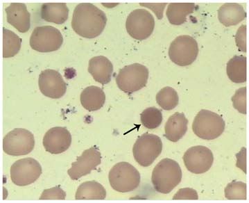
3254 LIU et al: A NOVEL SPTB GENE MUTATION IN HEREDITARY SPHEROCYTOSIS
brown in color, kaolin stools or torsion spasm were observed. of the chest and abdomen were within their normal ranges.
The patient's father had a history of anemia, and received Ultrasonography indicated no abnormalities in the liver, gall-
cholecystectomy due to gallstones. The patient's mother had bladder, spleen, kidneys or brain.
no history of haematological system diseases. Upon admission, the patient received phototherapy. Type O
On physical examination, the body temperature of the patient washed red blood cells were supplemented to correct the
was 36.5˚C, the respiration rate was 50 breaths/min, the pulse anemia, together with supporting therapy. The whole treat-
was 150 beats/min and the blood pressure was 65/35 mmHg. ment duration was four days. The jaundice showed remission,
The blood oxygen saturation level was 94%. According to the and the anemia was corrected. Finally, the patient exhibited
standard of growth curves for Chinese children and adolescents a satisfactory outcome. In the 9‑month follow‑up, the patient
which contains weight, length/height, head circumference, received erythrocytes supplementation due to anemia at
weight‑for‑length/height and body mass index aged 0‑18 years, months 3 and 7, respectively. No jaundice, splenectasis or liver
the child's nutrition and development were normal (8). The dysfunction were observed.
results of neonatal behavioral neurological assessment showed
that the child's mental response was satisfactory. Ochrodermia Genetic analysis. The genomic DNA was extracted from
was observed across the whole body. Estimation of bilirubin the patient's and her parents' peripheral blood using the
according to the location of jaundice on the skin, the bilirubin QIAamp blood DNA mini kit (QiagenGmbH) following the
value of the child was close to 5‑10 mg/dl. No edema was manufacturer's protocol. The exome sequencing kit (xGen®
observed. The doctors in the department of neonatology carried Exome Research Panel; Integrated DNA Technologies, Inc.)
out physical examination on the patient and no positive signs were was used for the preparation of the sequencing library. The
indicated, such as lassitude, feeding difficulty and hepatospleno- generated library was analyzed on the NextSeq 500 analyzer
megaly. For the blood routine examination, the hemoglobin level (Illumina, Inc.) for the sequencing of the exons of the 302
was 51 g/l (normal range, 110‑160 g/l), the mean corpuscular genes associated with hematological system disease related
volume (MCV) was 81.5 fl (normal range, 86‑100 fl), the mean genes (such as ANK1, SPTA1, EPB42, SLC4A1 and SPTB).
hemoglobin was 29.3 pg (normal range, 26‑31 pg), the mean The mean sequencing depth was 100X. Sequencing data was
hemoglobin concentration was 384 g/l (normal range, 210‑370 fl) processed using Burrows‑Wheeler‑Aligner forhg19 reference
and the red cell distribution width was 53.8 fl (37‑50 fl). The sequence (9) alignment and a Genomic‑Analysis‑Toolkit
results of cell counting demonstrated that a total of 10 interme- (V4.0.6.0) for variant calling. Variants annotation was
diate erythroblasts and 3 acidophilic erythroblasts were identified performed using Annovar (V20180118) (10).
per 100 leukocytes. These were counted manually using an According to the high‑throughput sequencing results, the
Olympus CX21 optical microscope (100X oil immersion objec- SPTB mutation was verified using Sanger sequencing based on
tive; Olympus Optical Co, Ltd.). The proportion of reticulocyte the neonatal and parental DNA samples. Specific primers were
was 12.3%. The number of leukocyte was 11.03x10⁹/l. The designed using the Primer Premier 5.0 software (PREMIER
proportion of lymphocytes, neutrophils, monocytes, eosinophils Biosoft) for the coding region of the exon of the target gene
and basophils was 60, 31, 7, 1 and 1%, respectively. The platelet (forward, 5'‑CCG C TC ATG G AA T CC C AC‑3'; reverse,
count was 333x1011/l. The concentration of C‑reactive protein was 5'‑GGAGTAGTGCCTCCTCCCTG‑3'). PCR amplification
EXPERIMENTAL AND THERAPEUTIC MEDICINE 20: 3253-3259, 2020 3255
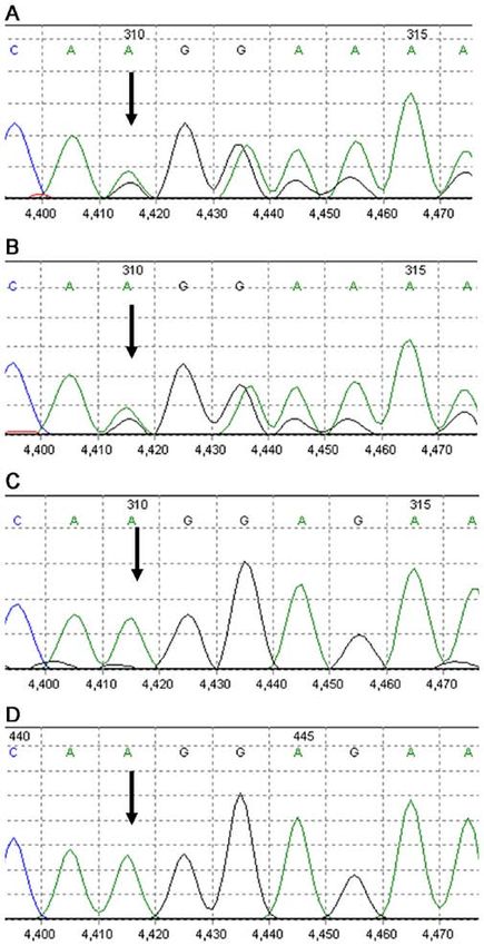
Figure 1. Morphology of erythrocytes in the blood smear (Wright‑Giemsa
stain; magnification, x1000). The spherocyte is indicated by the black arrow.
Figure 2. Family genetic pedigree. The mutation c.3737delA of SPTB gene
was identified in the proband (indicated by arrow) and her father. Sanger
sequencing of the mother showed wild-type.
pathogenic mutation. Sequencing data of the SPTB gene in the
patient and her father are presented in Fig. 3. The patient was
diagnosed with type 2 HS, which was divided into autosomal
dominant (AD) inheritance.
Prediction of protein structure. The SWISS‑MODEL
homology modeling server (https://www.swissmodel.expasy.
org/) was used to predict and compare the spatial structure of
the wild‑type protein and the protein encoded by the mutated
genes. Meanwhile, the effects of the SPTB gene mutation on Figure 3. Gene sequencing results of the mutation c.3737delA of SPTB
coding protein were justified, in order to speculate the patho- gene in the (A) child, (B) her father, (C) her mother and the (D) referencing
sequences.
genicity of the new protein.
Fig. 4A indicates the results of the prediction of the
tertiary structure of the SPTB protein (817‑1265) using the
SWISS‑MODEL software. The predicted template was some patients with moderate manifestations are not diagnosed
Protein Data Bank ID number 4uxv.1.A. Fig. 4B summarizes in clinical settings (6). Due to this, the number of patients may
the prediction data of the protein sequence with a mutation be higher than expected (11).
on residue 1246. Compared with the tertiary structure of The pathogenesis of HS may be associated with the defi-
wild‑type protein, the c.3737delA (p.Lys1246fs) mutation ciency of a variety of membrane proteins of the erythrocytes,
resulted in premature termination of the protein, triggering the including ankyrin‑1, band 3, SPTB, α‑spectrin and protein 4.2,
loss of the subsequent α‑spiral. which result in the decline of the surface area of the eryth-
rocyte membrane in patients with HS (12). Meanwhile, the
Discussion damage of erythrocytes with poor deformation capacity in the
spleen of individuals with HS is a major cause of hemolysis (8).
HS, which affects many individuals worldwide, exhibits a prev- According to the genetic deficiency of erythrocyte membrane
alence of 27.6 per million within the Chinese population (2). In proteins, HS is divided into five types (Table I), among which
neonates aged3256 LIU et al: A NOVEL SPTB GENE MUTATION IN HEREDITARY SPHEROCYTOSIS
to assist the diagnosis (14,17). In the present study, the patient's
father exhibited anemia with a family history of cholelithiasis,
which was clinically manifested as delayed remission of
jaundice and severe anemia. Finally, diagnosis of HS was
confirmed based on laboratory findings and the results of the
genetic analysis.
HS is a rare disease of genetic deficiency that lacks appro-
priate treatment options. Currently, its treatment is mainly
focused on the control of its severity (2). Phototherapy, which
lowers the bilirubin in the neonatal HS, is considered to be the
major treatment therapy in the early post‑partum period (16).
Moreover, treatment should begin immediately to those with a
Figure 4. SPTB protein structure prediction using the SWISS‑MODEL soft- high level of bilirubin or a higher medium than the risk zone
ware. (A) normal protein tertiary structure. (B) Tertiary structure of mutated (>75th percentile zone). Furthermore, according to the guide-
protein. lines proposed by the American Academy of Pediatrics (3),
further blood exchange transfusion is required. In cases of
signs of anemia, blood transfusion may also be required. Since
The typical features of HS include anemia, jaundice, erythropoiesis is damaging at a certain post‑partum period
splenomegaly and ceticulocytosis (2). The severity of HS is (1‑4 weeks) (16), single administration of erythropoietin could
divided into asymptomatic state, mild, moderate and severe, be used, or utilized simultaneously with blood transfusion.
according to the degree of anemia (14). The majority of Folic acid supplementation should be considered for those
patients exhibit mild HS, and up to 20‑30% present with a with moderate and severe HS in order to prevent the complica-
purely compensated hemolysis due to the balance between tions associated with folic acid deficiency (16). Splenectomy
reticulocyte production and red cell destruction (15). is not recommended for ~12 months after delivery (16).
Approximately 50% of neonates with HS are anemia‑free at Splenectomy is effective for treating moderate and severe
post‑natal week one, and rare cases exhibit splenomegaly (16). HS, however, it may lead to trauma, decline of immunity,
Jaundice is the most common manifestation for neonatal and pulmonary hypertension that is induced by arterial and
HS (3,15,17). Neonatal jaundice usually occurs within a few venous thrombosis (5,14). Total splenectomy may therefore be
post‑natal days. The hemoglobin concentration would be in more effective than partial splenectomy (2,21‑24). Individual
the normal range, cases may develop transient or even severe follow‑up schemes should be established for children with HS,
anemia within a few post‑natal weeks, due to inadequate which are based on the severity of anemia and the monitoring
compensation of the splenic filtration function caused by the of the growth and development (16). Meanwhile, care should
lack of appropriate reticulocytes (7). Most of these condi- be taken regarding iron overload in the children who undergo
tions exhibit remission within 12 months post‑partum (14). persistent blood transfusion (3).
In the present study, the neonate with HS exhibited delayed The SPTB gene, which encodes for the β subunit of spec-
remission of jaundice and severe anemia without kernicterus. trin, is a member of the spectrin gene family. It is localized
The patient was followed up for 9 months, and blood transfu- on 14q23.3 with a length of 100 kb, consisting of 35 exons,
sion was required to correct the anemia. and its encoded proteins form the cytoskeletal superstructure
Clinical manifestations, family history and peripheral of the erythrocyte plasma membrane (25). Upon binding
blood smear findings are relied upon in the diagnosis of HS. with ankyrin, spectrin serves a crucial role in the formation
For the blood smear, patients with HS exhibit alternations of and stability of the erythrocyte membrane. The SPTB gene
spherocyte proportion that are associated with the severity mutation is associated with type II spherocytosis, hereditary
of anemia, as well as presence of mushroom‑shaped eryth- elliptocytosis and hemolytic anemia of neonates (26,27).
rocytes, poikilocytosis and acanthocyte (18). According to In general, the AD pattern was reported in 75% of patients
the HS diagnosis guidelines that are proposed by the British with HS, while in the remaining 25%, the AR pattern was
Committee for Standards in Haematology (6), additional tests exhibited, or the disease was due to denovo mutations (5).
are not recommended for patients with HS and with typical Moreover, the inheritance patterns of some SPTB mutations
clinical manifestations and laboratory findings. As the clinical are unknown (28). In the present study, the patient's father
manifestations in the neonatal patients with HS are not typical, carried the similar mutation of the SPTB gene, which was
and some patients usually present spherocytes, the diagnosis not identified in the patient's mother. The patient's father
of HS in these patients is still difficult (14). A parental history had a history of anemia and cholelithiasis. Therefore, it was
of HS has been reported in the majority (65%) of the neonates proposed that this novel mutation in SPTB gene was of AD
with HS (16). Therefore, determining the parental history inheritance, Therefore, the risk of HS was speculated to be up
of anemia and/or the family history of anemia, jaundice, to 50% in the second child of this family.
splenectomy or early‑stage cholelithiasis in these neonates In general, the common mutation types of SPTB gene
with jaundice is crucial for the diagnosis of HS in clinical include nonsense mutations, frame‑shifting mutations and
practice (3). In addition, the eosin‑5‑maleimide binding test, splice site mutations, which give rise to mRNA defects and
osmotic fragility test, osmotic gradient ektacytometry, AGLT truncated β ‑spectrin (29). In this case, a novel mutation of
and pink test all contributed to the diagnosis of HS in clinical c.3737delA (p.Lys1246fs) in the SPTB gene was identified as
practice (11,19,20). For some patients, a genetic test is required a frameshift mutation, which led to premature termination ofEXPERIMENTAL AND THERAPEUTIC MEDICINE 20: 3253-3259, 2020 3257 Table I. Correlation between the gene and phenotype of the HS. Gene Percentage Type Gene location Protein Genetic type (%) Severity Type 1, OMIM:182900 ANK1 (612641) 8p11.21 Ankyrin‑1 Autosomal dominant 40‑50 Mild and moderate Type 2, OMIM: 616649 SPTB (182870) 14q23.3 β‑spectrin Autosomal dominant 15‑30 Mild and moderate Type 3, OMIM: 270970 SPTA1 (182860) 1q23.1 α‑spectrin Autosomal recessive
3258                            LIU et al: A NOVEL SPTB GENE MUTATION IN HEREDITARY SPHEROCYTOSIS

Availability of data and materials                                          12. Farias MG: Advances in laboratory diagnosis of hereditary sphe-
                                                                                rocytosis. Clin Chem Lab Med 55: 944‑948, 2017.
                                                                            13. He BJ, Liao L, Deng ZF, Tao YF, Xu YC and Lin FQ: Molecular
The datasets used and/or analyzed during the current study                      genetic mechanisms of hereditary spherocytosis: Current
are available from the corresponding author on reasonable                       perspectives. Acta Haematol 139: 60‑66, 2018.
                                                                            14. Wang X, Liu A, Lu Y and Hu Q: Novel compound heterozygous
request.                                                                        mutations in the SPTA1 gene, causing hereditary spherocytosis
                                                                                in a neonate with Coombsnegative hemolytic jaundice. Mol Med
Authors' contributions                                                          Rep 19: 2801‑2807, 2019.
                                                                            15. King MJ, Garcon L, Hoyer JD, Iolascon A, Picard V, Stewart G,
                                                                                Bianchi P, Lee SH and Zanella A; International Council for
YL and JZ collected and analyzed the data, and wrote the                        Standardization in Haematology: ICSH guidelines for the labo-
manuscript. LS, CS and NL collected the clinical data. JS and                   ratory diagnosis of nonimmune hereditary red cell membrane
                                                                                disorders. Int J Lab Hematol 37: 304‑325, 2015.
YF predicted the protein structure. GL and JS participated in               16. Christensen RD, Yaish HM and Gallagher PG: A pediatrician's
making substantial contributions to the conception and design,                  practical guide to diagnosing and treating hereditary spherocy-
drafting and revising the important intellectual content of                     tosis in neonates. Pediatrics 135: 1107‑1114, 2015.
                                                                            17. Christensen RD, Nussenzveig RH, Yaish HM, Henry E,
manuscript All authors read and approved the final manuscript.                  Eggert LD and Agarwal AM: Causes of hemolysis in neonates
                                                                                with extreme hyperbilirubinemia. J Perinatol 34: 616‑619, 2014.
Ethics approval and consent to participate                                  18. Da Costa L, Galimand J, Fenneteau O and Mohandas N:
                                                                                Hereditary spherocytosis, elliptocytosis, and other red cell
                                                                                membrane disorders. Blood Rev 27: 167‑178, 2013.
The study protocols were approved by the Ethical Committee                  19. L l a u d e t‑ Pl a n a s E , Vive s ‑ C o r r o n s J L , R i z z ut o V,
of Tianjin Medical University General Hospital.                                 Gómez‑Ramírez P, Sevilla Navarro J, Coll Sibina MT,
                                                                                García‑Bernal M, Ruiz Llobet A, Badell I, Velasco‑Puyó P, et al:
                                                                                Osmotic gradient ektacytometry: A valuable screening test for
Patient consent for publication                                                 hereditary spherocytosis and other red blood cell membrane
                                                                                disorders. Int J Lab Hematol 40: 94‑102, 2018.
                                                                            20. Park SH, Park CJ, Lee BR, Cho YU, Jang S, Kim N, Koh KN,
Consent for publication was obtained from the patient's family.                 Im HJ, Seo JJ, Park ES, et al: Comparison study of the eosin‑5'‑ma-
                                                                                leimide binding test, flow cytometric osmotic fragility test, and
Competing interests                                                             cryohemolysis test in the diagnosis of hereditary spherocytosis.
                                                                                Am J Clin Pathol 142: 474‑484, 2014.
                                                                            21. Baloira A, Bastos M, Pousada G and Valverde D: Pulmonary
The authors declare that they have no competing interests.                      arterial hypertension associated with hereditary spherocytosis
                                                                                and splenectomy in a patient with a mutation in the BMPR2 gene.
                                                                                Clin Case Rep 4: 752‑755, 2016.
References                                                                  22. Bader‑Meunier B, Gauthier F, Archambaud F, Cynober T,
                                                                                Mielot F, Dommergues JP, Warszawski J, Mohandas N and
 1. Tole S, Dhir P, Pugi J, Drury LJ, Butchart S, Fantauzzi M, Langer JC,       Tchernia G: Long‑term evaluation of the beneficial effect of
    Baker JM, Blanchette VS, Kirby‑Allen M and Carcao MD:                       subtotal splenectomy for management of hereditary spherocy-
    Genotype‑phenotype correlation in children with hereditary sphe-            tosis. Blood 97: 399‑403, 2001.
    rocytosis. Br J Haematol: May 20, 2020 (Epub ahead of print).           23. Abdullah F, Zhang Y, Camp M, Rossberg MI, Bathurst MA,
 2. Wang C, Cui Y, Li Y, Liu X and Han J: A systematic review                   Colombani PM, Casella JF, Nabaweesi R and Chang DC:
    of hereditary spherocytosis reported in Chinese biomedical                  Splenectomy in hereditary spherocytosis: Review of 1,657
    journals from 1978 to 2013 and estimation of the prevalence of              patients and application of the pediatric quality indicators.
    the disease using a disease model. Intractable Rare Dis Res 4:              Pediatr Blood Cancer 52: 834‑837, 2009.
    76‑81, 2015.                                                            24. Guizzetti L: Total versus partial splenectomy in pediatric
 3. Suzuki H, Kiryluk K, Novak J, Moldoveanu Z, Herr AB,                        hereditary spherocytosis: A systematic review and meta‑analysis.
    Renfrow MB, Wyatt RJ, Scolari F, Mestecky J, Gharavi AG and                 Pediatr Blood Cancer 63: 1713‑1722, 2016.
    Julian BA: The pathophysiology of IgA nephropathy. J Am Soc             25. Fan LL, Liu JS, Huang H, Du R and Xiang R: Whole exome
    Nephrol 22: 1795‑1803, 2011.                                                sequencing identified a novel mutation (p.Ala1884Pro) of
 4. Da Costa L, Suner L, Galimand J, Bonnel A, Pascreau T,                      β ‑spectrin in a Chinese family with hereditary spherocytosis.
    Couque N, Fenneteau O and Mohandas N; Society of Hematology                 J Gene Med 21: e3073, 2019.
    and Pediatric Immunology (SHIP) group; French Society of                26. Boguslawska DM, Heger E, Machnicka B, Skulski M,
    Hematology (SFH): Diagnostic tool for red blood cell membrane               Kuliczkowski K and Sikorski AF: A new frameshift mutation
    disorders: Assessment of a new generation ektacytometer. Blood              of the β‑spectrin gene associated with hereditary spherocytosis.
    Cells Mol Dis 56: 9‑22, 2016.                                               Ann Hematol 96: 163‑165, 2017.
 5. Perrotta S, Gallagher PG and Mohandas N: Hereditary spherocy-           27. Shin S, Jang W, Kim M, Kim Y, Park SY, Park J and Yang YJ:
    tosis. Lancet 372: 1411‑1426, 2008.                                         Targeted next‑generation sequencing identifies a novel nonsense
 6. Bolton‑Maggs PH, Langer JC, Iolascon A, Tittensor P and                     mutation in SPTB for hereditary spherocytosis: A case report of
    King MJ; General Haematology Task Force of the British                      a Korean family. Medicine (Baltimore) 97: e9677, 2018.
    Committee for Standards in Haematology: Guidelines for the              28. van Vuren A, van der Zwaag B, Huisjes R, Lak N, Bierings M,
    diagnosis and management of hereditary spherocytosis‑2011                   Gerritsen E, van Beers E, Bartels M and van Wijk R: The
    update. Br J Haematol 156: 37‑49, 2012.                                     complexity of genotype‑phenotype correlations in hereditary
 7. Manciu S, Matei E and Trandafir B: Hereditary spherocy-                     spherocytosis: A Cohort of 95 Patients: Genotype‑phenotype
    tosis‑diagnosis, surgical treatment and outcomes. A literature              correlation in hereditary spherocytosis. Hemasphere 3: e276,
    Review. Chirurgia (Bucur) 112: 110‑116, 2017.                               2019.
 8. Zong XN and Li H: Construction of a new growth references for           29. Maciag M, Plochocka D, Adamowicz‑Salach A and Burzynska B:
    China based on urban Chinese children: Comparison with the                  Novel beta‑spectrin mutations in hereditary spherocytosis
    WHO growth standards. PLoS One 8: e59569, 2013.                             associated with decreased levels of mRNA. Br J Haematol 146:
 9. Li H and Durbin R: Fast and accurate short read alignment with              326‑332, 2009.
    Burrows‑Wheeler transform. Bioinformatics 25: 1754‑1760, 2009.          30. Barcellini W, Bianchi P, Fermo E, Imperiali FG, Marcello AP,
10. Wang K, Li M and Hakonarson H: ANNOVAR: Functional                          Vercellati C, Zaninoni A and Zanella A: Hereditary red cell
    annotation of genetic variants from high‑throughput sequencing              membrane defects: Diagnostic and clinical aspects. Blood
    data. Nucleic Acids Res 38: e164, 2010.                                     Transfus 9: 274‑277, 2011.
11. Andolfo I, Russo R, Gambale A and Iolascon A: New insights on           31. Iolascon A and Avvisati RA: Genotype/phenotype correlation
    hereditary erythrocyte membrane defects. Haematologica 101:                 in hereditary spherocytosis. Haematologica 93: 1283‑1288,
    1284‑1294, 2016.                                                            2008.
EXPERIMENTAL AND THERAPEUTIC MEDICINE 20: 3253-3259, 2020 3259
32. Waterhouse A, Bertoni M, Bienert S, Studer G, Tauriello G, 35. Xue J, He Q, Xie X, Su A and Cao S: Erratum to clinical utility
Gumienny R, Heer FT, de Beer TAP, Rempfer C, Bordoli L, et al: of targeted gene enrichment and sequencing technique in the
SWISS‑MODEL: Homology modelling of protein structures and diagnosis of adult hereditary spherocytosis. Ann Transl Med 7:
complexes. Nucleic Acids Res 46: W296‑W303, 2018. S391, 2019.
33. Park J, Jeong DC, Yoo J, Jang W, Chae H, Kim J, Kwon A, 36. Qin L, Nie Y, Zhang H, Chen L, Zhang D, Lin Y and Ru K:
Choi H, Lee JW, Chung NG, et al: Mutational characteristics Identification of new mutations in patients with hereditary
of ANK1 and SPTB genes in hereditary spherocytosis. Clin spherocytosis by next‑generation sequencing. J Hum Genet 65:
Genet 90: 69‑78, 2016. 427‑434, 2020.
34. Wang R, Yang S, Xu M, Huang J, Liu H, Gu W and Zhang X:
Exome sequencing confirms molecular diagnoses in 38 Chinese This work is licensed under a Creative Commons
families with hereditary spherocytosis. Sci China Life Sci 61: Attribution-NonCommercial-NoDerivatives 4.0
947‑953, 2018. International (CC BY-NC-ND 4.0) License.You can also read