Protein losing enteropathy in Yorkshire Terriers - Retrospective study in 31 dogs - Revue de ...
←
→
Page content transcription
If your browser does not render page correctly, please read the page content below
2                                                                                                             BOTA (D.) AND COLLABORATORS

Protein losing enteropathy in Yorkshire Terriers –
Retrospective study in 31 dogs
D. BOTA1, A. LECOINDRE2, A. POUJADE3, M. CHEVALIER4, P. LECOINDRE2, F. BAPTISTA5, E. GOMES1,
J. HERNANDEZ1*

1
  Centre Hospitalier Vétérinaire Fregis, 43 av. Aristide Briand, 94110 Arcueil, France
2
  Clinique des Cerisioz, Route de Saint-Symphorien-d’Ozon 69800 Saint-Priest France
3
  Laboratoire d‘Anatomie Pathologique Vétérinaire du Sud-Ouest, 129, route de Blagnac 31201 Toulouse cedex 2, France
4
  Laboratoire Biomnis, 17/19 avenue Tony Garnier, 69007 Lyon, France
5
  StemCell2Max Biocant Park Nucleo 04, Lote 02 3060-197 Cantanhede Portugal

*Corresponding author: jhernandez@fregis.com

SUMMARY                                                                             RESUME

Yorkshire Terriers are reportedly predisposed to PLE. The aim of our                Entéropathie exsudative chez le Yorkshire Terrier – Étude retrospective
retrospective study was to describe the clinical, laboratory, ultrasonographic,     de 31 cas
endoscopic and histological findings of Yorkshire Terriers with PLE and
to compare potential prognostic factors to those recently published. A              Selon la littérature, les Yorkshire Terriers sont prédisposés au développement
retrospective study was performed using clinical reports from two private           d’entéropathies exsudatives. Le but de notre étude rétrospective est de
practices in France from 2010-2012. For each case, the age, sex, clinical           décrire les caractéristiques cliniques, de laboratoire, échographiques,
signs, serum albumin, abdominal ultrasound findings, gross endoscopic               endoscopiques et histologiques de cas d’entéropathies exsudatives chez le
appearance, histopathological reports, and outcome at 1 month and 1 year            Yorkshire Terrier et de comparer les facteurs pronostiques à ceux récemment
were recorded. Females were over-represented (20/31). The sole clinical             publiés. L’étude a été réalisée en utilisant les dossiers médicaux issus de 2
finding in more than 40% of cases was ascites. Hyperechoic striations were          centres privés de référés en France de 2010 à 2012. Pour chaque cas, l’âge, le
frequently seen on abdominal ultrasound (16/25). Multiple white villi on            sexe, les signes cliniques, l’albuminémie, les observations échographiques,
gross endoscopic examination was observed in all the 22 dogs with intestinal        l’aspect endoscopique duodénal, les lésions histologiques intestinales
lymphangiectasia on histopathology. Lacteal and crypt dilation were common          ainsi que l’évolution à 1 mois et à 1 an ont été analysés. Les femelles sont
histopathological lesions (24/31 and 27/31 cases, respectively). Yorkshire          surreprésentées (20/31). L’ascite est l’observation clinique la plus fréquente
terriers are susceptible to a potentially severe form of PLE characterized          (40% des cas). Des striations hyperéchogènes dans la muqueuse intestinale
by abdominal effusion due to hypoalbuminaemia, and histologically by                grêle sont fréquemment observées au cours de l’examen échographique
crypt lesions, lacteal dilation, and inflammation. The absence of diarrhoea         (16/25). Des villosités blanchâtres ont été observées au cours de l’examen
cannot be used to rule out PLE. Abdominal ultrasound and gross endoscopy            endoscopique sur tous les cas où une lymphangiectasie intestinale a été
appearance are useful for detecting intestinal lymphangiectasia. Outcome is         identifiée à l’examen histopathologique (22 cas). La dilatation des chylifères
variable and some dogs completely fail to respond to therapy.                       et des cryptes intestinales est une observation histologique fréquente (24
                                                                                    et 27 cas respectivement). Les Yorkshire Terriers présentent une forme
Keywords : Crypt dilation, lymphangiectasia, protein                                potentiellement grave d’entéropathie exsudative caractérisée par des
                                                                                    épanchements cavitaires liée à l’hypoalbuminémie et histologiquement par
losing enteropathy, Yorkshire Terrier                                               une dilatation des chylifères et des cryptes intestinales et de l’inflammation.
                                                                                    L’absence de diarrhée ne peut être utilisée pour exclure la maladie. L’examen
                                                                                    échographique et l’endoscopie sont des examens efficaces permettant
                                                                                    d’identifier les lésions de lymphangiectasie. L’évolution des Yorkshire Terriers
                                                                                    atteints est variable avec quelques chiens qui ne répondent pas au traitement.

                                                                                    Mots-clés : Dilatation des cryptes intestinales,
                                                                                    lymphangiectasie, entéropathie exsudative, Yorkshire
                                                                                    Terrier

Introduction                                                                        peripheral oedema, dyspnoea secondary to pleural effusion
                                                                                    or pulmonary thrombosis, and muscle tremors and seizure
    Protein-losing enteropathy (PLE) is caused by a group of                        activity due to hypocalcaemia and hypomagnesaemia [4, 5,
intestinal pathologies that are responsible for nonselective                        9]. Diagnosis involves identification of hypoalbuminaemia,
protein loss resulting in hypoproteinaemia [12]. Common                             exclusion of renal protein loss by urinalysis and urine protein-
causes of PLE are inflammatory bowel disease (IBD),                                 to-creatinine ratio, and exclusion of hepatic dysfunction by
intestinal lymphoma, and intestinal lymphangiectasia                                normal pre and post-prandial serum bile acids. Detection
(IL) [4]. Intestinal infectious diseases, gastrointestinal                          of enteric protein loss by measuring faecal α1-proteinase
haemorrhage, and intestinal crypt lesions may also induce                           inhibitor is not routinely performed [10]. Hyperechoic
PLE [4].                                                                            intestinal mucosal striations on ultrasound are described
                                                                                    as highly specific for lacteal dilation [6]. Ultimately,
    Clinical signs of PLE include diarrhoea, vomiting, weight                       intestinal biopsies are needed to identify gastrointestinal
loss, abdominal distension secondary to ascites and/or                              histopathologic lesions that define the underlying aetiology
                                                                                    of the PLE [7].

                                                                                                                             Revue Méd. Vét., 2016, 167, 1-2, 2-9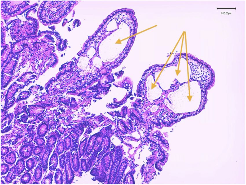
PROTEIN LOOSING ENTEROPATHY IN YORKSHIRE TERRIER                                                                                   3

   Yorkshire Terriers are reportedly predisposed to PLE,              using a random study number and both pathologists reviewed
being ten times more likely to contract the disease than other        all the slides. Each pathologist was blinded to the original
breeds [4]. Recently, clinical features, intestinal histopathology,   histopathology results and to any clinical information. A
and outcome were described in a retrospective study of                standard form was completed for each dog, including quality,
30 Yorkshire Terriers [12]. Females were overrepresented.             morphologic features (villous stunting, epithelial injury,
A third of the dogs in this study did not have a history of           crypt lesion, lacteal dilation, and mucosal fibrosis), and
diarrhoea. Predictors of death within 4 months of diagnosis           inflammation (intraepithelial lymphocytes, lamina propria
were a history of vomiting, monocytosis, low BUN, degree of           lymphocytes and plasma cells, lamina propria eosinophils,
hypoalbuminaemia, and intestinal villous blunting.                    lamina propria neutrophils and other). Inadequate biopsy
                                                                      quality was a criterion for exclusion. Morphological features
    The aim of our retrospective study was to describe                and inflammation were graded as normal, mild, moderate,
the clinical, laboratory, ultrasonographic, endoscopic,               and marked. Diagnosis of intestinal lymphangiectasia was
histological findings, treatment and outcome of Yorkshire             based on the pathologist’s ability to constantly identify the
Terriers with PLE and to compare potential prognostic                 lumen of dilated lacteals. Only cases with consensual final
factors to those recently published.                                  diagnoses between both pathologists were included.

Material and Methods                                                      Clinical signs and measurement of serum albumin
                                                                      at 1 month after discharge were recorded. All referring
   Medical records from two referral practices in France              veterinarians or owners were contacted 1 year after diagnosis
(Centre Hospitalier Vétérinaire Frégis (CHVF), and the                to determine the outcome (animal alive or deceased,
Clinique Vétérinaire des Cerisioz (CVC)) were reviewed for            albuminemia, with or without treatment).
Yorkshire Terriers diagnosed with PLE from January 2010 to
January 2012.                                                             Treatment options were different between the 2
                                                                      centers. At the CHVF, all 19 dogs were started on a low-fat
    The inclusion criteria were as follows: Yorkshire Terriers        hyperdigestible diet (Hills i/D Low Fat® Sophia-Antipolis,
with hypoalbuminemia < 25 g/l (reference range for albumin            France), metronidazole (Flagyl®, Sanofi-Aventis, Gentilly,
30-40 g/l), urine dipstick negative for proteinuria or urine          France 15mg/kg twice daily), and prednisolone (Megasolone®,
protein-to-creatinine ratio < 0.5, normal pre and post                Merial, Lyon, France, 1-2 mg/kg/day). At the CVC, all 12 dogs
prandial serum bile acids, and histopathological intestinal           were started on the same diet, metronidazole, prednisolone,
lesions characteristic of diseases recognised to cause PLE. All       ciclosporine (Atopica®, Novartis, Huningue, France, 5 mg/
dogs must have had a negative faecal flotation test within 1          kg/day), and marbofloxacin (Marbocyl®, Vetoquinol, Lure,
month of presentation or been treated for intestinal parasites        France, 2mg/kg/day). These differences were only due to the
before presentation. Dogs with evidence of protein-losing             different protocols used at each institution.
nephropathy or impaired liver function were excluded.
Dogs with gastrointestinal parasites or haemorrhage,                  Statistical analysis
parvovirus infection, neoplasia, and those that had received
glucocorticoids or other immunosuppressive treatment                      Statistical analysis was performed using SAS v. 9.1.3. (SAS
within 2 weeks of the intestinal biopsy were excluded. Cases          Institute Inc., Cary, NC, USA). The descriptive statistical
where tissue biopsy depth was inadequate for assessing                analysis was expressed as a mean ± SD for age, sex,, frequency
intestinal crypts were also excluded.                                 for clinical signs, serum albumin, abdominal ultrasound
                                                                      findings, and histological lesions. Multivariate logistic
    Clinical information collected for each dog included:             regression analyses were used to determine the variables
age, sex, previous treatment, physical examination findings           associated with outcome (alive or deceased) at 1 month
(namely the presence/absence of diarrhoea and abdominal               and 1 year after diagnosis, respectively. The identification
distension), serum total protein and albumin at admission,            of the significant variables and interactions (P < 0.05) was
abdominal ultrasonographic findings, histopathological                performed by backward elimination.
lesions and treatment.
                                                                          The candidate explanatory variables were the following:
    A board-certified radiologist reviewed all available              sex (male/female), age (10yrs), clinical signs
captured static images of abdominal ultrasound studies.               (presence or absence of vomiting, diarrhea, abdominal
Endoscopy was performed using an Olympus GIF-180 and                  distention), ultrasound findings (presence or absence of
biopsies using disposable “alligator swing-jaw fenestrated”           small intestinal hyperechoic mucosal striations and/or
(2.45 mm) biopsy forceps. The minimum number of biopsies              ascites), histopathological findings (presence or absence of
taken per segment was 6. Histopathologic evaluation was               lymphangiectasia and/or crypt distension) and treatment
based upon review of all endoscopic biopsies under the                (the use of steroids with and without ciclosporine) were
recommendations of the World Small Animal Veterinary                  determined as categorical variables, with serum albumin
Association Gastrointestinal Standardization Group (2008)             concentration as a continuous variable.
by 2 pathologists (AP and MC) [1]. The slides were relabelled

Revue Méd. Vét., 2016, 167, 1-2, 2-9
4 BOTA (D.) AND COLLABORATORS
A Fisher’s exact test was performed to test the association in 16 animals. Fourteen animals had ascites and small
between: 1) treatment and albumin at 1 month (≤ 15 g/l intestinal hyper echoic striations on abdominal ultrasound.
and > 15 g/l); 2) treatment and histopathological findings Other intestinal ultrasonographic findings included mucosal
and 3) treatment and ultrasound findings. The identification speckling in 8 dogs, increased intestinal wall thickness in 5
of the significant variables and interactions (P < 0.05) was dogs and reduced intestinal peristalsis in 2 dogs.
performed by backward elimination. Statistical significance
was set at a P-value < 0.05 A strong association was found between the presence of
hyper echoic striation on ultrasound lesions and the presence
Results of intestinal lymphangiectasia on histopathology (p=0.02).
Medical records of 66 Yorkshire terriers with suspected UPPER GASTROINTESTINAL ENDOSCOPY
PLE were reviewed. Nine dogs were excluded because of
absence of intestinal biopsies. Fifteen dogs were excluded Duodenal endoscopy was performed in all 31 dogs.
because of absence of serum bile acids test results or Abnormalities affecting the duodenal mucosa included
abnormal urine protein-to-creatinine ratios. Eleven dogs friability in 25 dogs and multiple white villi in 22 dogs. All this
were excluded because of inadequate biopsy quality. A total 22 dogs had confirmed lymphangiectasia on histopathology.
of 31 dogs fulfilled the inclusion criteria. Nineteen dogs were
diagnosed at the CHVF and 12 at the CVC. GASTROINTESTINAL HISTOPATHOLOGY
CLINICAL FINDINGS Intestinal biopsies were obtained from all 31 dogs by
intestinal endoscopy. The mean number of duodenal biopsies
Descriptive results are presented in Table I. The mean was 7. Tissue quality was classified as adequate in all cases
age at presentation was 7.7 years ± 3.0 years. Twenty dogs (Tables III-V).
were females and 11 were males. Presenting complaints
included small bowel diarrhoea (18), abdominal distension Lacteal dilation was identified in 24 and was classified
(13), and vomiting (6). Median duration of clinical signs as mild in 20 and moderate in 4 dogs (Fig. 1). Crypt
before presentation was 1 month (3 days-6 months). dilation was identified in 27 and classified as mild in 19
Previous medications included maropitant, loperamide, dogs, moderate in 6, and marked in 2 dogs (Fig. 2). The
and antibiotics (cefovexin, marbofloxacin, metronidazole). presence of lacteal dilation on histopathology was strongly
Physical examination revealed abdominal distension associated with an albumin at admission of < 15 g/l (p=0.02).
consistent with ascites in 13 dogs. Dyspnoea and/or muffled Lymphoplasmacytic inflammation was detected within the
lung sounds were not recorded in any of the dogs. One dog lamina propria of 27 dogs. Only 2 dogs had some degree of
presented with signs of dehydration and one with peripheral neutrophilic inflammation.
ventral oedema.
LABORATORY FINDINGS
Abnormal leucograms were rare: 1 dog had a mild mature
neutrophilia and 2 dogs had thrombocytosis.
Hypoglobulinaemia (reference range 15-35 g/l) was
found in 6/31 dogs (Table II).
Cholesterol was measured in 12 dogs and was below the
reference range in 10 dogs (reference range 125-300 mg/dl,
median 93,47 mg/dl). Serum total calcium was measured in
12 dogs and was low in all the dogs (reference range 9-11.5
mg/dl) serum ionized calcium concentrations were not
measured in any of the dogs. Clinical signs compatible with
hypocalcaemia (tremors, tetany, seizures) were not reported
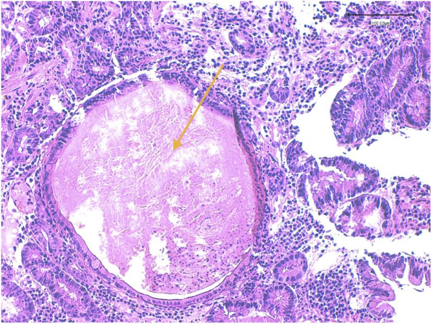
in any of the cases. Cobalamin was measured in 12 dogs and Figure. 1: Dilated lacteal vessels (arrow). Hematoxylin and Eosin stain.
40X. Courtesy of Dr Poujade, LAPVSO.
hypocobalaminaemia was found in 3 dogs (reference range
200-600 ug/ml).
ABDOMINAL ULTRASOUND FINDINGS
Abdominal ultrasonography was performed in 25 dogs.
Small intestinal hyperechoic mucosal striations were visible
Revue Méd. Vét., 2016, 167, 1-2, 2-9PROTEIN LOOSING ENTEROPATHY IN YORKSHIRE TERRIER 5
Dog Sex Age Diarrhea Ascites Albumin AUS Lymph. Cryptitis F. up
1 FS 7 + - 19 + + +
2 F 9 - + 14 - + -
3 F 8 - + 18 + + +
4 FS 6 + + 13 - + -
5 M 4 + - 18 - + + D
6 F 5 + + 12 - - + LFU
7 F 4 + + 15 + - +
8 M 2 - + 10 - - +
9 F 6 + - 23 - - +
10 F 8 + + 7 + + -
11 M 8 + + 9 - + + D
12 MC 11 + - 23 + + +
13 MC 14 + - 18 - - + D
14 FS 11 + + 19 - + + D
15 M 3 + + 14 - + + D
16 F 8 - + 15 + + +
17 FS 10 - + 8 + + +
18 M 7 + + 12 + + -
19 M 4 - + 10 + + +
20 F 13 - + 10 - + +
21 F 7 - + 7 - - +
22 F 10 - + 14 + - + D
23 F 12 + + 6 + + +
24 F 5 + + 8 + + + D
25 F 6 - + 8 - + + D
26 M 7 - + 12 - + +
27 M 11 + + 12 - + +
28 F 7 - + 7 + + +
29 M 8 - + 10 + + + D
30 F 8 + + 7 + + + D
31 F 11 + + 9 + + +
F female; M male; + present; - absent ; AUS abdominal ultrasound; Lymph lymphangiectasia; Cryp cryptitis; F. up follow up after 1 year. LFU - lost to follow
up;
Table I: Results for age, clinical signs, albumin at admission, presence of hyperechoic mucosal striations on abdominal ultrasound, presence of
lymphangiectasia and/or cryptitis on histopathology and follow up (dead or alive).
Variable Median (for all dogs) Median (CHVF/CVC) Range (CHVF / CVC)
Albumin (RR 30-40 g/l) 12.4 g/l 14.5 g/l (CHVF) 8-23 (CHVF)
9.0 g/l (CVC) 7-14 (CVC)
Table II: Albumin variation between the 2 practices (CHVF/CVC)1.
1
CHVF Centre Hospitalier Vétérinaire Frégis; CVC: Clinique Vétérinaire des Cerisioz
PLE Protein Loosing Enteropathy
Revue Méd. Vét., 2016, 167, 1-2, 2-96 BOTA (D.) AND COLLABORATORS
of >18 months. Reasons for euthanasia included: fungal
osteomyelitis (1), signs related to the enteropathy (diarrhoea,
weight loss, ascites) (6), suspected lymphoma (1), and
pleural effusion (1). One dog died of unknown causes.
The dog with fungal osteomyelitis had been living on the
Ivory Coast and had received prednisolone (0.5mg/kg/d)
and cclosporine (5mg/kg/d). Among the 6 dogs that were
euthanized for causes related to the disease, 1 dog developed
hypocalcaemia (total calcium 36 mg/dl, RR: 90-120) and
seizures, and was euthanized. The remaining five continued
to show clinical signs despite medical treatment and the
owners elected for euthanasia. Detailed histopathology from
these 6 dogs is presented in Table VI. One dog was diagnosed
with pleural effusion and the owner requested euthanasia
without performing further examination. One dog was
Figure 2: Intestinal crypt distension (arrow). Hematoxylin and Eosin stain. euthanized one year after diagnosis with persistent GI signs
40X. Courtesy of Dr Poujade, LAPVSO.
and ultrasonographic findings compatible with multicentric
TREATMENT lymphoma (weight loss, vomiting, diarrhoea, thickened
small intestine and lymph node enlargement at abdominal
Treatment options were different between the 2 centers. At ultrasound) but the diagnosis could not be confirmed. One
the CHVF, all 19 dogs were started on a low-fat hyperdigestible dog (number 15) discontinued treatment for 6 months and
diet metronidazole and prednisolone. Ciclosporine was similar clinical signs recurred: diarrhoea and abdominal
added to the treatment in 4 dogs where treatment did not distention due to ascites. Hypoalbuminaemia was present (18
control clinical signs. At the CVC, all 12 dogs were started g/l) and upper GI endoscopy revealed similar lesions to those
on the same low-fat hyperdigestible diet , metronidazole, found previously. Histopathologic lesions were unchanged
prednisolone, ciclosporine and marbofloxacin. These (lymphoplasmacytic inflammation with lacteal dilation and
differences were only due to the different protocol used at crypt lesions).
each institution.
There was no statistically significant difference
Clinical signs, serum albumin concentration, abdominal between mortality at 1 month and at 1 year in dogs with
ultrasound findings and histopathology results did not lymphangiectasia and/or crypt lesions (p>0.05).
statistically differ between the 2 treatment groups.
DISCUSSION
Other initial therapies included colloid perfusion (8) This study reports the clinical, ultrasonographic,
and vitamin B12 injections (3). The increase on albumin at endoscopic, clinicopathological, and GI histopathological
1 month (> 20 g/L) was strongly correlated with the use of findings of 31 Yorkshire Terriers with a diagnosis of PLE
prednisolone and metronidazole alone (p=0,004), compared Yorkshire Terriers of all age groups were affected with
to the use of prednisolone, ciclosporine, metronidazole and a predominance of females. Small bowel diarrhoea and
marbofloxacin. abdominal distension due to ascites were common clinical
findings. Near 50% of the dogs in this study did not have a
OUTCOME history of diarrhoea. Therefore, PLE should not be excluded
in Yorkshire terriers without diarrhoea. Vomiting was less
Clinical signs, abdominal ultrasound findings, frequent than in a previous study and it was not identified as
gastrointestinal histopathology results, and serum albumin a negative prognostic indicator [12].
at admission and at 1 month were not correlated with the
outcome at 1 year. Outcome and survival varied widely: some dogs achieved
prolonged survival and remission of clinical signs, whereas
At 1 month, 2/31 dogs had died, 3 were lost to follow- others failed to respond. More than 50% of dogs with long-
up, and 26 were re-evaluated. Six of those 26 dogs had an term follow-up had a prolonged response to treatment with
albumin of less than or equal to 15 g/l, and 20 had an albumin complete resolution of clinical signs. In Simmerson’s study
concentration of more than 15 g/l. [12], predictors of death within 4 months of diagnosis
were a history of vomiting, monocytosis, low BUN, degree
At 1 year, 10/31 dogs had died. Four dogs died within 2 of hypoalbuminemia, and intestinal villous blunting. In
months, 4 dogs died between 2 and 6 months, and 2 dogs this study, none of the studied variables was identified as a
died between 6 month and 1 year after diagnosis. Eighteen prognostic indicator. There was a strong association between
dogs were alive at the time of writing with a median survival albumin > 20 g/l at 1 month and the use of prednisolone and
metronidazole alone, compared to the use of prednisolone
Revue Méd. Vét., 2016, 167, 1-2, 2-9PROTEIN LOOSING ENTEROPATHY IN YORKSHIRE TERRIER 7
Villous Epithelial Crypt Lacteal Mucosal fibrosis
stunting injury dilation dilation
Normal 12 8 4 7 27
(4/8) (3/5) (0/4) (2/6) (9/18)
Mild 16 23 19 20 4
(6/10) (8/15) (8/11) (7/13) (2/2)
Moderate 3 0 6 4 0
(1/2) (3/3) (2/2)
Marked 0 0 2 0 0
(0/2)
Table III: Morphologic changes identified on duodenal mucosa of Yorkshire terrier with PLE (CVC/CHVF)
Intraepithelial Lamina propria Lamina propria Lamina propria Other
lymphocytes lymphocytes and eosinophils neutrophils
plasma cells
Normal 6 0 30 29 30
(2/4) (10/20) (9/20) (10/20)
Mild 21 14 1 2 1
(7/14) (5/9) (1/0) (2/0) (1/0)
Moderate 3 16 0 0 0
(1/2) (6/10)
Marked 1 1 0 0 0
(1/0) (0/1)
Table IV: Types and degrees of inflammatory cells identified on duodenal mucosa of Yorkshire terrier with PLE (number of cases at CVC/ numberr of
cases at CHVF)
Normal tissue 0
Lymphoplasmocytic inflammation 31 (11/20)
Eosinophilic inflammation 0
Neutrophilic inflammation 2 (2/0)
Lymphangiectasia 24 (9/15)
Mucosal atrophy/fibrosis (non inflammatory) 0
Other : crypt dilation 27 (11/16)
Table V: Final histopathologic diagnosis (number of cases at CVC/ number of cases at CHVF)
Dog VS EI CD LD MF IL LpLP LpE LpN
14 0 + + ++ 0 + + 0 0
15 0 + + + 0 + ++ 0 0
22 0 + + 0 0 0 + 0 0
24 + + +++ +++ 0 +++ ++ 0 +
25 0 + + ++ 0 + ++ 0 0
30 + + ++ + 0 + ++ 0 0
VS villous stunting; EI epithelial injury; CD crypt distention; LD lacteal dilation; MF mucosal fibrosis; IL intraepithelial lymphocytes; LpLP lamina propria
lymphocytes and plasma cells; LpE lamina propria eosinophils; LpN lamina propria neutrophils; normal = 0; mild = + ; moderate = ++; marked = +++
Table Vi : Morphologic features and inflammation identified on duodenal mucosa from Yorkshire terrier who died from causes related to the disease.
Revue Méd. Vét., 2016, 167, 1-2, 2-98 BOTA (D.) AND COLLABORATORS
and metronidazole combined with ciclosporine and reviewed and it could be part of the limitations. Other
marbofloxacin (p=0.004). The impact of this higher limitation of the study is the absence of ileum biopsies.
albuminaemia at 1 month in the CHVF group on survival Despite the fact that these dogs had small intestinal diarrhea,
could not be established. only upper gastro intestinal endoscopy was performed and
none had lower gastro intestinal endoscopy. The lack of
There was a strong correlation between the presence ileal biopsies may have introduced bias on the severity of the
of hyperechoic mucosal striations on ultrasound and the disease and distribution of the lesions. Treatment protocols
presence of intestinal lymphangiectasia on histopathology could not be compared, as allocation was not randomized. In
(p=0.02) This finding has been reliably associated with PLE this study, treatment options were a board certified internal
in dogs and could be used as an indicator of intestinal protein medicine specialist decision. Other protocols are also
losses in cases of hypoalbuminaemia in Yorkshire terriers described in literature and should be considered in this breed
[13]. Only 2 dogs with hyperechoic mucosal striations as well: a recent study in dogs with PLE [2] showed that a
did not show intestinal lymphangiectasia on duodenal chlorambucil-prednisolone protocol was efficient in treating
histopathology. This may be due to the heterogeneity of these animals.
lacteal dilation distribution within the mucosal layer.
In conclusion, Yorkshire terriers are susceptible to a
All dogs with multiple white villi on gross endoscopic potentially severe form of PLE characterized by abdominal
evaluation of duodenal mucosa were confirmed to have effusion due to hypoalbuminaemia, and histologically by
intestinal lymphangiectasia on histopathology. crypt lesions, lymphangiectasia, and inflammation. Females
are over-represented. The absence of diarrhoea cannot
Hypocalcaemia and hypomagnesaemia have been be used to rule out PLE. Abdominal ultrasound and gross
described in Yorkshire terriers with PLE [5]. Poor intestinal endoscopy appearance are useful for detecting intestinal
absorption of vitamin D and calcium, loss of vitamin D lymphangiectasia. Outcome is variable and some dogs
binding protein and increased leakage of calcium and completely fail to respond to therapy.
magnesium into the intestinal lumen are the main suspected
reasons for this. In this study, only 12/31 animals underwent References
total serum calcium analysis. All 12 dogs were found to have
low total calcium although only 1 dog developed clinical signs 1. - CRAVEN M., DUHAMEL G.E., SUTTER N.B. and
compatible with hypocalcaemia (seizures). Unfortunately, SIMPSON K.W.: Absence of bacterial association in
measurement of ionized calcium was not available. Yorkshire Terriers with protein-losing enteropathy and
cystic intestinal crypts. J. Vet. Intern. Med., 2009, 23,
Crypt dilation was present in 27/31 dogs and lacteal 757.
dilation in 24/31 dogs. More than 20% of the dogs in this 2. - DANDRIEUX J.R., NOBLE P.J.M., SCASE T.J., CRIPPS
study lacked lymphatic dilation. Therefore, PLE in Yorkshire P.J., GERMAN A.J.: Comparison of a chlorambucil-
Terriers may be a more complex disease than previously prednisolone combination with an azathioprine-
thought. Intestinal lymphangiectasia in this breed may be due prednisolone combination for treatment of chronic
to another underlying pathology. Simmerson hypothesized enteropathy with concurrent protein-losing enteropathy
that crypt lesions could be the primary lesions but, in this in dogs: 27 cases (2007-2010). J. Am. Vet. Med. Assoc.,
study, lymphangiectasia and crypt lesions (crypt distension) 2013, 242, 1705-14.
coexisted in only two thirds of cases [12]. Inflammation is 3. - DAY M.J., BILZER T., MANSELL J., WILCOCK B.,
another potential initiating lesion. Sample bias introduced HALL E.J., JERGENS A., MINAMI T., WILLARD,
by endoscopic biopsies and regional distribution of lesions is M., WASHABAU R., World Small Animal Veterinary
possible. Quality bias is less likely as only cases with adequate Association Gastrointestinal Standardization Group.:
biopsy quality were included in this study. Histopathological standards for the diagnosis of
gastrointestinal inflammation in endoscopic biopsy
Crypt lesions are much more common in this study than samples from the dog and cat: a report from the World
in previous reports [8]. This may be due to improved biopsy Small Animal Veterinary Association Gastrointestinal
technique over time. It has been suggested that crypt dilation Standardization Group. J. Comp. Pathol., 2008, 138
is not associated with bacterial colonization in this breed Suppl 1: S1–S43.
[1]. The lack of association between outcome and the use of 4. - DOSSIN O., LAVOUE R.: Protein-losing enteropathies
marbofloxacin argues in favour of a non-septic origin. It has in dogs. Vet. Clin. North Am. Small Anim. Pract., 2011,
been hypothesised that crypt lesions represent a degenerative 41, 399–418.
process and it is not known whether they are a cause of PLE 5. - KIMMEL S.E., WADDELL L.S., MICHEL K.E.:
or markers of another underlying lesion. Hypomagnesemia and hypocalcemia associated with
protein-losing enteropathy in Yorkshire terriers: five
The limitations of this study include its retrospective cases (1992-1998). J. Am. Vet. Med. Assoc., 2000, 217,
nature and the lack of uniformity in blood work and follow- 703–706. .
up. For instances, video or images of the scoping were not
Revue Méd. Vét., 2016, 167, 1-2, 2-9PROTEIN LOOSING ENTEROPATHY IN YORKSHIRE TERRIER 9
6. - KULL P.A., HESS R.S., CRAIG L.E., SAUNDERS 10. - MURPHY K.F., GERMAN A.J., RUAUX C.G., STEINER
H.M., WASHABAU R.J.: Clinical, clinicopathologic, J.M., WILLIAMS D.A., HALL E.J.: Fecal alpha1-
radiographic, and ultrasonographic characteristics of proteinase inhibitor concentration in dogs with chronic
intestinal lymphangiectasia in dogs: 17 cases (1996- 1998). gastrointestinal disease. Vet. Clin. Pathol., 2003, 32, 67–72.
J. Am. Vet. Med. Assoc., 2001, 219, 197–202. 11. - PETERSON P.B., WILLARD M.D.: Protein-losing
7. - LARSON R.N., GINN J.A., BELL C.M., DAVIS M.J., FOY enteropathies. Vet. Clin. North Am. Small Anim. Pract.,
D.S.: Duodenal endoscopic findings and histopathologic 2003, 33, 1061–1082.
confirmation of intestinal lymphangiectasia in dogs. J. Vet. 12. - SIMMERSON S.M., ARMSTRONG P.J., WUNSCHMANN
Intern. Med., 2012, 26, 1087–1092. A., JESSEN C.R., CREWS L.J., WASHABAU R.J.: Clinical
8. - LECOINDRE P., CHEVALLIER M., GUERRET S.: features, intestinal histopathology, and outcome in
Protein-losing enteropathy of non neoplastic origin in the protein-losing enteropathy in Yorkshire Terrier dogs. J.
dog: a retrospective study of 34 cases. Schweiz. Arch. Für Vet. Intern. Med., 2014, 28, 331–337.
Tierheilkd., 2010, 152, 141–146. 13. - SUTHERLAND-SMITH J., PENNINCK D.G., KEATING
9. - MELLAMBY R.J., MELLOR P.J., ROULOIS A., BAINES J.H., WEBSTER C.R.L.: Ultrasonographic intestinal
E.A., MEE A.P., BERRY J.L., HERRTAGE M.E.: hyperechoic mucosal striations in dogs are associated with
Hypocalcaemia associated with low serum vitamin D lacteal dilation. Vet. Radiol. Ultrasound., 2007, 48, 51–57.
metabolite concentrations in two dogs with protein-losing
enteropathies. J. Small Anim. Pract., 2005., 46, 345–351.
Revue Méd. Vét., 2016, 167, 1-2, 2-9You can also read