Uncoventional approaches and volumes in Rectal Cancer - Gemelli ART
←
→
Page content transcription
If your browser does not render page correctly, please read the page content below
28° Residential Course

   Modern Radiotherapy and unconventional treatments:
         fractionations, volumes and new drugs

                         Rome 8-10 October 2018

Uncoventional approaches and volumes in
             Rectal Cancer

     Maria Antonietta Gambacorta
           Policlinico Gemelli IRCCS
Università Cattolica del Sacro Cuore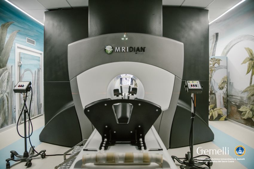
Modern Radiotherapy and unconventional treatments:
      fractionations, volumes and new drugs

  Clinical Trial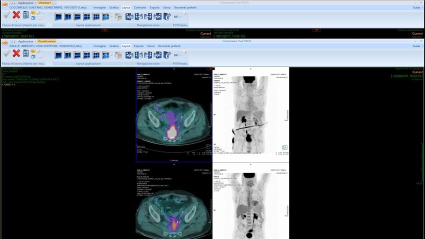
Uncoventional approaches and volumes in Rectal Cancer

 • Locally advanced rectal cancer

 • Local relapse
Conventional treatment in LARC
                                                                                    C.J.H. van de Velde et al. / European Journal of Cancer xxx (201
20                                 C.J.H. van de Velde et al. / European Journal of Cancer xxx (2013) xxx–xxx

                                                                                 TREATMENT MODALITIES: cT3 (MRF+) N0 -2-M0 or cT4 an
                                                       TREATMENT MODALITIES: cT3 (MRF -) N1-2 M0

                   CLINICAL                                      cT3 (MRF-) N1-2 M0                            cT3 (MRF+) N0-2-M0 or cT4 any N M0
                                                                        CLINICAL
                   STAGE                                            Rectal cancer                                         Rectal cancer
                                                                              STAGE
                                                                                                                                       1
                                                           1                                                   2
                                                                              MDT                   PREOPERATIVE    PREOPERATIVE
                   MDT                             PREOPERATIVE
                                                  RT SHORT COURSE             PRIMARY             RT CHEMOTHERAPY RTCHEMOTHERAPY
                   PRIMARY                                                    TREATMENT                             LONG COURSE
                   TREATMENT                                                                        LONG COURSE
                 STANDARD
                                                          2-3 DAYS                                      6-8 WEEKS                 6-8 WEEKS
                                                                                                                                      2
                                                          TME                                               TME                   At least TME,              3     +/
                                                                                                                                  if necessary
                   PATHOLOGY                                         CRM+                                           CRM+                                         Brach
                   REPORT
                                              CRM-                                               CRM-
                                                                     +/- N+                                         +/- N+
                                                                              MDT                                   4                                    5
                                            1.1                 1.2           1.3         2.1         2.2           2.3          2.4
                   POSTOPERATIVE                  FU                                 FU                               FU
                                                               ADJUVANT                                             ADJUVANT          ADJUVANT
                   MDT DECISION
                  OPTIONAL                                      CHEMO                             ADJUVANT                             CHEMO     Follow up
                                                                                                   CHEMO             CHEMO
                                                                                                ACCORDING TO
                                                                                                NOMOGRAM*           FU Follow up
                      *                                                                                             CRM
                                                                                                                    MRF = Mesorectal Fascia

                                     Fig.rectal
                                          9. Treatment
                                                cancer Sixmodalities:
                                                           decisions incT3
                                                                        the (MRF+)
                                                                            algorithm any N M0    or cT4
                                                                                                      largeany N Moderate    consensus
                                                                                                                                 with redwas
                                                                                                                                          linina
Fig. 8. Treatment strategy: cT3 N+, M0
                                                                              Mod from Van de Velde C et al. Eur J Can 2013
                                                                                      did not achieve       consensus, indicated
being no consensus for decision 2.2 adjuvant chemotherapy according to the nomogram; minimum consensus for decision 1.2, 1.3, 2.3 and 2.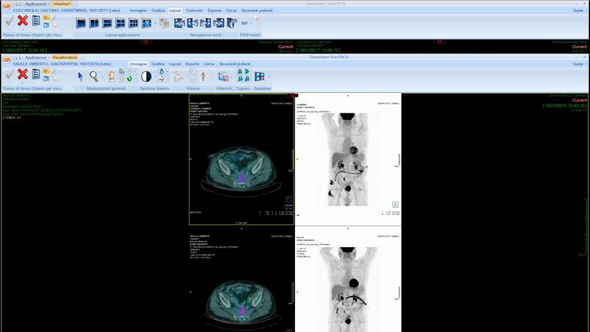
Conventional Volume in LARC                                     Radiotherapy and Oncology 120 (2016) 195–201

                                                                Contents lists available at ScienceDirect

                                                          Radiotherapy and Oncology
                                                     journal homepage: www.thegreenjournal.com

Rectal cancer guidelines

International consensus guidelines on Clinical Target Volume
delineation in rectal cancer
Vincenzo Valentini a, Maria Antonietta Gambacorta a,⇑, Brunella Barbaro b, Giuditta Chiloiro a, Claudio Coco c,
Prajnan Das d, Francesco Fanfani e, Ines Joye f, Lisa Kachnic g, Philippe Maingon h, Corrie Marijnen i,
Samuel Ngan j, Karin Haustermans f
a
  Università Cattolica del Sacro Cuore, Radiation Oncology Department; b Università Cattolica del Sacro Cuore, Department of Radiological Sciences; c Università Cattolica del Sacro
Cuore, Department of Surgical Science, Rome, Italy; d University of Texas MD Anderson Cancer Center, Department of Radiation Oncology, Houston, USA; e University G. D’Annunzio,
Gynecologic Oncology Department of Medicine and Aging Sciences, Chieti, Italy; f KU Leuven – University of Leuven, Department of Oncology and University Hospitals Leuven,
Radiation Oncology, Belgium; g Boston Medical Center, Department of Radiation Oncology, USA; h Centre Georges-François Leclerc, Department of Radiation Oncology, Dijon, France;
i
  Leiden University Medical Center, Department of Radiation Oncology, The Netherlands; j Peter MacCallum Cancer Centre, Division of Radiation Oncology and Cancer Imaging,
Melbourne, Australia

a r t i c l e         i n f o                           a b s t r a c t

Article history:                                        Introduction: The delineation of Clinical Target Volume (CTV) is a critical step in radiotherapy. Several
Received 23 May 2016                                    guidelines suggest different subvolumes and anatomical boundaries in rectal cancer (RC), potentially
Received in revised form 7 July 2016                    leading to a misunderstanding in the CTV definition. International consensus guidelines (CG) are needed
Accepted 23 July 2016
                                                        to improve uniformity in RC CTV delineation.
Available online 12 August 2016
                                                        Material and methods: The 7 radiation oncologist experts defined a roadmap to produce RC CG. Step 1:
                                                        revision of the published guidelines. Step 2: selection of RC cases with different clinical stages. Step 3:
Keywords:
                                                        delineation of cases using Falcon following previously published guidelines. Step 4: meeting in person
Consensus guidelines
Rectal cancer
                                                        to discuss the initial delineation outcome, followed by a CTV proposal based on revised and if needed,
Clinical Target Volume delineation                      adapted anatomical boundaries. Step 5: peer review of the agreed consensus. Step 6: peer review meeting
                                                        to validate the final outcome. Step 7: completion of RC delineation atlases.
                                                        Results: A new ontology of structure sets was defined and the related table of anatomical boundaries was
                                                        generated. The major modifications were about the lateral lymph nodes and the ischio-rectal fossa delin-
                                                        eation. Seven RC cases were made available online as consultation atlases.
                                                        Conclusion: The definition of international CG for RC delineation endorsed by international experts might

                                                                                                               Valentini V. et al. Radiother Oncol 2016
                                                        support a future homogeneous comparison between clinical trial outcomes.
                                                               ! 2016 Elsevier Ireland Ltd. All rights reserved. Radiotherapy and Oncology 120 (2016) 195–201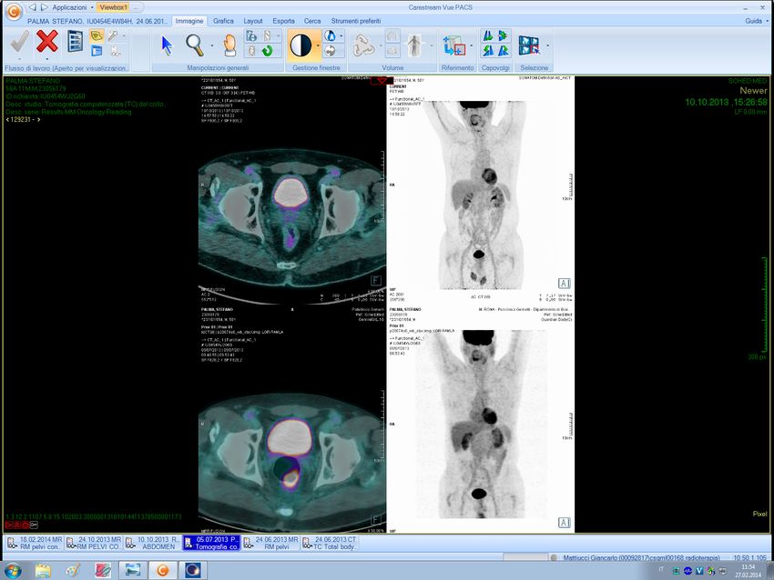
Clinical trial in LARC

                 Dose escalation à cCR
Clinical Trial   (intermediate)

                 Total Neoadjuvant Therapy
                 (advanced)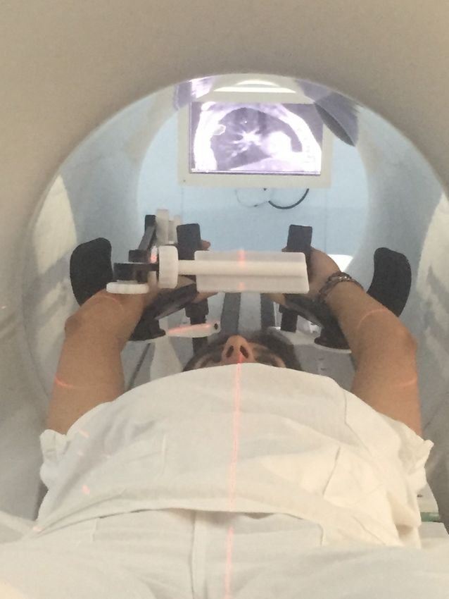
Dose Escalation Clinical Trial

                      cT3 or cT2 low

                                 Long Course
                                    RT-CT

Pucciarelli et al, S. et al Dis Colon Rectum 2013
Gérard JP et al. J Clin Oncol 2014
Vuong T et al. Semin Colon Rectal Surg 2010
Maas M et al J Clin oncol 2011
Appelt A. Lancet Oncol 2015                         cCR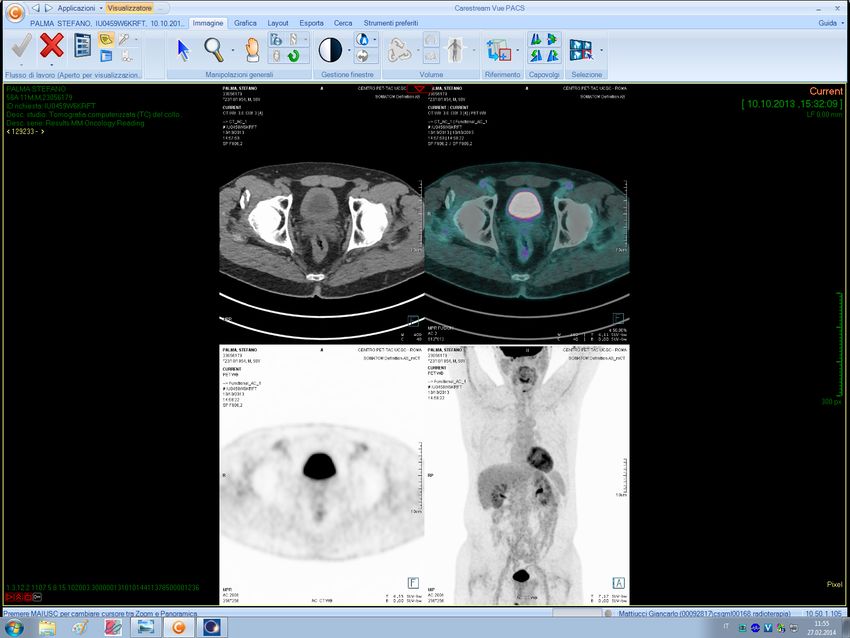
Dose escalation

       D50 TRG1            à 92.0 Gy
       D50 TRG1-2 à 72.1 Gy

      Appelt AL et al. Int. J Radiat Oncol Biol Phys 2013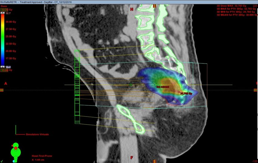
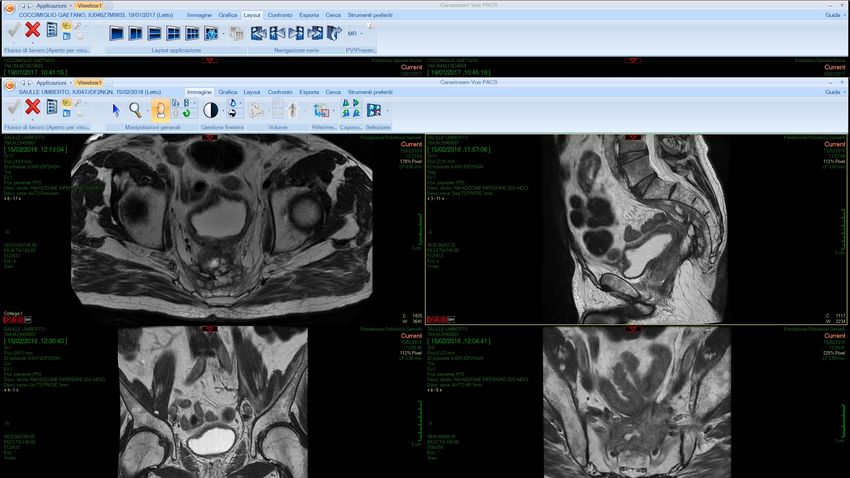
GTV identification
…during treatment

Tumor:
  – Moves
                     NOT easely VISIBLE on CBCT
  – Regresses

            SURROGATE VOLUMES:
                Mesorectum
                 Rectal wall
Rectal motion during neo-adjuvant radiochemotherapy for r
Target modification
The time-trend analysis revealed a significant 350
correlation (p ! 0.05) between volume and time in
rectal envelope (cm3)
300
6/10 patients, as shown in Figure 1.
On average, the Spearman’s rank correlation coef- 250
ficient R was "0.84 (p # 0.0005, 95% confidence inter-
val (CI) "0.62; "0.94); of interest, R was correlated 200
with the rectal volume at the first MVCT (p # 0.03).
150
The time-trend was significant in the first part of
Mesorectum (CT scan) the treatment (R # "0.98, 95% CI "0.92; "0.997,
p # 0.005) but not when considering the second part
100
1322 E. Ippolito et al. (R # "0.42, 95% CI "0.85; 0.34 p # 0.24), confirm- 50
1 2 3 4 5 6
ing that the rectal volume reduction occurred in the
patient
first fractions.
By fitting the data with a polynomial curve, as Figure 2. The volumes of the rectal enve
shown in Figure 1, the average reduction was found (dark), second half (grey) and total treatme
for each patient.
to be around 35 cm3 in the first 9 fractions (4 cm3/
fraction). When considering the envelope volumes,
the values referring to the first part of the treatment Margins for rectal motion
were always larger than those referring to the second
half (Figure 2) with average values equal to 129 $ A summary of the results is shown
76 cm3 and 87 $ 23 cm3 (p # 0.002), respectively. Table II.
When considering the median
whole treatment as the reference co
Contour agreement analysis using DSC 10 mm and 15 mm included about
Mean DSC values were found to increase after the envelope for seven and 10 patie
Figure 1a. Mesorectum motion observed in one patient during the course of radiotherapy: Front view.
first few fractions (Figure 3). The mean DSC refer- Instead, when considering the seco
ring to the first fraction was significantly worse than ment, margins of 5 mm and 7mm w
Ippolito E et al - Acta Oncol – 2009 eight and 10 patients, respectively;
CTV and its motion to be safely applied. IGRT, PET to demonstrate tumour response during a
providing an exact knowledge of anatomy during the radiotherapy course, can furthermore improve rectal the mean DSC value referring to all the other frac-
course of treatment, permits adjustments to improve cancer treatment. tions (0.67 $ 0.09 vs. 0.75 $ 0.03; p % 0.00001, t-test). of the rectal envelope was included
Future developments will probably involve the use
accuracy in dose delivery. Also IGRT taking advan-
tage of more reliable imaging techniques such us of new PET tracers in order to identify new boost Similarly, the average value referring to the first four of 12 mm and 5 mm of the media
ultrasmall superparamagnetic iron oxide (USPIO) areas within the CTV and the use of PET to monitor fractions was worse than the remaining fractions considering the whole or the second
Rectal wall
enhanced MRI to detect node involvement or FDG- the dose deposition during treatment [54] leading to
(0.70 $ 0.04 vs. 0.75 $ 0.03; p % 0.00001, t-test). ment respectively.
(CBCT)
al motion during neo-adjuvant radiochemotherapy for rectal cancer 321 When considering the first MVC
rectum as the reference contours,
significant 350 were adequate compared to the exp
Time-trend
200
median contour of the whole treat
and time in
rectal envelope (cm3)
180 This result is not surprising and
300
160
elation coef- 250 140
fidence inter- 120 0.80
as correlated 200
cm3
100
0.75
(p # 0.03).
Average DSC
80
150
first part of 60
0.70
92; "0.997, 100
Figure 1b. Mesorectum motion observed in one patient during the course of radiotherapy: Back view. 40 0.65
second part 20
4), confirm- 50 0.60
0
1 2 3 4 5 6 7 8 9 10
urred in the 1 2 3 4 5 6 7 8 9 10 11 12 13 14 15 16 17 18 0.55
patient Fraction 1 2 3 4 5 6 7 8 9 10 11 1
fraction
al curve, as Black: first half of the treatment
Figure 2. The volumes of the rectal envelope of the first half
Figure 1. Time-trend analysis of rectal volume variation during
treatment. Thin dotted lines (& black squares): patients with Figure 3. The mean similarity coefficients (D
n was found (dark), second half (grey) and total treatment (white) are plotted
Grey: second half of the treatment
significant trend (p % 0.05); grey squares: patients without trend; agreement between the rectum at each kth
continuous thick black line: average values; thick black dotted line: others N-1 fractions are shown: the values
for each patient.
ons (4 cm3/
pe volumes, White: entire treatment Maggiulli E et al. Acta Oncol 2012
polynomial fit of the average trend. after the first few fractions.MRI-Guided RT Direct TUMOR visualization: § During each fraction § Throughout the treatment § By Rad Onc § By patient § Gated dose delivering
Rectal Cancer Neoadjuvant
Treatment
• 16 Patients affected by LARC undergoing neoadjuvant
chemoradiotherapy (CRT) with MRIdian.
• Long course CRT was prescribed according to a Simultaneous
Integrated Boost (SIB) delivery protocol.
55 Gy in fractions of 2.2 Gy to PTV1 (GTV)
45 Gy in fractions of 1.8 Gy to PTV2 (pelvic subsites)
• Clinical restaging was assessed 6-8 weeks after the end of
CRT by digital rectal exam (DRE) and restaging MRI.
• Surgery was planned at 10 weeks
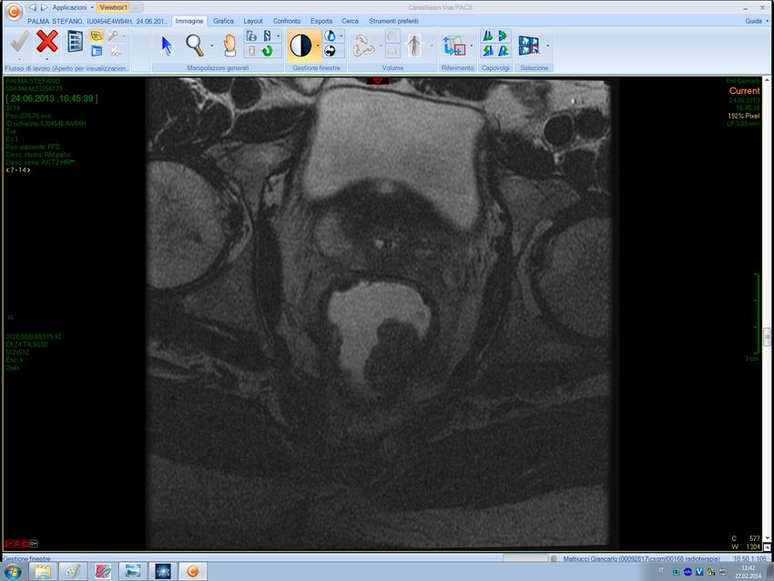
Boldrini L et al. submitted 2018Imaging protocol
1. MRI scan acquisition at 6 time-points simulation and every 5
fractions: t0 -t5- t10- t15-t20-t25
2. GTV was delineated and measured on each MR
3. Radiomics features were extracted from each 6 MRI
4. Delta radiomics (t0 vs t5, t0 vs t10…t0 vs t25) were correlated with cCR
Boldrini L et al. submitted 2018Imaging protocol t0 t5 t10 t15 t20 t25
Volumetric changes during CRT

        cCR rate: 31%
                        Boldrini L et al. submitted 2018
Radiomics features and cCR prediction Feature type t0Gy t11Gy t22Gy t33Gy t44Gy t55Gy (S) Min - 0,009 0,025 - 0,024 - (S) Range - - - - 0,038 0,019 (S) Energy - 0,025 0,002 0,009 0,006 0,028 (M) Surface - - - - 0,003 0,019 (M) Volume - 0,049 0,003 - 0,006 0,028 (M) Areavolume - - 0,003 0,013 0,006 - (M) L major - - - - 0,028 - (M) L least - 0,037 0,001 0,006 0,002 0,013 (M) Compactness 1 0,038 - - - - - (M) Compactness 2 0,038 - - - - - (M) Sphdispr 0,038 - - - - - (M) Sphericity 0,038 - - - - - (M) Asphericity 0,038 - - - - - (F) MedianFD - 0,038 - - - - (F) MinFD - - 0,013 - - - (T) glnu 0,027 0,038 0,001 0,013 0,003 0,038 (T) sre - 0,019 0,019 0,028 - - (T) lre - 0,019 0,019 0,038 - - (T) hgre - - 0,038 - 0,013 0,009 (T) srhge - - 0,038 - 0,013 0,009 (T) lrhge - - 0,038 - 0,013 0,009 (T) rlnu - - 0,028 - 0,013 - (T) rlnu norm - - 0,019 0,028 - - (T) rperc - 0,019 0,019 0,028 - - (T) rlvar - - 0,019 0,038 0,018 - 6 simulation features 57 ‘delta’ features showed a p value in discriminating cCR DELTA Least Axis Lenght t10/t0 (22 Gy) Boldrini L et al. submitted 2018
Volumetric changes during CRT

                         cCR

Delta Least axis (t10/t0) and cCR rate
                          Boldrini L et al. submitted 2018
Clinical trial in LARC
Dose escalation
• Boost is delivered on the tumor mass
• Boost in the second half of the treatment
– Tumor shrinks in the first 2 weeks of treatment
– Then it remains ‘stable’
Clinical Trial • MRI-guided RT
– Tumor is visible
– Delta radiomics in the first part of the treatment may
predict cCRUnconventional BT treatment in LARC
Brachytherapy in RC
– Palliative
– Boost after ERT
– Neoadjuvant
Clinical TrialReview paper
all the complete set of clips was lost before the end of the Details of the treatment planning procedure have
course of treatment on only two occasions. Although we been described previously [6,7]. Prior to CT simulation,
could have opted to carry out the based on bony anato- an initial antero-posterior (AP) scout view of the patient
High-dose-rate pre-operative endorectal
my, in both patients we inserted new clips and repeated
the treatment planning for the remaining treatment frac-
tions.
lying in supine position is performed in order to visual-
ize the endorectal radio-opaque clips. The endorectal ap-
plicator is introduced using lubrication with the patient
brachytherapy for patients with rectal cancer
An intracavitary mould applicator (Elekta AB, Stock- lying in the lateral decubitus position. The patient is then
holm, Sweden) of cylindrical shape (27 cm long and 2 cm repositioned in the supine position, and a plate with the
in diameter) is used in our study. As shown in Figure 3, mounted hydraulic locking clamp is slid under the pa-
eight catheter channels are distributed equally over the tient’s pelvis and the Oncosmart intracavitary mould is
circumference 1
Té Vuong, MD, FRCP , Slobodan Devic, PhD, FCCPM
of the applicator in equal angular 2
incre- latched onto the clamp (Elekta AB, Stockholm, Sweden).
ments, and a central lumen is also available for inser- Repeated AP and lateral scout views are then taken and
1Department
tion of Radiation Oncology,
an additional 2Department
central catheter. of MedicalisPhysics,
The applicator Jewish General
examined. Hospital, adjustments
When necessary, McGill University, Montreal,
are made to Québec, Canada
made of a pliable silicon rubber material, which allows the cephalic orientations of the applicator relative to the
Abstract
T3 and low T2Nx
an easy insertion and navigation through the rectum and
sigmoid colon. Figure 3 also indicates schematically the
radio-opaque clip locations.
Following the CT simulation, the acquired images
rectal cancer patients
convention we are using with respect to the loading of
High-dose-rate endorectal
are sent to a dedicated virtual simulation image process-
the channels (from 1 to 8) as brachytherapy (HDREBT)
well as the catheters loaded ingis workstation.
an image guided brachytherapy
The tumor treatment for patients with
(GTV) and intramesorectal
rectal cancer. ItTREATMENT
is based on tumor imaging with magnetic resonance in particular, which is used to choose eligible
patients and improve tumor visualization. Treatment planning is performed using 3D CT simulation and treatment
planning. The treatment is given on an outpatient basis and requires minimal local anesthesia. The validation of the
technique was carried out through a preoperative study and is now explored as part of a radical treatment for early
rectal cancer or as a boost modality.
We describe technical aspects of the HDREBT and we discuss the ongoing institutional review board approved
studies exploring the clinical applications of this treatment modality for patients with rectal cancer: 1) as a neoadjuvant
Day 1 Day 4 Day 5 Day 6 Day 7 Day 8 After
treatment for patients with operable
(Fri) rectal
(Fri) tumor;
(Tue) 2)(Wed)
as a option
(Thu) to improve 6-8
(Fri) local control
weeks Time in patients with newly diag-
nosed rectal cancer but with previous pelvic radiation.
MRI CT Rx #1 Rx #2 Rx #3 Rx #4 Surgery
J Contemp Brachytherapy 2015; 7, 2: 183-188
US Endoscopy
scan DOI: 10.5114/jcb.2015.51402
+
Radio-opaque
Key words: brachytherapy, endorectal,
clips insertion image guidance, pre-operative, rectal cancer.
Fig. 1. Time scheme for preoperative endorectal high-dose-rate brachytherapy; the days of the week in parentheses represent
26Gy
Purpose the mostin 4 fractions
suitable ofa logistical
days not only from 6.5 Gy pointaof week;
view but also dose
forevidenceprescribed
a reliable of necrotic
reproduction to
of theor the
daily TUMORnodes larger than
extramesorectal
dose distribution
1 cm were selected for participation in the study. Over the
Contact X-ray therapy for treatment of rectal cancer, last 15 years techniques for imaging, treatment planning
introduced by Papillon [1] in the early 1970s, is highly Vuong
and dose T et al. have
delivery J Contemp Brachytherapy
evolved into 2015
the currently usedBrachytherapy: volumes Té Vuong, Slobodan Devi
186
GTVà tumor
CTVàvisible pararectal nodes
Dose prescribed to the
188 Té Vuong, Slobodan Devic
CTV
100
5. Kaufman N, Nori D, Shank B et al. Remote afterloading intra-
luminal brachytherapy in the treatment of rectal, rectosig- 80
moid, and anal cancer: a feasibility study. Int J Radiat Oncol
Biol Phys 1989; 17: 663-668.
Volume (%)
6. Vuong T, Belliveau P, Michel R et al. Conformal preoperative 60
endorectal brachytherapy treatment for locally advanced
rectal cancer. Dis Colon Rectum 2002; 45: 1486-1495.
40
7. Vuong T, Devic S, Moftah B et al. High dose rate endorec-
tal brachytherapy in the treatment of locally advanced rectal
carcinoma: Technical aspects. Brachytherapy 2005; 4: 230-235. 20
8. Devic S, Vuong T, Moftah B et al. Image guided high dose
rate endorectal brachytherapy. Med Phys 2007; 34: 4451-4458.
9. Devic S, Vuong T, Evans M et al. Endorectal high dose rate 0
0 1000 2000
brachytherapy quality assurance. J Oncol 2008; 58: 53e-54e. Dose (CGy)
10. Vuong T, Richard C, Niazi T et al. High dose rate endorectal
CTV Bladder RT Femur LT Femur Bone ma
brachytherapy for patients with curable rectal cancer. Semin
Fig. 4. Dose distribution obtained by treatment planning system
Colon Rectal Cancer Surg 2010; 21: 115-119.
Vuong T et al. J Contemp Brachytherapy 2015Brachytherapy: clinical results
Time of accrual 1989-2005
N° of patients 483 43 post op RTCT for pN+
Age 68.2 (28-90)
Stage T3-low T2, Nx
Post-op CT 5-FU at discretion of the MO up to 2005 FOLFOX after 2005
FUP 63 months
pCR 27%
pN+ 30%
LR 4.8%
DFS 65.5%
OS 72.8%
Vuong T et al. J Contemp Brachytherapy 2015Unconventional BT volume in LARC
Brachytherapy in RC
• High pCR rate
• Same outcome as standard RTCT
• No concomitant CT
• No pelvic nodes irradiation
Clinical trial
Clinical trial
•
•
No bone marrow irradiation
Fractionated doses < 10 Gy
Possible immune system contribution?Uncoventional approaches and volumes in Rectal Cancer • Locally advanced rectal cancer • Local relapse
Low conventional palliative volume

                 Re-treatment
                 R0 resection ≈ 20%
                 Good symptoms control: 80-100%
                 Duration: 8-10 month
                 Dose: 30-40 Gy hyperfractionated
                 Volume: Involved field

Clinical trial

                             Guren MG et al. Radiother Oncol 2014
Unconventional palliative treatment
High burden ‘PITH’
tumor mass Partial
Irradiation
Tumor
Hypoxia
Clinical Trial‘PITH’ in Rectal Cancer: case report 70 years old, male, ECOG 0 2017 UN-RESECTABLE-PREIRRADIATED Local Relapse (presacral space, iliac L vessels, SB, SV, prostate, mucinous) NOT SUITABLE FOR RE-IRRADIATION Uncontrolled PAIN 2015 Adenoca cT4bN2, low rectum RTCT à APR à CT
Role of radiotherapy in «high burden» disease

In vitro studies
Irradiation of the HYPOXIC-HYPOVASCOLARIZED
part of the tumor TRIGGERS tumor regression

                          Radiation Induced Bystander Effect (RIBE):
                          Irradiating only a part of the tumor induces
                          rergression of the whole tumor

                          Radiation Induced Abscopal Effect (RIAE):
                          tumor regression in untreated distant sites

                                                                Tubin S et al. Acta Oncol 2017
                                                   Tubin S 27° Residential Course, Rome 2017
Role of radiotherapy in «high burden» metastatic disease
RT 3: PERSONAL EXPERIENCE WITH THE USE OF UNCOVENTIONAL
RADIOTHERAPY FOR LARGE TUMORS: DEVELOPMENT OF A NEW
HERAPY TECHNIQUE FOR THE PARTIAL TUMOR IRRADIATION EXPLOITING
THE BYSTANDER EFFECT
RESULTS:
Bystander Tartget Volume (BTV)
GTV (bulky)
GTV mean volume: 230.9 cm³ (range:132.5–306.8)
GTV mean diameter: 8.1 cm (range: 7-11)
Average GTV SUVmax: 19.9 (range:15.2-27.8)
BTV
BTV mean volume: 70.6 cm³ (range:42.6–95.5)
(30% of the GTV´s)
Average BTV SUVmax: 2.7 (range:1-3)
(15% of the GTV´s)
Tubin S.
KABEG Klinikum Klagenfurt, AUT
E-Mail: s.tubin@kabeg.at
Hypometabolic-hypovascularized part of the tumor
PETàSUV < 3
No margin
DOSE: 10 or 12 Gy X 1 to 70%-isodose (Dmax 14.5 and 18 Gy, respectively) to the BTV
Tubin S et al. Acta Oncol 2017
Tubin S 27° Residential Course, Rome 2017Role of radiotherapy in «high burden» metastatic disease
25 consecutive symptomatic, oligometastatic
patients with bulky tumors were included in
this prospective clinical study
RESULTS:
• Median follow-up was 8 months (range: 2-15).
• Significant bystander and/or abscopal effects: in all patients
• Mean time to the induction of the effects: 2-3 weeks
• Overall response rates for symptom relief: 100%
• Overall mass response: 100%
• Average tumor shrinkage: 60% (50-80%)
• Disease-specific survival: 96% Tubin S et al. Acta Oncol 2017
• Acute or late toxicity: no patients Tubin S 27° Residential Course, Rome 2017PITH in Rectal Cancer: case report
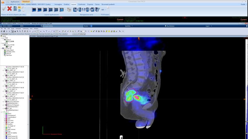
GTV BTV Treatment plan
PET MR 10Gy @ 70% isodose
(hypovascolarized
region of the tumor)PITH in Rectal Cancer: case report
Uncontrolled PAIN relief NO PAIN
PAIN VAS 2 No drug intake
opioid No acute effect No late effects
DIAGNOSIS 6 weeks after RT 3 month after RTUnconventional palliative volume
PITH BTV
Partial irradiation of tumor hypoxic/
hypovascularized area
single 10 Gy dose (@70% isodose)
may generate response unirradiated T,
symptoms relief, without toxicity
Clinical Trial A new option to exploreConclusions: locally advanced RC
Preop RTCT à pelvic volume
186 Té Vuong, Slobodan Devic
BT in intermediate RCà GTV
100
80
Volume (%)
60
40
20
0
0 1000 2000
Dose (CGy)
CTV Bladder RT Femur LT Femur Bone marrow
Fig. 4. Dose distribution obtained by treatment planning system
Dose escalationà
GTV delineation
Clinical Trial strict IGRT protocols Daily radiograph Reference DRR
role for hybrid MR machines
Fig. 5. Daily longitudinal treatment adjustment
Journal of Contemporary Brachytherapy (2015/volume 7/number 2)Conclusions: Local recurrence
Re-irradiationà involved field on LR
High burden diseaseà BTV
Clinical TrialConclusions
Preop RTCT à pelvic volume
Re-irradiationà involved field on LR
186 Té Vuong, Slobodan Devic
BT in intermediate RCà GTV
100
80
Volume (%)
60
40
20
High burden diseaseà BTV
0
0 1000 2000
Dose (CGy)
CTV Bladder RT Femur LT Femur Bone marrow
Fig. 4. Dose distribution obtained by treatment planning system
Dose escalationà
GTV delineation
Clinical Trial strict IGRT protocols
role for hybrid MR machines
Daily radiograph Reference DRR
Fig. 5. Daily longitudinal treatment adjustmentYou can also read