Is Platelet-rich Plasma Injection more Effective than Steroid Injection in the Treatment of Chronic Plantar Fasciitis in Achieving Long-term Relief?
←
→
Page content transcription
If your browser does not render page correctly, please read the page content below
3-OA1-188_OA1 11/27/19 11:10 AM Page 8

            Malaysian Orthopaedic Journal 2019 Vol 13 No 3                                                                                                       Soraganvi P, et al
            doi: http://doi.org/10.5704/MOJ.1911.002

                     Is Platelet-rich Plasma Injection more Effective than
                     Steroid Injection in the Treatment of Chronic Plantar
                           Fasciitis in Achieving Long-term Relief?

                           Soraganvi P, MS Ortho, Nagakiran KV, DNB Ortho, Raghavendra-Raju RP, MS Ortho,
                            Anilkumar D, MS Ortho, Wooly S, MS Ortho, Basti BD*, MD, Janakiraman P*, MD
              Department of Orthopaedics, PES Institute of Medical Sciences and Research Kuppam Campus, Kuppam, India
                *Department of Community Medicine, PES Institute of Medical Sciences and Research Kuppam Campus,
                                                            Kuppam, India

                               This is an open-access article distributed under the terms of the Creative Commons Attribution License, which permits unrestricted use,
                                                      distribution, and reproduction in any medium, provided the original work is properly cited

                                                                          Date of submission: 27th September 2018
                                                                             Date of acceptance: 31st May 2019

            ABSTRACT                                                                                  follow-up. Mean AOFAS score in Group A improved from
                                                                                                      54 to 90.03 and in Group B from 55.63 to 74.67 at six
            Introduction: Plantar fasciitis is characterised by pain in the
                                                                                                      months’ follow-up. The improvements observed in VAS and
            heel, which is aggravated on weight bearing after prolonged
                                                                                                      AOFAS were statistically significant. At the end of six
            rest. Many modalities of treatment are commonly used in the
                                                                                                      months’ follow-up, plantar fascia thickness had reduced in
            management of plantar fasciitis including steroid injection.
                                                                                                      both groups (5.78mm to 3.35mm in Group A and 5.6 to 3.75
            Many studies show that steroid injection provides pain relief
                                                                                                      in Group B) and the difference was statistically significant.
            in the short term but not long lasting. Recent reports show
                                                                                                      Conclusion: Local injection of platelet-rich plasma is an
            autologous platelet-rich plasma (PRP) injection promotes
                                                                                                      effective treatment option for chronic plantar fasciitis when
            healing, resulting in better pain relief in the short as well as
                                                                                                      compared with steroid injection with long lasting beneficial
            long term. The present study was undertaken to compare the
                                                                                                      effect.
            effects of local injection of platelet-rich plasma and
            Corticosteroid in the treatment of chronic plantar fasciitis.
                                                                                                      Key Words:
            Materials and methods: Patients with the clinical diagnosis
                                                                                                      platelet-rich plasma, plantar fascia, steroid injection,
            of chronic plantar fasciitis (heel pain of more than six weeks)
                                                                                                      plantar fasciitis
            after failed conservative treatment and plantar fascia
            thickness more than 4mm were included in the study.
            Patients with previous surgery for plantar fasciitis, active
                                                                                                      INTRODUCTION
            bilateral plantar fasciitis, vascular insufficiency or
            neuropathy related to heel pain, hypothyroidism and diabetes                              Plantar fasciitis is a common pathological condition
            mellitus were excluded from the study. In this prospective                                affecting the hindfoot and can often be a challenge for
            double-blind study, 60 patients who fulfilled the criteria were                           clinicians to treat successfully1. It is an overuse injury from
            divided randomly into two groups. Patients in Group A                                     repetitive microtrauma that leads to inflammation and local
            received PRP injection and those in Group B received steroid                              tissue damage2. Treatment options include non-surgical
            injection. Patients were assessed with visual analog scale                                management, like non-steroidal anti-inflammatory drug
            (VAS) and American Orthopedic Foot and Ankle Society                                      (NSAID) prescription, physiotherapy, night splints and
            (AOFAS) score. Assessment was done before injection, at                                   steroid injection, and surgical intervention1. The treatment of
            six weeks, three months and six months follow-up after                                    plantar fasciitis may require a combination of treatment
            injection. Plantar fascia thickness was assessed before the                               modalities, rather than administering only one treatment at a
            intervention and six months after treatment using                                         time3. There is no single treatment which has been proven as
            sonography.                                                                               a gold standard for the treatment of chronic plantar fasciitis.
            Results: Mean VAS in Group A decreased from 7.14 before                                   Traditionally, local injection of steroid was used widely for
            injection to 1.41 after injection and in Group B decreased                                chronic plantar fasciitis treatment. Cochrane review on the
            from 7.21 before injection to 1.93 after injection, at final                              use of corticosteroid for plantar fasciitis showed

            Corresponding Author: Nagakiran KV, Department of Orthopaedics, PES Institute of Medical Sciences and Research Kuppam Campus, NH
            219, Kuppam, Andhra Pradesh 517425, India
            Email: prasad.doct@yahoo.co.in

             8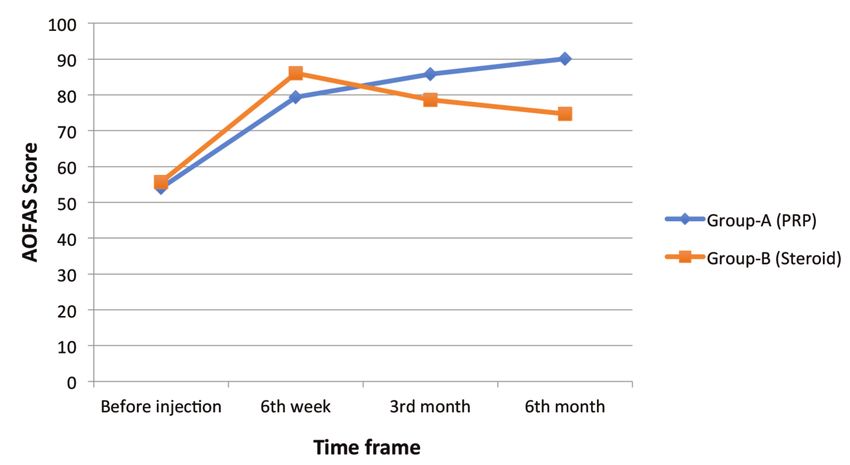
3-OA1-188_OA1 11/27/19 11:10 AM Page 9

                                                                                                         A Prospective Double Blind Study

            improvement in symptoms at one month, which did not last           The carbon paper ensures written matter on envelope is
            long4. In recent years, platelet-rich plasma (PRP) is being        transferred to treatment allocation paper inside envelope.
            used successfully for the treatment of various chronic             After preparation all enveloped were sealed. These 60
            tendinitis, including chronic plantar fasciitis. Earlier results   prepared envelopes were shuffled thoroughly and later
            of using the PRP to treat plantar fasciitis have been              marked with a serial number over it. All these envelopes
            favorable, but there is a paucity of literature where the          were placed in a container in numerical order.
            effectiveness of steroid injection is compared to PRP in
            chronic plantar fasciitis treatment.                               The envelopes were allocated sequentially and participant’s
                                                                               name and other details were entered on the front of the
            In this study, we compared the efficacy of PRP and steroid         envelope before opening the seal. All patients received
            injection in the treatment of chronic plantar fasciitis and also   treatment as indicated inside the envelope (treatment A –
            analysed the effect of PRP and steroid injection on the            PRP or treatment B - Steroid). A colleague who was not
            thickened plantar fascia.                                          involved in the study did the opening of the sealed envelope
                                                                               and administration of appropriate injection. This method was
                                                                               followed to eliminate bias in the study.
            MATERIALS AND METHODS
                                                                               Blood was drawn from patients in both groups for blinding
            This study was a randomised double-blind study done at PES
                                                                               purpose and a screen was used while giving injection so the
            Institute of Medical Sciences and Research, Kuppam,
                                                                               patients were blinded from the type of treatment they were
            Andrapradesh, India, from September 2014 to September
                                                                               receiving.
            2016. Patients were diagnosed as having plantar fasciitis
            based on history and clinical examination. The study
                                                                               This study included 60 patients with chronic plantar fasciitis.
            subjects included all patients with a clinical diagnosis of
                                                                               Patients in Group A received PRP (3ml) injection and Group
            plantar fasciitis (heel pain lasting more than six weeks) with
                                                                               B patients received a steroid injection (Depomedrol 80mg
            sonographic evidence (plantar fascia thickness of more than
                                                                               (2ml) + 0.5ml xylocaine 2%). Treatment with NSAIDs was
            4mm). Patients with previous surgery for plantar fasciitis,
                                                                               discontinued one week before the injection in both groups.
            active bilateral plantar fasciitis, presence of vascular
                                                                               All patients in both groups were advised on plantar fascia
            insufficiency or neuropathy associated with heel pain,
                                                                               stretching exercise.
            hypothyroidism and diabetes mellitus were excluded from
            the study. Ethical clearance was obtained from the institution
                                                                               For PRP preparation, blood was drawn from the cubital vein
            for the study and consent from all patients participating in
                                                                               into six vacutainer tubes, which contained 0.35ml of 3.2%
            the study.
                                                                               sodium citrate. Vacutainer was centrifuged at 1200 rpm for
                                                                               10 min in a routine 380 R centrifuge model. Following
            The sample size was calculated using a formula based on
                                                                               centrifugation three layers were identified, of which, the
            means and standard deviation as in the study by Jain et al
                                                                               bottom layer consisted of red blood cells, the intermediate
            using stat software5. In our study it was found to be 54 with
                                                                               layer of white blood cells, and upper layer of plasma,
            27 for each study group. We have rounded it off to 60 with
                                                                               platelets, and some white blood cells. The concentrate in the
            30 in each group (PRP and steroid groups).
                                                                               upper layer was carefully collected with a 10cc syringe. The
                                                                               collected volume ranged from 1 to 1.25ml in each vacutainer.
            We applied the randomisation method by using sequentially
                                                                               Approximately, 1ml of the upper layer of the sample that
            numbered, opaque, sealed envelope technique (SNOSE)6.
                                                                               underwent the first spin step was collected and transferred to
            For preparation of envelope, we took 60 identical letter sized
                                                                               one empty 6ml tube. This tube was centrifuged again for 10
            envelopes, aluminium foil and single sided carbon paper.
                                                                               min at speed of 2400 rpm (second spin). The upper half of
            Aluminium foil was cut into 60 sheets that are same width as
                                                                               the plasma volume, platelet poor plasma (PPP), was
            envelope and twice its height, so that when folded it will be
                                                                               removed. The remaining volume of PRP was used for
            same size as envelope. Single sided carbon paper was cut
                                                                               injection.
            into 60 envelope size sheets. Sixty sheets of standard size
            papers are taken and each one marked as treatment A (PRP –
                                                                               Random PRP samples were sent for estimation of platelet
            30 papers) or treatment B (Steroid – 30 papers) by writing on
                                                                               count by autoanalyser. Majority of the samples had platelet
            it. These papers are folded to fit the envelope. One sheet of
                                                                               count of more than 1,000,000/ul in 5ml volume, which was
            carbon paper was placed on the top of the folded paper so
                                                                               five times the baseline.
            that carbon side is facing paper. One sheet of aluminium foil
            was folded over both side of carbon and treatment paper
                                                                               Before administration of the PRP or steroid injection, all
            combination. This completed insert was placed in to
                                                                               patients underwent a random blood sugar level assessment.
            envelope with carbon paper closest to the front of envelope.
                                                                               The participants were appropriately counselled before the
            The aluminium foil ensures the envelope is opaque and
                                                                               injection. Injections were given under aseptic condition.
            cannot be read by holding it up against strong light source.

                                                                                                                                            9
3-OA1-188_OA1 11/27/19 11:10 AM Page 10
Malaysian Orthopaedic Journal 2019 Vol 13 No 3 Soraganvi P, et al
Table I: Comparison of the characteristics of both groups
PRP Group (29) STEROID Group (28)
Male 14 (48%) 12 (57%)
Female 15 (52%) 16 (43%)
Age 40.27yrs (mean, SD – 8.03) 39.35yrs (mean, SD-12.5)
Right Side 14 (48%) 15 (54%)
Left Side 15 (52%) 13 (46%)
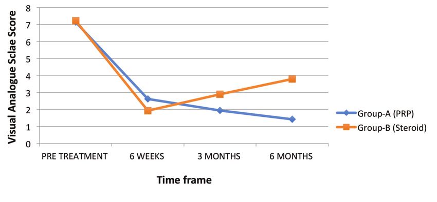
Table II: Mean VAS score in both groups
VAS Group A (PRP) Group B (Steroid) P value (at end of 6 months follow-up)
Pre-Treatment 7.137 7.214
6 Weeks 2.62 1.928
3 Months 1.931 2.89
6 Months 1.413 3.7853-OA1-188_OA1 11/27/19 11:10 AM Page 11
A Prospective Double Blind Study
Fig. 1: Representation of VAS scores before treatment and at interval of six weeks, three months and six months after treatment.
Fig. 2: AOFAS before treatment and at different follow-up visits in both groups.
up, and 2.89 at three months and 3.76 at six months follow- following the injection, Group A had significant reduction
up (Fig. 1). The difference between the two groups was (mean 3.35mm, 35.45%) in the thickness of plantar fascia as
statistically significant at six weeks (p3-OA1-188_OA1 11/27/19 11:10 AM Page 12
Malaysian Orthopaedic Journal 2019 Vol 13 No 3 Soraganvi P, et al
Many treatment modalities have been in practice, among study16-18. In this technique, fascia is injected at multiple sites
which corticosteroid injections have been extensively used, through a single skin portal16-18. The injection was
but only seemed to be useful in the short term and only to a administered at the point of maximum tender points.
small degree8. Potential complications associated with Benefits of injection under ultrasound guidance are doubtful.
steroid injection raise concern about benefit against the risk Ultrasound-guided administration of injection was used in
involved in steroid injection. Histological studies have some studies21,22. However, studies have reported no
indicated plantar fasciitis as a degenerative disorder, hence significant difference between ultrasound-guided injection
prostaglandin mediated anti-inflammatory action of steroid and injection at the tender spot23.
is unclear. However, inhibition of fibroblast proliferation and
expression of ground substance proteins by corticosteroids There are various methods of preparation of platelet-rich
may be the possible explanation for the beneficial effect of plasma. In each method of preparation, platelet concentration
steroid injection9. varies. In present literature, there is paucity of information
regarding the more superior type of method of preparation.
Various studies have shown that platelet-rich plasma Also, the effectiveness of leukocyte-reduced or rich PRP
injection as an effective treatment option for chronic plantar preparation is debatable24,25. In our study, leucocytes were not
fasciitis10-19. Plantar fasciitis is considered a degenerative filtered from PRP. Activation of PRP initiated during the
tissue condition due to micro-tear in fascia rather than preparation process by degranulation of platelets26. Adding
inflammation. This results in denaturation of collagen and thrombin or calcium chloride activates PRP. Spontaneous
angiofibroblastic hyperplastic tissue is seen in histology7. platelet activation occurs after exposure to native collagen27.
PRP is rich in growth factors like transforming growth factor, Presently, available literature lack evidence of the most
vascular endothelial growth factor, and platelet-derived suitable method of activation of PRP. The beneficial effect of
growth factor and inflammatory mediators like cytokines and activating PRP before the injection is not supported by all
interleukins, such as interleukin 4, 8, 13, interferon-α, and studies. All patients in our study received freshly prepared
tumor necrosis factor-α7. The concentration of these factors PRP. We have not used any agent to activate PRP.
is low in the plantar fascia due to hypovascularity and
hypocellularity7. PRP delivers growth factors along with Jain et al in their study comparing single injection of PRP
platelets directly to the site of the lesion, since all these and steroid injection in chronic plantar fasciitis, found no
factors affect healing stages necessary to reverse chronic significant difference in functional outcome in both groups at
plantar fasciitis7. Alpha particles of platelets release stored six months follow-up28. Similar results were also observed in
platelet-derived growth factors after stimulation. It increases other studies29, whereas many studies have shown the long-
fibroblast migration and proliferation and improves collagen lasting beneficial effects of PRP when compared to steroid
deposition, which promotes angiogenesis and fiber repair7. injection with improved AOFAS score and VAS score5,10,30.
Literature on treatment options show a variable outcome In our study, we observed that in both PRP and steroid
when PRP and steroid injection are used in the treatment of injection group, VAS and AOFAS score improved after one
chronic plantar fasciitis. Some studies found PRP to be more injection and improvement in pain and AOFAS score was
effective whereas others did not find a significant difference more in the steroid group compared to PRP group at first
in the outcome10,12,13. When steroid injection was compared follow-up visit. On later follow-up both VAS and AOFAS
with autologous blood injection in a study by Lee et al, they score in PRP group continued to improve and at the end of
found that the corticosteroid group had significantly lower six months follow-up the PRP group showed better
VAS than autologous blood group14. Monto et al comparing improvement compared to steroid group and improvement in
PRP and corticosteroid injection in the treatment of failed score was statistically significant. The decline in pain and
non-surgical treatment of plantar fasciitis, concluded that a function scores of steroid group after six weeks suggest that
single injection of PRP improved pain and function more steroid injection is more effective only for short-term relief.
than steroid injection and beneficial effects sustained for a The mechanism of reduction in pain and improvement in the
longer time10. function after PRP injection is not clear. PRP contains
hepatocyte growth factor (HGF) along with other growth
In our study, we compared the effectiveness of PRP and factors. The anti-inflammatory action of HGF is mediated by
steroid injection in patients with chronic plantar fasciitis disrupting the nuclear factor kappa B (NF-kB)
where other conservative treatments had failed. We adopted transactivating activity, which results in decreased
PRP preparation as per Amanda et al recommendations20. expression of COX-1 and COX-2 genes. By this action, HGF
Two spin method showed higher growth factor levels and is known to protect tissues from inflammatory damages.
higher platelet counts. In our study, most of the samples had Thus, the anti-inflammatory action of PRP is through HGF.
platelet counts of more than 1,000,000/ul in 5ml volume, This explains the initial improvement in VAS score and
which is five times the baseline. Peppering technique of reduction in pain following PRP injection31.
injection was found to be more effective and was used in our
123-OA1-188_OA1 11/27/19 11:10 AM Page 13
A Prospective Double Blind Study
Studies have shown a significant reduction in plantar fascia mechanism of action of PRP. However, the results of PRP
thickness after PRP injection18,20. In our study, plantar fascia injection as a biological modality of treatment in orthopedic
thickness in both the PRP group and corticosteroid group conditions are encouraging.
were comparable prior to injection. However, at six months
follow-up, the PRP group had a significant reduction
(35.45%) in the thickness of plantar fascia compared to CONCLUSION
corticosteroid group (29.16%). The difference between the
Local injection of platelet-rich plasma is an effective
two groups was statistically significant.
treatment option for chronic plantar fasciitis when compared
to steroid injection and beneficial effects are long-lasting.
Limitation of this study is the variability of platelet
concentration among different patients. Lack of
standardisation in preparation, concentration of platelets and
CONFLICT OF INTERESTS
dosage were barriers for critical evaluation. Further basic
The authors declare that there is no conflict of interest.
research is necessary in this field for understanding the exact
REFERENCES
1. Cornwall MW, McPoil TG. Plantar fasciitis: Etiology and treatment. J Ortho Sports Phys Ther. 1999 Dec; 29(12): 756-60.
2. Young CC, Rutherford DS, Niedfelt MW. Treatment of plantar fasciitis. Am Fam Physician. 2001 Feb; 63(3): 467-74, 477-8.
3. Wolgin M, Cook C, Graham C, Mauldin D. Conservative treatment of plantar heel pain: long-term follow-up. Foot & Ankle
Int.1994 Mar; 15(3): 97-102.
4. Crawford F, Thomson C. Interventions for treating plantar heel pain. Cochrane Database Syst Rev. 2003; (3): CD000416.
5. Jain K, Murphy PN, Clough TM. Platelet-rich plasma versus corticosteroid injection for plantar fasciitis: A comparative study.
Foot (Edin). 2015 Dec; 25(4): 235-7.
6. Sampoornam W. Nurse Researcher and Allocation Concealment. J Psychiatr Nurs. 2012; 1(3): 81-5.
7. Yang WY, Han YH, Cao XW, Pan JK, Zeng LF, Lin JT et al. Platelet-rich plasma as a treatment for plantar fasciitis: A meta-
analysis of randomized controlled trials. Medicine (Baltimore). 2017 Nov; 96(44): e8475.
8. Acevedo AJ, Beskin J. Complications of plantar fascia rupture associated with corticosteroid injection. Foot Ankle Int. 1998, 19:
91-7.
9. McMillan AM, Landorf KB, Gilheany MF, Bird AR, Morrow AD, Menz HB. Ultrasound guided corticosteroid injection for
plantar fasciitis: randomized controlled trial. BMJ. 2012; 344: e3260.
10. Monto RR. Platelet-rich plasma efficacy versus corticosteroid injection treatment for chronic severe plantar fasciitis. Foot Ankle
Int. 2014; 35: 313-8.
11. Shetty VD, Dhillon M, Hegde C, Jagtap P, Shetty S. A study to compare the efficacy of corticosteroid therapy with platelet-rich
plasma therapy in recalcitrant plantar fasciitis: a preliminary report. Foot Ankle Surg. 2014; 20(1): 10-3.
12. Say F, Gurler D, Inkaya E, Bulbul M. Comparison of platelet-rich plasma and steroid injection in the treatment of plantar fasciitis.
Acta Orthop Traumatol Turc. 2014; 48(6): 667-72.
13. Aksahin E, Dogruyol D, Yuksel HY, Hapa O, Dogan O, Celebi L, et al. The comparison of the effect of corticosteroids and
platelet-rich plasma (PRP) for the treatment of plantar fasciitis. Arch Orthop Trauma Surg. 2012; 132(6): 781-5.
14. Lee TG, Ahmad TS. Intralesional autologous blood injection compared to corticosteroid injection for treatment of chronic plantar
fasciitis: a prospective, randomized, controlled trial. Foot Ankle Int. 2007; 28: 984-90.
15. Martinelli N, Marinozzi A, Carnì S, Trovato U, Bianchi A, Denaro V. Platelet-rich plasma injections for chronic plantar fasciitis.
Int Orthop. 2013; 37(5): 839-42.
16. Kumar V, Millar T, Murphy PN, Clough T. The treatment of intractable planter fasciitis with platelet-rich plasma injection. Foot
(Edinb). 2013; 23(2-3): 74-7.
17. Wilson JJ, Lee KS, Miller AT, Wang S. Platelet-rich plasma for the treatment of chronic planter fasciopathy in adults: a case
series. Foot Ankle Spec. 2014; 7(1): 61-7.
133-OA1-188_OA1 11/27/19 11:10 AM Page 14
Malaysian Orthopaedic Journal 2019 Vol 13 No 3 Soraganvi P, et al
18. Ragab EM, Othman AM. Platelets rich plasma for treatment of chronic plantar fasciitis. Arch Orthop Trauma Surg. 2012; 132(8):
1065-70.
19. Barrett SL, Erredge SE. Podiatry Today. November Issue 11. Pennsylvania; Healthcare Made Practical: 2004. Volume 17, Growth
Factors For Chronic Plantar Fasciitis?; p. 36-42.
20. Amanda GMP, José FSDL, Ana AR, Angela CML, William DB, Maria HAS. Relevant Aspects of Centrifugation Step in the
Preparation of Platelet-Rich Plasma. ISRN Hematol. 2014; 176060:8. doi: 10.1155/2014/176060
21. Tsai WC1, Wang CL, Tang FT, Hsu TC, Hsu KH, Wong MK. Treatment of proximal plantar fasciitis with ultrasound-guided
steroid injection. Arch Phys Med Rehab. 2000; 81: 1416-21.
22. Cunnane G, Brophy DP, Gibney RG, FitzGerald O. Diagnosis and treatment of heel pain in chronic inflammatory arthritis using
ultrasound. Semin Arthritis Rheum. 1996; 25(6): 383-9.
23. Kane D1, Greaney T, Shanahan M, Duffy G, Bresnihan B, Gibney R, et al. The role of ultrasonography in the diagnosis and
management of idiopathic plantar fasciitis. Rheumatology (Oxford). 2001; 40(9): 1002-8.
24. Russell RP, Apostolakos J, Hirose T, Cote MP, Mazzocca AD. Variability of platelet-rich plasma preparations. Sports Med
Arthrosc Rev. 2013; 21(4): 186-90.
25. Arnoczky SP1, Sheibani-Rad S. The basic science of platelet-rich plasma (PRP): what clinicians need to know. Sports Med
Arthrosc. 2013; 21(4): 180-5.
26. Wasterlain AS, Braun HJ, Dragoo JL. Contents and formulations of platelet-rich plasma. Operative Techniques in Orthopaedics.
2012; 22(1): 33-42.
27. Matteo DB, Filardo G, Kon E, and Marcacci M. Platelet-rich plasma: evidence for the treatment of patellar and Achilles
tendinopathy—a systematic review. Musculoskeletal Surgery. 2015 Apr; 99(1): 1-9.
28. Jain SK, Suprashant K, Kumar S, Yadav A, Kearns SR. Comparisom of plantar fasciitis injected with platelet-rich plasma vs
corticosteroids. Foot Ankle Int. 2018 Jul; 39(7): 780-6.
29. Olivo CA, Rodriguez JE, Cavazos RL, Cavazos FV, Mendia MS, Lemus OM. Plantar fasciitis-a comparison of treatment with
intralesional steroids versus platelet-rich plasma a randomized, blinded study. J Am Podiatr Med Assoc. 2017; 107(6): 490-6.
30. Vahdatpour B, Kianimehr L, Moradi A, Haghighat S. Beneficial effects of platelet-rich plasma on improvement of pain severity
and physical disability in patients with plantar fasciitis: a randomized trial. Adv Biomed Res. 2016; 28; 5: 179.
31. Bendinelli P, Matteucci E, Dogliotti G, Corsi MM, Banfi G, Maroni P, et al. Molecular basis of anti-inflammatory action of
platelet-rich plasma on human chondrocytes: mechanisms of NF-κB inhibition via HGF. J Cell Physiol. 2010; 225(3): 757-66.
14You can also read