THE THERAPEUTIC POTENTIAL OF LOSARTAN IN LUNG METASTASIS OF COLORECTAL CANCER
←
→
Page content transcription
If your browser does not render page correctly, please read the page content below
EXCLI Journal 2020;19:927-935 – ISSN 1611-2156
                                        Received: February 23, 2020, accepted: June 25, 2020, published: June 29, 2020

                                                 Original article:

        THE THERAPEUTIC POTENTIAL OF LOSARTAN IN LUNG
              METASTASIS OF COLORECTAL CANCER
Milad Hashemzehi1,2*, Niloufar Naghibzadeh2*, Fereshteh Asgharzadeh 1,2*, Asma
Mostafapour3, Seyed Mahdi Hassanian3, Gordon A. Ferns5, William C. Cho6,
Amir Avan2,3,4,#, Majid Khazaei2,3,#
1
    Department of Medical Physiology, Faculty of Medicine, Mashhad University of
    Medical Sciences, Mashhad, Iran
2
    Student Research Committee, Faculty of Medicine, Mashhad University of Medical
    Sciences, Mashhad, Iran
3
    Metabolic Syndrome Research Center, Mashhad University of Medical Sciences,
    Mashhad, Iran
4
    Department of Medical Genetics and Molecular Medicine, Faculty of Medicine,
    Mashhad University of Medical Sciences, Mashhad, Iran
5
    Brighton & Sussex Medical School, Division of Medical Education, Falmer, Brighton,
    Sussex BN1 9PH, UK
6
    Department of Clinical Oncology, Queen Elizabeth Hospital, Kowloon, Hong Kong, China

* These authos contributed equally as first author.

# Corresponding authors: Majid Khazaei, MD, Ph.D., and Amir Avan, Ph.D. Metabolic
  Syndrome Research Center, Mashhad University of Medical Sciences, Mashhad, Iran;
  Tel: +98 513 8002298; E-mail: Khazaeim@mums.ac.ir; avana@mums.ac.ir;

http://dx.doi.org/10.17179/excli2020-2093

This is an Open Access article distributed under the terms of the Creative Commons Attribution License
(http://creativecommons.org/licenses/by/4.0/).

                                                      ABSTRACT
Colorectal cancer (CRC) is a common cancer with a high incidence rate. Components of the renin-angiotensin
system (RAS) have been reported to be dysregulated in several malignancies including CRC. Here, we have ex-
plored the potential anti-metastatic effects of a RAS inhibitor, losartan, in an experimental model of lung metas-
tasis in CRC. A murine model of lung metastasis of CRC was used, which involved the intravenous injection of
CT26 cells via a tail vein. Four experimental groups comprised: an untreated group; a group that received 5-FU
which was administered intraperitoneally; a losartan group and a combination group that received 5-FU plus losar-
tan . We evaluated the anti-inflammatory effects of losartan by histopathological method, and the measurement of
oxidative or antioxidant markers including malondialdehyde (MDA) and total thiol (T-SH) tissue levels, superox-
ide dismutase (SOD) and catalase activity. We found that losartan inhibited lung metastasis of CRC and there was
a reduction of the IL-6 expression level in the tissue sample. It was also associated with reduced levels of the anti-
angiogenic factor vascular endothelial growth factor (VEGF). Furthermore, we found that losartan induced oxida-
tive stress as assessed by an elevation of MDA level, reduction of T-SH, SOD and catalase activities in lung tissue.
Our findings demonstrated that losartan ameliorates angiogenesis, inflammation and the induction of oxidative
stress via angiotensin II type I receptor (AT1R). This may shine some lights on targeting the RAS pathway as a
potential therapeutic approach in the treatment of metastatic CRC patients.

Keywords: Renin-angiotensin system, losartan, colorectal cancer, lung metastasis

                                                            927
EXCLI Journal 2020;19:927-935 – ISSN 1611-2156
                              Received: February 23, 2020, accepted: June 25, 2020, published: June 29, 2020

             INTRODUCTION                                   MATERIAL AND METHODS
     As one of the most common malignan-             Reagents and chemicals
cies, colorectal cancer (CRC) constitutes ap-           5-fluorouracil (5-FU), hematoxylin-eosin
proximately 9 % of all cancers and is the            (H&E), losartan, and superoxide dismutase
fourth most common cause of cancer-related           (SOD), total thiol (T-SH), catalase (CAT) and
morbidity and mortality globally (Haggar and         malondialdehyde (MDA) assays were pur-
Boushey, 2009; Siegel et al., 2017). Colon           chased from Sigma–Aldrich Chemical Co. (St
cancer usually metastasizes to the liver and         Louis, MO, USA). Streptomycin (50 μg/mL),
lung (Mun et al., 2019). Although the pro-           penicillin (50 IU/mL), fetal bovine serum
gress of CRC screening and surveillance have         (FBS) and Dulbecco modified Eagle medium
contributed to the reduction in colon cancer         (DMEM) were purchased from Gibco
mortality, tumor recurrence and metastasis re-       (Gaithersburg, MD, USA). 5-FU and losartan
main a common cause of treatment failure.            were dissolved in normal saline and distilled
Therefore, it is important to explore new ther-      water, respectively.
apies for CRC.
     There is growing evidence that compo-           In vitro model
nents of the renin-angiotensin system (RAS)              The CT‐26 cell line was purchased from
are expressed in different types of cancer, in-      Pasteur Institute, Tehran, Iran. It was grown
cluding colon cancer (Godugu et al., 2013).          in DMEM supplemented with heat-inacti-
Not only involved in body fluid homeostasis,         vated FBS (10 %) and streptomycin/penicillin
this system also has been reported to regulate       (1 %). The cells were incubated at 37 °C with
tumor progression (Wang et al. 2008; Ager et         5 % CO2.
al., 2008). Angiotensin converting enzyme
(ACE) is an important component of this sys-          In vivo model
tem, and is involved in the production of an-             Twenty-four male inbred BALB/c mice
giotensin II (Smith and Missailidis, 2004).          (6–8 weeks old / 20-25 g) were purchased
Angiotensin II is a key factor in the renin-an-      from the Pasteur Institute (Tehran, Iran). The
giotensin system that also gears with cell pro-      animals were kept under standard conditions
liferation, angiogenesis and migration of tu-        which were approved by the Institute Animal
mor cell through the activation of angiotensin       Ethics Committee of the Mashhad University
type-1 receptor (AT-1) (Neo et al., 2007;            of Medical Sciences (humidity of 54 ± 2 %,
Uemura et al., 2003). Some studies showed            temperature 22 ± 2 °C and 12 h light/dark cy-
that the use of renin-angiotensin system inhib-      cle). Mice were injected with 2x10⁶ CT26
itors (RASIs) can reduce tumor development           cells by intravenous tail injection and were
in several cancer types (Deshayes and                then treated from 24 hours after the cell injec-
Nahmias, 2005). Previous studies have                tion for 10 days (Li et al., 2017). The animals
demonstrated that ACE and ARB reduce liver           were randomly divided into four groups as
metastasis from colon cancer by reducing tu-         follows (n = 6 in each group): 1) a control
mor growth, angiogenesis yet induce apopto-          group, 2) a 5-FU only group (treated with
sis of cancer cells (Neo et al., 2007; Koh et        5 mg/kg every other day, intraperitoneally
al., 2014). Interestingly, it has also been          (ip)) (Marjaneh et al., 2018), 3) a losartan
shown that these drugs can reduce the risk of        group (treated with 90 mg/kg/day, ip), 4) a
cancer incidence and improve outcomes in             combination group (treated with 5mg/kg 5-
cancer patients (Mc Menamin et al., 2012). In        FU every other day, ip and losartan 90
this study, we have evaluated the effects of         mg/kg/day, ip). Finally, the animals were sac-
losartan, as an angiotensin receptor blocker,        rificed and the lungs were removed. The lung
on lung metastasis of CRC in an experimental         tissues were weighed, a part of the tissue was
animal model.                                        fixed for histological evaluation and another

                                                928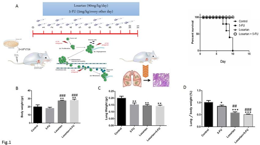
EXCLI Journal 2020;19:927-935 – ISSN 1611-2156
Received: February 23, 2020, accepted: June 25, 2020, published: June 29, 2020
part was put into Bouin's solution for evalua- measured in tissue based on Ellman method
tion of appearance symptoms. The rest of the using the following formula: The rate of thiol
lung was stored at -70 °C for the examination groups = (A2-A1-B) × (1.07.05)/ 13.6
of oxidant, antioxidant agents and inflamma- Measurement of SOD
tory cytokines. The measurement is consisted of SOD
generation through pyrogallol autoxidation
Histological evaluation and inhibition of MTT superoxide-dependent
The lung tissue was removed and washed revival to formazan. The reaction was stopped
with physiological saline and a portion of that by the addition of dimethyl sulfoxide
was fixed in 10 % formalin. Tissue samples (DMSO), which helps to dissolve formazan
were embedded in paraffin, after dehydration, and produce a stable color. Overall, the appro-
5 μm thick sections were cut using a micro- priate amount of homogenized tissue, pyro-
tome. H&E stained sections were examined
gallol, and MTT was appended into the wells
under light microscopy (×40 magnification). and kept for 5 mins at 37 ° C in the dark. After
that, it was quenched using DMSO, and anal-
Evaluation of the metastatic microscopic
ysis was performed at max= 630 nm (refer-
nodules in the lung
ence) and 570 nm (Madesh and Balasubrama-
The isolated lung tissue was placed into
nian, 1998).
Bouin's solution, the metastatic nodules were
examined using a stereomicroscope. The Measurement of CAT enzyme activity
number of metastatic microscopic nodules in The activity of the CAT in tissue was
different groups in the lung of mice was eval- measured by the Aebi method at the absorp-
uated. tion spectrum of max= 240 nm. Based on
this protocol, we used hydrogen peroxide (30
Evaluation of oxidant and antioxidant mM) and phosphate buffer (50 mM). The re-
agents action began with the addition of hydrogen
The fresh lung tissue was stored at -80 °C, peroxide and the change in absorbance was
and 50 mg of tissue was homogenized in PBS measured by spectrophotometer for 1 min at
(pH 7.4), then, the homogenized solution was max= 240 nm (Aebi, 1984).
centrifuged for 10 mins and the total thiol
group, CAT, SOD, and MDA were measured. ELISA
Evaluation of MDA Measurements of VEGF/VEGFR-1, IL-6
MDA concentrations were measured as a in lung tissue were performed using Zellbio
lipid peroxidation biomarker in lung tissue. 2 ELISA kits (ZellBio GmbH, Berlin, Ger-
mL of thiobarbituric acid solution (TBA) was many) according to the manufacturer's in-
added to 1 mL of a color with absorption at struction.
max= 535 nm (10 % solution of homoge-
nized lung tissue solution. The solution was In silico and heat map analysis of
incubated for 45 mins in a boiling water-bath, losartan response signature
We applied Autodock and LigPlot+ soft-
then it was centrifuged. MDA reacts with
ware or R software to evaluate the association
TBA and generates Janero, 1990).
of losartan with oxidant/antioxidant factors,
Measurement of total thiol groups IL-6, and VEGF/VEGFR1 markers.
DTNB reagent was used to measure thiol
groups. This reagent reacted with the SH Statistical analysis
groups and it would produce a yellow com- All data are shown as mean ± SEM and
plex (nitro-mercapto benzoate anion), that analyzed by one way ANOVA test followed
had an absorption at max= 412 nm (Sadegh- by LSD as the post hoc test. The data were
nia et al., 2013). The total thiol content was
929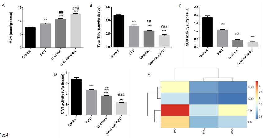
EXCLI Journal 2020;19:927-935 – ISSN 1611-2156
                                Received: February 23, 2020, accepted: June 25, 2020, published: June 29, 2020

calculated using SPSS v.20 statistical soft-           ence (Figure 1C). In comparison to the con-
ware (IBM, Chicago, IL, USA). The statisti-            trols, the percentage of the lung to body
cal difference was significant at P < 0.05.            weight was lower in the 5-FU group (P <
                                                       0.05), as well as losartan and combination
                  RESULTS                              groups (P < 0.001 for both). This proportion
                                                       in losartan and losartan+5-FU treated groups
Effect of losartan on survival, body weight
                                                       was statistically reduced compared with the 5-
and lung weight
                                                       FU group (P < 0.01, P < 0.001 respectively)
    Losartan alone and losartan plus 5-FU
                                                       (Figure 1D).
treatment groups increased the survival of the
treated animals compared to the control and
5-FU groups (Figure 1A). We also assessed              Histological and macroscopic assessment
                                                           Intravenous injection of CT26 cells into
body weight at the end of the experiment. We
                                                       mice led to the development of metastatic
observed a slightly decreased body weight in
                                                       nodules in the lung tissue, but after treatment
the 5-FU group, but it was not statistically sig-
                                                       with 5-FU, losartan and the combination of
nificant. On the contrary, the body weights of
                                                       losartan with 5-FU, metastatic nodules were
the losartan and losartan+5-FU treated groups
                                                       significantly reduced (P < 0.001). Losartan
were significantly greater than those of the
untreated group (P < 0.01) and the 5-FU                and losartan+5-FU treatment reduced the
group (P < 0.001) (Figure 1B). Furthermore,            number of metastatic nodules in lung tissue
there was a significant reduction of lung              compared to the treatment with 5-FU (P <
weight in the 5-FU, losartan, and losartan+5-          0.001) (Figure 2A-C). Histological images
FU groups compared to the control group (P             show the number of microscopic nodules and
< 0.01), but in the losartan and combination           percentage of metastatic area. These results
group, compared with the 5-FU only group,              showed that there was a significant decrease
there was no statistically significant differ-         of microscopic nodules in 5-FU, losartan and
                                                       co-administration of losartan with 5-FU treated

Figure 1: The treatment plan and survival rate of mice (A), Body weight (gr) (B), Lung weight (gr) (C),
Lung to body weight ratio ( %) (D). Data are shown as mean ± SEM (n = 6 per group). * P < 0.05, ** P
< 0.01 and *** P < 0.001 compared with control group, ## P < 0.01 and ### P < 0.001 compared with
5-FU group.

                                                  930
EXCLI Journal 2020;19:927-935 – ISSN 1611-2156
Received: February 23, 2020, accepted: June 25, 2020, published: June 29, 2020
Figure 2: The number of macroscopic metastatic lung nodules (A-C). ***P < 0.001 compared to control
group and ### P < 0.001 compared to 5-FU.
groups, compared to control (P < 0.001). The 0.01). The levels of SOD and CAT activities
number of microscopic nodules was signifi- were significantly lower in all treated groups
cantly lower in losartan and losartan+5-FU compared to the control group (P < 0.001 both
groups compared to the 5-FU group( P < of groups) (Figure 4C-E). CAT activity levels
0.001) (Figure 3A and B). Furthermore, his- in the losartan and combination of losartan
tological evaluation also showed that 5-FU, with 5-FU groups were significantly lower
losartan and losartan+5-FU reduced the per- compared to the 5-FU group (P < 0.01 and P
centage of metastatic area in tissue samples, < 0.001, respectively). Furthermore, the total
compared to control group (P < 0.001). We thiol content in all the treated groups was sig-
also observed that percentage of metastatic nificantly lower than in the untreated group (P
area was reduced in losartan and losartan+5- < 0.001 both groups). It has also been shown
FU treated groups compared to the 5-FU that total thiol content was significantly de-
group (P < 0.001) (Figure 3C). creased in losartan and its combination with
5-FU than the 5-FU group (P < 0.01 and P <
Oxidative and antioxidative parameters in 0.001, respectively) (Figure 4B and E).
lung tissue
Statistically, we found that losartan and its IL-6, VEGF and VEGFR-1 levels
combination with 5-FU increased MDA con- The level of tissue IL-6 in the losartan and
centration, compared to the 5-FU treated losartan+5-FU treated groups was signifi-
group (P < 0.01 and P < 0.001, respectively) cantly lower than the control group (P <
(Figure 4A). The level of MDA in the 5-FU 0.001), but IL-6 level in losartan and its com-
group was higher than the control group (P < bination with 5-FU treated groups was not
931EXCLI Journal 2020;19:927-935 – ISSN 1611-2156
Received: February 23, 2020, accepted: June 25, 2020, published: June 29, 2020
Figure 3: H&E staining demonstrated microscopic finding metastatic lung and foci in lung that are rep-
resented with black asterisks and the number of metastatic microscopic nodules in the lung of mice (A-
B). The area of lung metastasis indicated was evaluated by an image analyzer (Image J, NIH, USA)
(C). ***P < 0.001 compared to control group and ### P < 0.001 compared to 5-FU.
Figure 4: The MDA concentrations (A), SOD activity (B), CAT activity (C), total thiol contents (D) asso-
ciation between losartan and oxidant/anti-oxidant markers in tissue (E). Data are shown as mean ± SEM
(n = 6 per group). ** P < 0.01 and *** P < 0.001 compared with control group, ## P < 0.01 and ### P <
0.001 compared with 5-FU group.
932EXCLI Journal 2020;19:927-935 – ISSN 1611-2156
Received: February 23, 2020, accepted: June 25, 2020, published: June 29, 2020
significantly different compared to 5-FU then increased VEGF expression (Fujita et al.,
treated group (Figure 5A and D). As shown in 2002). Previous studies have shown that RA-
Figure 5B-D, the levels of VEGF in the losar- SIs reduced cancer cell metastasis by inhibit-
tan group and the group treated with the com- ing angiogenesis through reduction of VEGF
bination of losartan and 5-FU were signifi- expression (Koh et al., 2014; Miyajima et al.,
cantly lower compared to the control group (P 2002). Consistent with these results, we have
< 0.01, P < 0.001 respectively). The VEGFR- demonstrated that losartan decreased angio-
1 level of all treated groups was not signifi- genesis through the reduction of VEGF level.
cantly different, compared to the control. ROS production is related to a reduction
of cell viability. It also induces cell apoptosis
DISCUSSION (Ahmadian et al., 2017; Eftekhari et al.,
2018). Piskounova et al. demonstrated that
To the best of our knowledge, this is the
oxidative stress suppressed metastasis of can-
first study showing the anti-metastatic poten-
cer cells (Piskounova et al., 2015). In line
tial of losartan in CRC, yet in which lung me-
with these results, Ahmadian et al. showed
tastasis is widely being reported (Chambers et
that Azilsartan, a novel blocker of AT1R, in-
al., 2002; Neo et al., 2010). It is well known
duced ROS formation and then activated
that angiogenesis is crucial in cancer progres-
sion and is involved in the development of apoptotic pathway (Ahmadian et al., 2018).
primary tumor, migration of tumor cells and Interestingly, our results demonstrate that in-
distant metastasis (Harlozinska, 2005). The hibitory effect of losartan on cancer cells me-
ATII/AT1R pathway induces the formation of tastasis correlates with oxidant/antioxidant
the new vessels. ATII activated MAPK and status in tumor tissue.
Figure 5: IL-6 level (A), VEGF level (B), VEGFR-1 level (C) and association between losartan and IL-
6, VEGF and VEGFR-1 levels (D). Data are shown as mean ± SEM (n = 6 per group). ** P < 0.01 and
*** P < 0.001 compared to control group.
933EXCLI Journal 2020;19:927-935 – ISSN 1611-2156
Received: February 23, 2020, accepted: June 25, 2020, published: June 29, 2020
There is some evidence that activation of Chambers AF, Groom AC, MacDonald IC. Dissemina-
AT1R in the tumor microenvironment is in- tion and growth of cancer cells in metastatic sites. Nat
Rev Cancer. 2002;2:563-72.
volved in progression of inflammation and
metastasis (Deshayes and Nahmias, 2005). Coulson R, Liew SH, Connelly AA, Yee NS, Deb S,
ATII also enhances the expression of inflam- Kumar B, et al. The angiotensin receptor blocker,
matory cytokines such as IL-6 by activating Losartan, inhibits mammary tumor development and
progression to invasive carcinoma. Oncotarget. 2017;
AT1R (Suzuki et al., 2003). Furthermore, 8:18640-56.
Coulson et al. also demonstrated losartan
could reduce tumor progression via down- Deshayes F, Nahmias C. Angiotensin receptors: a new
regulation of inflammatory cytokine such as role in cancer? Trends Endocrinol Metab. 2005;16:
IL-6 (Coulson et al., 2017). In addition, 293-9.
Sanchez et al. have shown that losartan de- Eftekhari A, Ahmadian E, Panahi-Azar V, Hosseini H,
creases inflammation through reduction of Tabibiazar M, Maleki Dizaj S. Hepatoprotective and
IL-1/-6 and TNF-α levels in LPS-induced in- free radical scavenging actions of quercetin nanoparti-
flammation (Sánchez-Lemus et al., 2009). For cles on aflatoxin B1-induced liver damage: in vitro/in
vivo studies. Artif Cells Nanomed Biotechnol.
us, our results also demonstrated that losartan 2018;46:411-20.
reduced IL-6 levels in tissue samples, com-
pared to the control group. Fujita M, Hayashi I, Yamashina S, Itoman M, Majima
In conclusion, our data has shown that M. Blockade of angiotensin AT1a receptor signaling
reduces tumor growth, angiogenesis, and metastasis.
losartan might reduce lung metastasis via
Biochem Biophys Res Commun. 2002;294:441-7.
down-regulation of angiogenic and inflam-
matory pathways in CRC; however, further Godugu C, Patel AR, Doddapaneni R, Marepally S,
extensive clinical studies may be warranted. Jackson T, Singh M. Inhalation delivery of Telmisartan
enhances intratumoral distribution of nanoparticles in
lung cancer models. J Control Release. 2013;172:86-
Funding 95.
This study was supported by grants
awarded by National Institute for Medical Re- Haggar FA, Boushey RP. Colorectal cancer epidemiol-
search Development, Grant No. 976853 ogy: incidence, mortality, survival, and risk factors.
Clin Colon Rectal Surg. 2009;22:191-7.
(Majid Khazaei).
Harlozinska A. Progress in molecular mechanisms of
Disclosure tumor metastasis and angiogenesis. Anticancer Res.
The authors have no conflicts of interest 2005;25:3327-33.
to declare. Janero DR. Malondialdehyde and thiobarbituric acid-
reactivity as diagnostic indices of lipid peroxidation
REFERENCES and peroxidative tissue injury. Free Radic Biol Med.
1990;9:515-40.
Aebi H. Catalase in vitro. Methods Enzymol. 1984;
105:121-6. Koh SL, Ager EI, Costa PL, Malcontenti-Wilson C,
Muralidharan V, Christophi C. Blockade of the renin-
Ager EI, Neo J, Christophi C. The renin-angiotensin angiotensin system inhibits growth of colorectal cancer
system and malignancy. Carcinogenesis. 2008;29: liver metastases in the regenerating liver. Clin Exp Me-
1675-84. tastasis. 2014;31:395-405.
Ahmadian E, Eftekhari A, Fard JK, Babaei H, Nayebi Li Y, Wang C, Li D, Deng P, Shao X, Hu J, et al. 1H-
AM, Mohammadnejad D, et al. In vitro and in vivo NMR-based metabolic profiling of a colorectal cancer
evaluation of the mechanisms of citalopram-induced CT-26 lung metastasis model in mice. Oncol Rep.
hepatotoxicity. Arch Pharm Res. 2017;40:1296-313. 2017;38:3044-54.
Ahmadian E, Khosroushahi AY, Eftekhari A, Farajnia Madesh M, Balasubramanian KA. Microtiter plate as-
S, Babaei H, Eghbal MA. Novel angiotensin receptor say for superoxide dismutase using MTT reduction by
blocker, azilsartan induces oxidative stress and NFkB- superoxide. Indian J Biochem Biophys. 1998;35: 184-
mediated apoptosis in hepatocellular carcinoma cell 8.
line HepG2. Biomed Pharmacother. 2018;99:939-46.
934EXCLI Journal 2020;19:927-935 – ISSN 1611-2156
Received: February 23, 2020, accepted: June 25, 2020, published: June 29, 2020
Marjaneh RM, Rahmani F, Hassanian SM, Rezaei N, Sadeghnia HR, Kamkar M, Assadpour E, Boroushaki
Hashemzehi M, Bahrami A, et al. Phytosomal curcu- MT, Ghorbani A. Protective effect of safranal, a con-
min inhibits tumor growth in colitis-associated colo- stituent of crocus sativus, on quinolinic acid-induced
rectal cancer. J Cell Physiol. 2018;233:6785-98. oxidative damage in rat hippocampus. Iran J Basic Med
Sci. 2013;16:73-82.
Mc Menamin ÚC, Murray LJ, Cantwell MM, Hughes
CM. Angiotensin-converting enzyme inhibitors and Sánchez-Lemus E, Benicky J, Pavel J, Larrayoz IM,
angiotensin receptor blockers in cancer progression Zhou J, Baliova M, et al. Angiotensin II AT1 blockade
and survival: a systematic review. Cancer Causes Con- reduces the lipopolysaccharide-induced innate immune
trol. 2012;23:221-30. response in rat spleen. Am J Physiol Regul Integr
Comp Physiol. 2009;296:1376-84.
Miyajima A, Kosaka T, Asano T, Asano T, Seta K, Ka-
wai T, et al. Angiotensin II type I antagonist prevents Siegel RL, Miller KD, Fedewa SA, Ahnen DJ, Meester
pulmonary metastasis of murine renal cancer by inhib- RGS, Barzi A, et al. Colorectal cancer statistics, 2017.
iting tumor angiogenesis. Cancer Res. 2002; 62:4176- CA Cancer J Clin. 2017;67:177-93.
9.
Smith GR, Missailidis S. Cancer, inflammation and the
Mun JG, Kee JY, Han YH, Lee S, Park SH, Jeon HD, AT1 and AT2 receptors. J Inflamm (Lond). 2004;1:3.
et al. Galla Rhois water extract inhibits lung metastasis
by inducing AMPK‑mediated apoptosis and suppress- Suzuki Y, Ruiz-Ortega M, Lorenzo O, Ruperez M,
ing metastatic properties of colorectal cancer cells. On- Esteban V, Egido J. Inflammation and angiotensin II.
col Rep. 2019;41:202-12. Int J Biochem Cell Biol. 2003;35:881-900.
Neo JH, Malcontenti-Wilson C, Muralidharan V, Uemura H, Ishiguro H, Nakaigawa N, Nagashima Y,
Christophi C. Effect of ACE inhibitors and angiotensin Miyoshi Y, Fujinami K, et al. Angiotensin II receptor
II receptor antagonists in a mouse model of colorectal blocker shows antiproliferative activity in prostate can-
cancer liver metastases. J Gastroenterol Hepatol. 2007; cer cells: a possibility of tyrosine kinase inhibitor of
22:577-84. growth factor. Mol Cancer Ther. 2003;2:1139-47.
Neo JH, Ager EI, Angus PW, Zhu J, Herath CB, Chris- Wang L, Cai SR, Zhang CH, He YL, Zhan WH, Wu H,
tophi C. Changes in the renin angiotensin system dur- et al. Effects of angiotensin-converting enzyme inhibi-
ing the development of colorectal cancer liver metasta- tors and angiotensin II type 1 receptor blockers on lym-
ses. BMC Cancer. 2010;10:134. phangiogenesis of gastric cancer in a nude mouse
model. Chin Med J (Engl). 2008;121:2167-71.
Piskounova E, Agathocleous M, Murphy MM, Hu Z,
Huddlestun SE, Zhao Z, et al. Oxidative stress inhibits
distant metastasis by human melanoma cells. Nature.
2015;527:186-91.
935You can also read