Utility of Magnetic Resonance Imaging for Differentiating Necrotizing Fasciitis from Severe Cellulitis: A Magnetic Resonance Indicator for ...
←
→
Page content transcription
If your browser does not render page correctly, please read the page content below
Journal of
           Clinical Medicine

Article
Utility of Magnetic Resonance Imaging for
Differentiating Necrotizing Fasciitis from Severe
Cellulitis: A Magnetic Resonance Indicator for
Necrotizing Fasciitis (MRINEC) Algorithm
Min-Chul Kim 1,† , Sujin Kim 2, *,† , Eun Been Cho 1,† , Guen Young Lee 2 , Seong-Ho Choi 1 ,
Seon Ok Kim 3 and Jin-Won Chung 1, *,†
 1    Division of Infectious Diseases, Department of Internal Medicine, Chung-Ang University Hospital,
      Seoul 06973, Korea; Kimminchulmd@caumc.or.kr (M.-C.K.); bin0508-1@caumc.or.kr (E.B.C.);
      tobeservant@cau.ac.kr (S.-H.C.)
 2    Department of Radiology, Chung-Ang University Hospital, Seoul 06973, Korea; netty0523@caumc.or.kr
 3    Departments of Clinical Epidemiology and Biostatistics, Asan Medical Center,
      University of Ulsan College of Medicine, Seoul 05505, Korea; chddk0707@naver.com
 *    Correspondence: jaywony@gmail.com (S.K.); drjwchung@cau.ac.kr (J.-W.C.);
      Tel.: +82-2-6299-2646 (S.K.); +82-2-6299-1402 (J.-W.C.)
 †    These authors contributed equally to this study.

 Received: 17 July 2020; Accepted: 18 September 2020; Published: 21 September 2020

 Abstract: We developed a new magnetic resonance indicator for necrotizing fasciitis (MRINEC)
 algorithm for differentiating necrotizing fasciitis (NF) from severe cellulitis (SC). All adults with
 suspected NF between 2010 and 2018 in a tertiary hospital in South Korea were enrolled. Sixty-one
 patients were diagnosed with NF and 28 with SC. Among them, 34 with NF and 15 with SC underwent
 magnetic resonance imaging (MRI). The MRINEC algorithm, a two-step decision tree including
 T2 hyperintensity of intermuscular deep fascia and diffuse T2 hyperintensity of deep peripheral
 fascia, diagnosed NF with 94% sensitivity (95% confidence interval (CI), 80–99%) and 60% specificity
 (95% CI, 32–84%). The algorithm accurately diagnosed all 15 NF patients with a high (≥8) laboratory
 risk indicator for necrotizing fasciitis (LRINEC) score. Among the five patients with an intermediate
 (6–7) LRINEC score, sensitivity and specificity were 100% (95% CI, 78–100%) and 0% (95% CI, 0–84%),
 respectively. Finally, among the 29 patients with a low (≤5) LRINEC score, the algorithm had a
 sensitivity and specificity of 88% (95% CI, 62–98%) and 69% (95% CI, 39–91%), respectively. The
 MRINEC algorithm may be a useful adjuvant method for diagnosing NF, especially when NF is
 suspected in patients with a low LRINEC score.

 Keywords: necrotizing fasciitis; nonnecrotizing soft tissue infection; magnetic resonance imaging;
 MRINEC algorithm; LRINEC score

1. Introduction
     Necrotizing fasciitis (NF) is a life-threatening skin and soft tissue infection that leads to extensive
tissue destruction and systemic toxicity [1]. Prompt and aggressive surgical debridement is crucial for
improving survival and reducing morbidity of patients with NF [2]. However, the diagnosis of NF has
remained challenging because the typical characteristics of NF such as bullae, necrosis, and hemorrhage
are absent in a significant number of NF cases [1,3,4]. The laboratory risk indicator for necrotizing
fasciitis (LRINEC) score, which is composed of white blood cells, hemoglobin, sodium, glucose,
creatinine, and C-reactive protein, has been widely used for differentiating NF from non-necrotizing

J. Clin. Med. 2020, 9, 3040; doi:10.3390/jcm9093040                                  www.mdpi.com/journal/jcm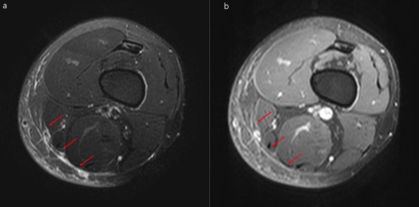
J. Clin. Med. 2020, 9, 3040                                                                        2 of 11

soft tissue infection (NNSTI) [5,6]. However, the LRINEC score should not be used to rule out NF since
the laboratory scoring might have suboptimal sensitivity [4,7]. Therefore, further testing might be
needed to resolve diagnostic uncertainty when NF is still suspected despite the LRINEC score having
been applied [2].
      The usefulness of magnetic resonance imaging (MRI) for diagnosing of NF has not been fully
evaluated. Previous studies suggested that MRI could be helpful in discriminating NF from NNSTI [8–12].
However, its sensitivity and specificity for diagnosing NF are ill defined [2,5]. Importantly, the impact
of MRI on the differentiation between NF and NNSTI has not been evaluated in conjunction with the
LRINEC score. We therefore investigated the difference in MRI findings between patients with NF and
those with NNSTI. Then, we newly developed a magnetic resonance indicator for necrotizing fasciitis
(MRINEC) algorithm to differentiate NF from NNSTI using MRI findings that were suggestive of NF.
Furthermore, we evaluated the diagnostic performance of the MRINEC algorithm in patients with
suspected NF, according to the classification based on their LRINEC score.

2. Materials and Methods

2.1. Study Population and Definition of Necrotizing Fasciitis
     Adult patients aged ≥18 years in Chung-Ang University Hospital, an 850-bed tertiary hospital
in Seoul, South Korea, who were suspected of having NF between November 2010 and July 2018
were retrospectively analyzed. Patients with mild cellulitis, pyomyositis involving primarily muscles
(without evidence of skin infection), and bone and joint infections were excluded. Diagnosis of
NF was established when (1) direct examination during surgical exploration or histopathology
of the surgical specimen revealed the infected fascia, or (2) when evidence of extensive soft tissue
destruction such as hemorrhagic bullae and necrosis was found on physical examination. The remaining
patients with NNSTI in whom there was no evidence of NF were diagnosed with severe cellulitis
(SC). Clinical characteristics and MRI findings from patients with NF and SC were compared.
The study protocol was approved by the Institutional Review Board of Chung-Ang University
Hospital (1909-001-16277). The need for informed consent was waived in view of the observational
nature of the study.

2.2. Analysis of Magnetic Resonance Imaging
     MRI was performed on 1.5-T (Magnetom Avanto; Siemens, Erlangen, Germany) or 3.0-T (Achieva;
Philips Medical Systems, Best, Netherlands and Skyra; Siemens, Erlangen, Germany) scanners. The MRI
protocol included the axial, sagittal, and coronal imaging planes, which were obtained using T1-
(T1WI) and T2- (T2WI) weighted imaging. Additionally, T1-weighted fat-suppressed fast spin-echo
contrast-enhanced images were obtained in the axial, sagittal, and coronal planes. The fields of view,
section thicknesses, and intersection gaps were variable for different anatomic sites of involvement.
Two musculoskeletal radiologists with 9 (S.K.) and 10 (G.Y.L.) years of experience, respectively,
who were blinded to the diagnoses of patients and their clinical characteristics, reviewed the MRIs
independently and reached a final decision regarding the findings by consensus.
     The MRIs of the enrolled patients were examined for the presence or absence of the following
findings: diffuse or localized T2 hyperintensity of deep peripheral fascia, thickness of deep peripheral
fascia, diffuse or localized T2 hyperintensity of intermuscular deep fascia, irregular or diffuse
fascial enhancement, myositis, intermuscular or subcutaneous abscess, and subcutaneous fat edema.
Deep peripheral fascia was defined as the peripheral investing layer of deep fascia that connects deep
adipose tissues and muscles [10]. Intermuscular deep fascia was defined as the intermuscular layer of
deep fascia that passes in between the muscles [10]. In addition, we defined “diffuse” hyperintensity
as a signal intensity change of ≥75% of fasciae surrounding the muscle. An abscess was defined as
a localized fluid collection with a low signal intensity on T1WI and a high signal intensity on T2WI,
with peripheral rim enhancement on contrast-enhanced images.
J. Clin. Med. 2020, 9, x FOR PEER REVIEW 3 of 11
J. Clin. Med. 2020, 9, 3040 3 of 11
2.3. Statistical Analysis
Categorical
2.3. Statistical data were compared using the χ² or Fisher’s exact test, and continuous variables
Analysis
were analyzed using the Mann–Whitney U test. Univariable and multivariable logistic regression
Categorical data were compared using the χ2 or Fisher’s exact test, and continuous variables
analyses were performed to identify predictive MRI findings for NF. The MRINEC algorithm for
were analyzed using the Mann–Whitney U test. Univariable and multivariable logistic regression
differentiating NF from SC was generated using a classification and regression tree analysis based
analyses were performed to identify predictive MRI findings for NF. The MRINEC algorithm for
on the MRI findings suggesting NF [13]. Diagnostic performance was expressed in terms of
differentiating NF from SC was generated using a classification and regression tree analysis based on
sensitivity, specificity, positive likelihood ratio, and negative likelihood ratio with a 95% confidence
the MRI findings suggesting NF [13]. Diagnostic performance was expressed in terms of sensitivity,
interval (CI). All reported p-values are two-sided, and p-values < 0.05 were considered statistically
specificity, positive likelihood ratio, and negative likelihood ratio with a 95% confidence interval
significant. Data manipulation and statistical analyses were conducted using R version 3.4.2 with
(CI). All reported p-values are two-sided, and p-values < 0.05 were considered statistically significant.
the package rpart.
Data manipulation and statistical analyses were conducted using R version 3.4.2 with the package rpart.
3. Results
3. Results
3.1. Clinical
3.1. Clinical Characteristics
Characteristics of
of Patients
Patients
In total,
In total,4528
4528 individuals
individuals were
were suspected
suspected of having
of having skin andskin
softand soft
tissue tissue from
infection infection from
November
November
2010 to July 2010 to Chung-Ang
2018 in July 2018 inUniversity
Chung-Ang University
Hospital (FigureHospital (Figure
1). Of these 1). Of4229,
patients, these patients,
whose 4229,
diagnosis
was established as mild cellulitis, were excluded. Additionally, 92 patients with pyomyositis andwith
whose diagnosis was established as mild cellulitis, were excluded. Additionally, 92 patients 178
pyomyositis
with bone andand joint178 with bone
infections were and joint along
excluded, infections
with were
29 who excluded, along with
were diagnosed 29 who were
with non-infectious
diagnosedThus,
diseases. with non-infectious diseases.
a total of 89 patients Thus,
with a total ofNF
suspected 89were
patients withanalyzed:
finally suspected61 NF were finally
patients were
analyzed: 61 patients were diagnosed with NF, and the
diagnosed with NF, and the remaining 28 were diagnosed with SC. remaining 28 were diagnosed with SC
Figure 1. Flow diagram of the study. Abbreviations: MRI = magnetic resonance imaging.
Figure 1. Flow diagram of the study. Abbreviations: MRI = magnetic resonance imaging.
The clinical and laboratory characteristics, microbiology, and outcomes of the patients with
The clinical and laboratory characteristics, microbiology, and outcomes of the patients with NF
NF and SC are shown in Table 1. Clinical characteristics such as discharge, bullae, necrosis,
and SC are shown in Table 1. Clinical characteristics such as discharge, bullae, necrosis,
petechiae/hemorrhage, altered mental status, and shock were more common in patients with NF than in
petechiae/hemorrhage, altered mental status, and shock were more common in patients with NF
those with SC. White blood cell counts and C-reactive protein levels were higher, and hemoglobin and
than in those with SC. White blood cell counts and C-reactive protein levels were higher, and
Na levels were lower, in the NF group than in the SC group. Regarding the microbiology, the causative
hemoglobin and Na levels were lower, in the NF group than in the SC group. Regarding the
pathogen was identified more often from blood and wound cultures in patients with NF than in those
microbiology, the causative pathogen was identified more often from blood and wound cultures in
with SC. Thirty-five (57%) patients with NF underwent surgical debridement, whereas none with SC
patients with NF than in those with SC. Thirty-five (57%) patients with NF underwent surgical
received surgical treatment. Additionally, patients with NF received longer antibiotic treatment than
debridement, whereas none with SC received surgical treatment. Additionally, patients with NF
those with SC. The mortality rate was higher in patients with NF than in those with SC. Patients with
received longer antibiotic treatment than those with SC. The mortality rate was higher in patients
NF had higher LRINEC scores than those with SC (p = 0.001). Notably, high LRINEC scores (≥8) were
with NF than in those with SC. Patients with NF had higher LRINEC scores than those with SC (p =J. Clin. Med. 2020, 9, 3040 4 of 11
more common in patients with NF than in those with SC (39% vs. 7%, p = 0.002), although 9 (15%) and
28 (46%) patients with NF had moderate (6–7) and low (≤5) LRINEC scores, respectively. The sensitivity
and specificity of high LRINEC scores (≥8) for the diagnosis of NF were 39% (95% CI, 27–53%) and
93% (95% CI, 77–99%), respectively.
Table 1. Clinical and laboratory characteristics, microbiology, and outcomes of patients with necrotizing
fasciitis and severe cellulitis.
Necrotizing Fasciitis Severe Cellulitis
Variables p-Value
(n = 61) (n = 28)
Male sex 31 (51) 17 (61) 0.39
Age, mean years ± SD 59 ± 16 59 ± 15 0.88
Acquisition site of infection 0.03
Community-acquired infection 41 (67) 25 (89)
Healthcare-associated infection 20 (33) 3 (11)
Infection site
Leg 41 (67) 20 (71) 0.69
Arm 13 (21) 4 (14) 0.43
Abdomen & pelvis 4 (7) 1 (4) >0.99
Others 3 (5) 3 (11) 0.37
Underlying disease
Diabetes 24 (39) 9 (32) 0.51
Trauma 22 (36) 4 (14) 0.04
Tinea pedis or onychomycosis 10 (16) 9 (32) 0.09
Malignancy 11 (18) 6 (21) 0.71
Liver cirrhosis 11 (18) 1 (4) 0.09
Peripheral artery disease 7 (12) 0 0.09
Chronic kidney disease 6 (10) 3 (11) >0.99
Lymphedema 3 (5) 1 (4) >0.99
Symptoms and signs
Pain and erythema 61 (100) 28 (100) >0.99
Fever 42 (69) 14 (50) 0.09
Discharge 27 (44) 4 (14) 0.006
Bullae 25 (41) 0 0.99
Laboratory findings
White blood cell, mean/mm3 ± SD 14,000 ± 8500 10,000 ± 3800 0.05
Hemoglobin, g/dL ± SD 11.4 ± 2.6 13.5 ± 1.9 0.001
Platelet, × 103 /mm3 ± SD 225 ± 131 217 ± 87 0.90
INR ± SD 1.3 ± 0.3 1.1 ± 0.2 0.07
Bilirubin, mg/dL ± SD 1.5 ± 2.9 0.9 ± 0.8 0.56
Creatinine, mg/dL ± SD 1.2 ± 1.0 1.0 ± 0.5 0.77
Na, mEq/L ± SD 135 ± 6 137 ± 4 0.02
Glucose, mg/dL ± SD 171 ± 124 153 ± 74 0.81
C-reactive protein, mg/dL ± SD 15.7 ± 11.4 9.4 ± 9.2 0.02
Procalcitonin, ng/mL ± SD 4.8 ± 5.3 6.7 ± 6.8 0.39
Lactate, mmol/L ± SD 2.5 ± 2.2 1.4 ± 0.2 0.54
LRINEC score ± SD 6.0 ± 3.6 3.1 ± 3.4 0.001
High risk (≥8) 24 (39) 2 (7) 0.002
Moderate risk (6–7) 9 (15) 4 (14) >0.99
Low risk (≤5) 28 (46) 22 (79) 0.004J. Clin. Med. 2020, 9, 3040 5 of 11
Table 1. Cont.
Necrotizing Fasciitis Severe Cellulitis
Variables p-Value
(n = 61) (n = 28)
Microbiology
Overall culture positivity 38 (62) 5 (18) 0.99
Group B and C streptococcus 4 (7) 1 (4) >0.99
Gram negative bacteria 18 (30) 1 (4) 0.006
Anaerobes 0 0
Surgical debridement 35 (57) 0Table 2. Magnetic resonance findings of necrotizing fasciitis and severe cellulitis.
Necrotizing Fasciitis Severe Cellulitis
Variables p-Value
(n = 34) (n = 15)
J. Clin. Med.from
Interval 9, 3040
2020, admission to MRI, 6 of 11
4±7 4±5 0.71
mean days ± SD
T2 hyperintensity of deep peripheral
Table 2. Magnetic resonance findings31of(91) 12 (80)cellulitis.
necrotizing fasciitis and severe 0.35
fascia
Diffuse 20 (59)
Necrotizing Fasciitis 3 (20)
Severe Cellulitis 0.01
Localized Variables 11 (32) (n = 34) 9 (n
(60) p-Value
0.07
= 15)
Thickness of superficial fascia, mm ±
Interval from admission to MRI, mean days ± SD 6.9 ± 4.8 4 ± 7 ±5
5.5 ±43.0 0.71
0.52
SD
T2 hyperintensity of deep peripheral fascia 31 (91) 12 (80) 0.35
T2 hyperintensity
Diffuse of intermuscular
28 (82) 20 (59) 3 (20)
6 (40) 0.01
0.006
deep Localized
fascia 11 (32) 9 (60) 0.07
Thickness
Diffuseof superficial fascia, mm ± SD 17 (50) 6.9 ± 4.8 (13)± 3.0
2 5.5 0.52
0.02
T2 hyperintensity
Localized of intermuscular deep fascia 11 (32) 28 (82) 6 (40)
4 (27) 0.006
0.75
Diffuse 17 (50)
Fascial enhancement 28/31 a (90) 12/14 b (13)
2
(86) 0.02
0.64
Localized 11 (32) 4 (27) 0.75
Irregular 6/31 a (19) a 0 b 0.16
Fascial enhancement 28/31 (90) 12/14 (86) 0.64
Diffuse
Irregular 22/31 a (71)
6/31 a (19) 12/14 b (86)
0 0.46
0.16
Myositis
Diffuse 24 (71)22/31 a (71) 6 (40)b (86)
12/14 0.04
0.46
Abscess
Myositis 17 (50) 24 (71) 1 (7)
6 (40) 0.004
0.04
Abscess
Intermuscular abscess 11 (32) 17 (50) 01 (7) 0.004
0.01
Intermuscular abscess
Subcutaneous 6 (18) 11 (32) 1 (7)0 0.01
0.41
Subcutaneous
Subcutaneous fat edema 30 (88) 6 (18) 1 (7)
14 (93) 0.41
>0.99
Subcutaneous fat edema 30 (88) 14 (93) >0.99
NOTE. Data are presented as the number (%) of patients unless otherwise indicated. Abbreviations:
NOTE. Data are presented as the number (%) of patients unless otherwise indicated. Abbreviations:
MRI
MRI == magnetic
magneticresonance
resonance imaging;
imaging; SDSD = standard
= standard deviation.
deviation.
a Thirty-one patients with necrotizing
a Thirty-one patients with necrotizing fasciitis
fasciitis
underwent underwent enhancedresonance
enhanced magnetic magneticimaging. b Fourteen
resonance imaging. b Fourteen patients with severe cellulitis
patients with severe cellulitis received enhanced
magnetic resonance
received enhancedimaging.
magnetic resonance imaging.
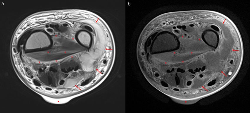
Figure 2. Representative
Representativemagnetic
magneticresonance
resonanceimages
imagesofof necrotizing
necrotizing fasciitis.
fasciitis. Necrotizing
Necrotizing fasciitis
fasciitis of
of the
the left in
left wrist wrist in a 71-year-old
a 71-year-old woman.
woman. Axial Axial T2
T2 weighted weighted
magnetic magnetic
resonance image resonance image (a) and
(a) and contrast-enhanced
contrast-enhanced
magnetic resonancemagnetic
image (b) resonance imagehyperintensity
showing diffuse (b) showing with
diffuse hyperintensity
irregular enhancementwithof irregular
the deep
enhancement
peripheral of and
fascia the intermuscular
deep peripheral
deepfascia
fasciaand intermuscular
(asterisk) deep
of the wrist. fascia (asterisk)
Additionally, there isofa lobulating
the wrist.
abscess in the ulnar
Additionally, there side
is a of the wristabscess
lobulating (arrows)inand
theaulnar
skin bulla
side (triangle).
of the wrist (arrows) and a skin bulla
(triangle).J. Clin. Med. 2020, 9, 3040 7 of 11
J. Clin. Med. 2020, 9, x FOR PEER REVIEW 7 of 11
Figure
Figure 3. 3. Representativemagnetic
Representative magneticresonance
resonance images
images ofof severe
severe cellulitis.
cellulitis.Severe
Severecellulitis
cellulitisof of
thethe
leftleft
thigh
thigh in in a 44-year-oldman.
a 44-year-old man.(a)
(a)Fat-suppressed
Fat-suppressed axial T2-weighted
T2-weightedmagnetic
magneticresonance
resonance image
image and a a
and
contrast-enhancedmagnetic
contrast-enhanced magneticresonance
resonance image
image (b) showing
showing localized
localizedhyperintensity
hyperintensity within
within thethe
deep
deep
peripheral
peripheral fascia
fascia (arrows)with
(arrows) withenhancement
enhancementin in the
the posteromedial
posteromedial thigh.
thigh.
Results of the univariate and multivariate logistic regression analysis of MRI findings
Table 3. NF
suggesting Multivariable
are shownlogistic
in Table regression analysis
3. The odds of predictive
ratios magnetic
(ORs) for diffuse T2resonance findings
hyperintensity of for
deep
necrotizing
peripheral fasciitis.
fascia, T2 hyperintensity of intermuscular deep fascia, diffuse T2 hyperintensity of
intermuscular deep fascia, and
Univariable Logistic Odds abscesses
Ratio were 5.1 (95%Multivariable
CI, 1.3–20.3) (p = 0.02), 6.4
Logistic Odds(95% CI, 1.7–24.6)
Ratio
p-Value p-Value
(p = 0.01), 5.4 (95%
Regression CI, 1.2–25.2)
Analysis. (95%(pCI)
= 0.03), and 9.6 (95% CI, 1.5–61.7)
Regression (p = 0.02), respectively.
Analysis (95% CI) From the
multivariate logistic regression
Diffuse T2 hyperintensity of analysis, diffuse
5.1 (1.3–20.3) 0.02
T2 hyperintensity of deepofperipheral
Diffuse T2 hyperintensity fascia (OR
4.4 (0.9–22.8)
4.4,
0.074
95%deep
CI, peripheral
0.9–22.8)fascia
(p = 0.074), diffuse T2 hyperintensity ofdeep peripheral fascia
intermuscular deep fascia (OR 5.5, 95% CI,
T2 hyperintensity of
0.9–33.8) (p = 0.065), and abscesses (OR 15.1,
6.4 (1.7–24.6) 0.0195% CI, 1.6–143.5) (p = 0.02) were suggestive of NF
intermuscular deep fascia
rather than of SC.
Diffuse T2 hyperintensity of Diffuse T2 hyperintensity of
5.4 (1.2–25.2) 0.03 5.5 (0.9–33.8) 0.065
intermuscular deep fascia intermuscular deep fascia
TableAbscess
3. Multivariable logistic regression 0.02
9.6 (1.5–61.7) analysis of predictive magnetic resonance
Abscess findings for0.02
15.1 (1.6–143.5)
necrotizing fasciitis.
Intermuscular abscess 15.2 (0.7–313.7) 0.078
Univariable Logistic Odds Abbreviations:
Ratio CI = confidence interval. Logistic
Multivariable Odds Ratio
p-Value p-Value
Regression Analysis. (95% CI) Regression Analysis (95% CI)
Diffuse T2
Diffuse T2 hyperintensity 5.1 4.4
3.3. Diagnostic Performance of the MRINEC Algorithm 0.02 hyperintensity of deep 0.074
of deep peripheral fascia (1.3–20.3) (0.9–22.8)
peripheral fascia
Using classification
T2 hyperintensity of and regression
6.4 tree analysis, we developed a novel MRINEC algorithm,
whichintermuscular
is a two-stepdeep
decision tree, including 0.01
fascia (1.7–24.6) the presence or absence of T2 hyperintensity of intermuscular
deep fascia (step one), and diffuse T2 hyperintensity of deep peripheral
Diffuse T2 fascia (step two) (Figure 4).
Diffuse T2 hyperintensity
The diagnostic performance of the 5.4
MRINEC algorithm hyperintensity
is shown in of
Table 4. Of 5.5
the 49 patients
of intermuscular deep 0.03 0.065 with
(1.2–25.2) intermuscular deep (0.9–33.8)
NF and SC who underwent MRIs, the overall sensitivity and specificity of the MRINEC algorithm
fascia
fascia
for differentiating NF from SC were 94% (95% CI, 80–99%) and 60% (95% CI, 32–84%), respectively.
9.6 15.1
The C-statistic Abscess
for this algorithm was 0.79 (95% 0.02 CI, 0.67–0.96).
AbscessThe MRINEC algorithm 0.02 correctly
(1.5–61.7) (1.6–143.5)
diagnosed all 15 patients with NF with a high LRINEC score. Among the five patients with an
15.2
Intermuscular abscess 0.078
intermediate LRINEC score, the (0.7–313.7)
MRINEC algorithm had a sensitivity of 100% (95% CI, 78–100%) and a
specificity of 0% (95% CI, 0–84%). Abbreviations:
Furthermore,CIthe= confidence
MRINEC algorithm
interval. differentiated NF from SC with
a sensitivity of 88% (95% CI, 62–98%) and a specificity of 69% (95% CI, 39–91%) among the 29 patients
3.3.low
with Diagnostic
LRINEC Performance
scores. of the MRINEC Algorithm
Using classification and regression tree analysis, we developed a novel MRINEC algorithm,
which is a two-step decision tree, including the presence or absence of T2 hyperintensity ofalgorithm correctly diagnosed all 15 patients with NF with a high LRINEC score. Among the five
patients with an intermediate LRINEC score, the MRINEC algorithm had a sensitivity of 100% (95%
CI, 78–100%) and a specificity of 0% (95% CI, 0–84%). Furthermore, the MRINEC algorithm
differentiated NF from SC with a sensitivity of 88% (95% CI, 62–98%) and a specificity of 69% (95%
CI,
J. 39–91%)
Clin. among
Med. 2020, 9, 3040the 29 patients with low LRINEC scores. 8 of 11
Figure 4. Magnetic resonance indicator for necrotizing fasciitis (MRINEC) algorithm for differentiating
Figure 4. fasciitis
necrotizing Magnetic
fromresonance indicator
severe cellulitis. for necrotizing
Abbreviations: MRI = fasciitis
magnetic(MRINEC) algorithm for
resonance imaging.
differentiating necrotizing fasciitis from severe cellulitis. Abbreviations: MRI = magnetic resonance
Table 4. Diagnostic performance of the magnetic resonance indicator for necrotizing fasciitis
imaging.
(MRINEC) algorithm.
Table 4. Diagnostic performance
NF by of the magnetic
SC by resonance indicator for necrotizing fasciitis
MRINEC/Patients
(MRINEC) algorithm. MRINEC/Patients Sensitivity, Specificity, PLR NLR
with NF with SC % (95% CI) % (95% CI) (95% CI) (95% CI)
(n = 34)NF by (n =SC15)
by
MRINEC/Patients MRINEC/Patients Sensitivity, % Specificity, PLR
2.4 NLR
0.1
Total (n = 49) 32/34with NF 9/15
with SC 94 (80–99)
(95% CI) 60 %
(32–84)
(95% CI) (95% CI) (95% CI)
(1.3–4.4) (0.02–0.4)
(n = 34) (n = 15)
High risk, LRINEC
15/15 0 100 (78–100) NA 2.4
1.0 0.1
NA
(n = (n
score ≥8Total 15)= 49) 32/34 9/15 94 (80–99) 60 (32–84)
(1.3–4.4) (0.02–0.4)
Intermediate
High risk, risk,
LRINEC score
LRINEC score
≥8 (n6–7
= 15) 3/3 15/15 0/20 100100 (78–100)
(29–100) NA
0 (0–84) 1.0
1.0 NA
NA
= 5)
(nIntermediate risk,
3/3 0/2 100 (29–100) 0 (0–84) 1.0 NA
LRINEC score 6–7 (n = 5)
Low risk,
2.8 0.2
LRINEC score
Low risk, 14/16 9/13 88 (62–98) 69 (39–91) 2.8 0.2
14/16 9/13 88 (62–98) 69 (39–91) (1.2–6.6) (0.05–0.7)
≤5 (n = 29)
LRINEC score ≤5 (n = 29) (1.2–6.6) (0.05–0.7)
Abbreviations:
Abbreviations:CI CI==confidence
confidenceinterval; MRINEC
interval; MRINEC= magnetic resonance
= magnetic indicator
resonance for necrotizing
indicator fasciitis;
for necrotizing
NF = necrotizing fasciitis; NLR = negative likelihood ratio; PLR = positive likelihood ratio; SC = severe cellulitis.
fasciitis; NF = necrotizing fasciitis; NLR = negative likelihood ratio; PLR = positive likelihood ratio;
SC = severe cellulitis.
4. Discussion
4. Discussion
We evaluated the utility of MRI for the diagnosis of NF, and assessed the diagnostic performance
of theWeMRINEC
evaluatedalgorithm for differentiating
the utility of MRI for NF the from SC. The
diagnosis of overall
NF, and sensitivity and
assessed specificity
the of
diagnostic
this algorithmoffor
performance thediagnosing NF were 94%
MRINEC algorithm for and 60%, respectively.
differentiating NF fromNotably,
SC. Thethe MRINEC
overall algorithm
sensitivity and
differentiated
specificity of this algorithm for diagnosing NF were 94% and 60%, respectively. Notably, low
NF from SC with a sensitivity of 88% and a specificity of 69% in patients with the
LRINEC
MRINECscores. Thus,
algorithm the MRINECNF
differentiated algorithm
from SCappeared to be useful
with a sensitivity of for
88%diagnosing NF, especially
and a specificity of 69% inin
cases in which
patients the differentiation
with low LRINEC scores. between
Thus,NFtheand SC basedalgorithm
MRINEC on clinicalappeared
and laboratory
to be findings
useful foris
difficult. Furthermore, the MRINEC algorithm may be useful for excluding a diagnosis of
diagnosing NF, especially in cases in which the differentiation between NF and SC based on clinical NF, given its
high
and sensitivity.
laboratory findings is difficult. Furthermore, the MRINEC algorithm may be useful for
Several
excluding a studies
diagnosishaveofpreviously
NF, givenevaluated MRI findings suggestive of NF rather than of NNSTI [8,9,11].
its high sensitivity.
Kim et al., showed that thick (≥3 mm) and extensive signal change of the deep fascia, focal or diffuse
non-enhancing fascia, and the involvement of three or more compartments were more frequent in
patients with NF than in those with NNSTI [8]. The MRINEC algorithm, in which T2 hyperintensity
of intermuscular deep fascia and diffuse T2 hyperintensity of deep peripheral fascia were included,
is consistent with the observations of the previous study. However, thickening and enhancement of
the fascia were not significantly different between NF and SC in the present study. Regarding fascial
enhancement, Schmid et al., reported that enhancement of deep fascia was found in all patients with
NF [11], whereas the absence of enhancement was an important MRI finding, indicating fascial necrosis in
Brothers et al.’s study [9]. This discrepancy could be because these earlier studies included only a limited
number of NF cases, thus the MRI findings of NF might not have been fully evaluated. Furthermore,J. Clin. Med. 2020, 9, 3040 9 of 11
previous studies have another important limitation, in that they have not evaluated the diagnostic utility of
the MRI findings in the context of the clinical judgment based on the LRINEC score. Recently, Yoon et al.,
reported that a new scoring system including thickening of the deep fascia ≥3 mm, multi-compartmental
involvement, and LRINEC score improved sensitivity and specificity for the diagnosis of NF compared
with the LRINEC score alone [14]. However, their study did not provide insights as to when MRI might
be advisable, although MRI might not be feasible in a considerable number of patients with NF due to the
aggressiveness of the disease. However, our MRINEC algorithm might provide additional benefits in
diagnostic performance, taking into consideration the LRINEC score classifications.
Imaging studies should not delay surgical intervention in patients in whom NF is strongly
suspected [1,2]. Given the rapidly deteriorating nature of NF, the LRINEC score is easily applicable
and could be a useful tool in differentiating NF from NNSTI. A recent systematic review showed
that an LRINEC score of ≥8 had a sensitivity and specificity of 41% and 95% for the diagnosis of
NF, respectively [4]. Considering the high specificity of the LRINEC score, it might be reasonable
for patients with suspected NF having high LRINEC scores to undergo surgical exploration without
additional imaging evaluation. However, the LRINEC score might not be sensitive enough to diagnose
NF [7,15–21]. Similarly, the sensitivity of LRINEC scores of ≥8 for the diagnosis of NF was low (39%)
in our study. The poor sensitivity of LRINEC could be attributable to the fact that laboratory findings
of patients with NF might be associated with the severity of the infection [1]. Early stages of NF
might affect the low LRINEC scores [17]. In addition, immunocompromised [15] and pediatric [21]
patients could have low LRINEC scores. Conversely, MRI is highly sensitive in the detection of
inflammation, fluid collection, and perfusion defects in soft tissue [10,12]. However, MRI alone
might overestimate the extent of deep fascial involvement, and thus the sensitivity of the MRI could
exceed its specificity [11]. Therefore, the MRINEC algorithm and LRINEC scoring might be mutually
complementary for differentiating NF from NNSTI.
Our study had a few limitations. First, approximately only half of enrolled patients with NF
and SC received MRI in the present study. This is partially because patients with strongly suspected
NF underwent surgical treatment without MRI, and further imaging evaluation was not required
in patients with SC who showed a favorable response to antibiotics. Thus, there could have been a
selection bias toward less aggressive cases of NF and more severe cases of SC. However, it would
be more likely to lead to a bias toward the null hypothesis. Second, given that a limited number
(57%) of NF patients received surgical treatment, accuracy of the diagnostic criteria for NF of this
study might be questioned. There have been no specific diagnostic criteria for NF due to a wide
range of clinical presentations of the disease [22]. However, inclusion of the NF cases that were not
confirmed in the operating room might affect the diagnostic performance of the MRINEC algorithm.
Therefore, the MRINEC algorithm needs to be further evaluated in surgically confirmed cases of NF.
Third, there might be a concern about the low specificity of the MRINEC algorithm in patients with an
intermediate LRINEC score. This might be because there were only two patients with SC having an
intermediate LRINEC score who received MRI. Finally, the MRINEC algorithm was not validated in
another cohort; therefore, further studies including larger numbers of patients with NF are needed to
determine the accuracy and generalizability of the MRINEC algorithm.
In conclusion, the MRINEC algorithm may be a useful adjuvant method for diagnosing NF,
especially when NF is still suspected in patients with low LRINEC scores.
Author Contributions: Conceptualization, M.-C.K., S.K., and G.Y.L.; methodology, S.O.K., S.-H.C., and M.-C.K.;
formal analysis, S.O.K. and M.-C.K.; investigation, S.-H.C., J.-W.C. and M.-C.K.; data curation, M.-C.K., E.B.C.,
S.K., and G.Y.L.; writing—original draft preparation, M.-C.K. and S.K.; writing—review and editing, M.-C.K.;
visualization, S.K., G.Y.L., and M.-C.K.; supervision, J.-W.C. and S.K. All authors have read and agreed to the
published version of the manuscript.
Funding: This research received no external funding.
Conflicts of Interest: The authors declare no conflict of interest.J. Clin. Med. 2020, 9, 3040 10 of 11
References
1. Stevens, D.L.; Bryant, A.E. Necrotizing Soft-Tissue Infections. N. Engl. J. Med. 2018, 378, 971. [CrossRef] [PubMed]
2. Stevens, D.L.; Bisno, A.L.; Chambers, H.F.; Dellinger, E.P.; Goldstein, E.J.; Gorbach, S.L.; Hirschmann, J.V.;
Kaplan, S.L.; Montoya, J.G.; Wade, J.C.; et al. Practice guidelines for the diagnosis and management of skin
and soft tissue infections: 2014 update by the Infectious Diseases Society of America. Clin. Infect. Dis. 2014,
59, e10–e52. [CrossRef]
3. McHenry, C.R.; Piotrowski, J.J.; Petrinic, D.; Malangoni, M.A. Determinants of mortality for necrotizing
soft-tissue infections. Ann. Surg. 1995, 221, 558–563. [CrossRef] [PubMed]
4. Fernando, S.M.; Tran, A.; Cheng, W.; Rochwerg, B.; Kyeremanteng, K.; Seely, A.J.E.; Inaba, K.; Perry, J.J.
Necrotizing Soft Tissue Infection: Diagnostic Accuracy of Physical Examination, Imaging, and LRINEC
Score: A Systematic Review and Meta-Analysis. Ann. Surg. 2019, 269, 58–65. [CrossRef] [PubMed]
5. Anaya, D.A.; Dellinger, E.P. Necrotizing soft-tissue infection: Diagnosis and management. Clin. Infect. Dis.
2007, 44, 705–710. [PubMed]
6. Wong, C.H.; Khin, L.W.; Heng, K.S.; Tan, K.C.; Low, C.O. The LRINEC (Laboratory Risk Indicator for
Necrotizing Fasciitis) score: A tool for distinguishing necrotizing fasciitis from other soft tissue infections.
Crit. Care Med. 2004, 32, 1535–1541. [CrossRef]
7. Burner, E.; Henderson, S.O.; Burke, G.; Nakashioya, J.; Hoffman, J.R. Inadequate Sensitivity of Laboratory
Risk Indicator to Rule Out Necrotizing Fasciitis in the Emergency Department. West. J. Emerg. Med. 2016, 17,
333–336. [CrossRef]
8. Kim, K.T.; Kim, Y.J.; Won Lee, J.; Kim, Y.J.; Park, S.W.; Lim, M.K.; Suh, C.H. Can necrotizing infectious
fasciitis be differentiated from nonnecrotizing infectious fasciitis with MR imaging? Radiology 2011, 259,
816–824. [CrossRef]
9. Brothers, T.E.; Tagge, D.U.; Stutley, J.E.; Conway, W.F.; Del Schutte, H., Jr.; Byrne, T.K. Magnetic resonance
imaging differentiates between necrotizing and non-necrotizing fasciitis of the lower extremity. J. Am.
Coll. Surg. 1998, 187, 416–421. [CrossRef]
10. Ali, S.Z.; Srinivasan, S.; Peh, W.C. MRI in necrotizing fasciitis of the extremities. Br. J. Radiol. 2014,
87, 20130560. [CrossRef]
11. Schmid, M.R.; Kossmann, T.; Duewell, S. Differentiation of necrotizing fasciitis and cellulitis using MR
imaging. AJR Am. J. Roentgenol. 1998, 170, 615–620. [CrossRef] [PubMed]
12. Malghem, J.; Lecouvet, F.E.; Omoumi, P.; Maldague, B.E.; Vande Berg, B.C. Necrotizing fasciitis: Contribution
and limitations of diagnostic imaging. Jt. Bone Spine 2013, 80, 146–154. [CrossRef] [PubMed]
13. Breiman, L.; Friedman, J.H.; Olshen, R.A.; Stone, C.J. Classification and Regression Trees; Chapman and
Hall/CRC: London, UK, 1984.
14. Yoon, M.A.; Chung, H.W.; Yeo, Y.; Yoo, H.J.; Kang, Y.; Chee, C.G.; Lee, M.H.; Lee, S.H.; Shin, M.J.
Distinguishing necrotizing from non-necrotizing fasciitis: A new predictive scoring integrating MRI in the
LRINEC score. Eur. Radiol. 2019, 29, 3414–3423. [CrossRef] [PubMed]
15. Hodgins, N.; Damkat-Thomas, L.; Shamsian, N.; Yew, P.; Lewis, H.; Khan, K. Analysis of the increasing
prevalence of necrotising fasciitis referrals to a regional plastic surgery unit: A retrospective case series.
J. Plast. Reconstr. Aesthet Surg. 2015, 68, 304–311. [CrossRef]
16. Wilson, M.P.; Schneir, A.B. A case of necrotizing fasciitis with a LRINEC score of zero: Clinical suspicion
should trump scoring systems. J. Emerg. Med. 2013, 44, 928–931. [CrossRef]
17. Swain, R.A.; Hatcher, J.C.; Azadian, B.S.; Soni, N.; De Souza, B. A five-year review of necrotising fasciitis in a
tertiary referral unit. Ann. R. Coll. Surg. Engl. 2013, 95, 57–60. [CrossRef]
18. Holland, M.J. Application of the Laboratory Risk Indicator in Necrotising Fasciitis (LRINEC) score to patients
in a tropical tertiary referral centre. Anaesth. Intensive Care 2009, 37, 588–592. [CrossRef]
19. van Stigt, S.F.; de Vries, J.; Bijker, J.B.; Mollen, R.M.; Hekma, E.J.; Lemson, S.M.; Tan, E.C. Review of 58
patients with necrotizing fasciitis in the Netherlands. World J. Emerg. Surg. 2016, 11, 21. [CrossRef]
20. Borschitz, T.; Schlicht, S.; Siegel, E.; Hanke, E.; von Stebut, E. Improvement of a Clinical Score for
Necrotizing Fasciitis: ‘Pain Out of Proportion’ and High CRP Levels Aid the Diagnosis. PLoS ONE
2015, 10, e0132775. [CrossRef]J. Clin. Med. 2020, 9, 3040 11 of 11
21. Putnam, L.R.; Richards, M.K.; Sandvall, B.K.; Hopper, R.A.; Waldhausen, J.H.; Harting, M.T.
Laboratory evaluation for pediatric patients with suspected necrotizing soft tissue infections: A case-control
study. J. Pediatric Surg. 2016, 51, 1022–1025. [CrossRef]
22. Gelbard, R.B.; Ferrada, P.; Yeh, D.D.; Williams, B.H.; Loor, M.; Yon, J.; Mentzer, C.; Khwaja, K.; Khan, M.A.;
Kohli, A.; et al. Optimal timing of initial debridement for necrotizing soft tissue infection: A Practice
Management Guideline from the Eastern Association for the Surgery of Trauma. J. Trauma Acute Care Surg.
2018, 85, 208–214. [CrossRef] [PubMed]
© 2020 by the authors. Licensee MDPI, Basel, Switzerland. This article is an open access
article distributed under the terms and conditions of the Creative Commons Attribution
(CC BY) license (http://creativecommons.org/licenses/by/4.0/).You can also read