Effects of Ultrasound-Guided Administration of Botulinum Toxin (IncobotulinumtoxinA) in Patients with Lateral Epicondylitis - MDPI
←
→
Page content transcription
If your browser does not render page correctly, please read the page content below
toxins
Article
Effects of Ultrasound-Guided Administration of
Botulinum Toxin (IncobotulinumtoxinA) in Patients
with Lateral Epicondylitis
Antonio Galván Ruiz 1,2, *, Gloria Vergara Díaz 1,3 , Beatriz Rendón Fernández 1 and
Carmen Echevarría Ruiz De Vargas 1,2
 1    Physical Medicine and Rehabilitation, Hospital Universitario Virgen del Rocío, 41013 Seville, Spain;
      glo.vergara@gmail.com (G.V.D.); rendin@Hotmail.com (B.R.F.); carmenerv@telefonica.net (C.E.R.D.V.)
 2    Pharmacology, Pediatrics and Radiology Department, University of Seville, 41004 Seville, Spain
 3    Motion Analysis Laboratory, Spaulding Rehabilitation Hospital, Department of Physical Medicine and
      Rehabilitation, Harvard Medical School, Boston, MA 02129, USA
 *    Correspondence: partinge@gmail.com; Tel.: +34-616-929-262

 Received: 3 October 2018; Accepted: 11 January 2019; Published: 15 January 2019

 Abstract: How effective and safe are incobotulinumtoxinA injections in adult patients with lateral
 epicondylitis refractory to other treatments? In this experimental study, ultrasound-guided
 incobotulinumtoxinA 10–30 U/muscle was injected into extensor carpi ulnaris, extensor digiti
 minimi, extensor digitorum longus and extensor carpi radialis brevis muscles. Pain (visual analogue
 scale [VAS], 0 to 10 [no pain to severe pain]) and upper-limb functionality (QuickDASH scale,
 0 to 100 [best to worst]), assessed at baseline, 1, 3 and 6 months post-treatment, were analysed
 using repeated-measures analysis of variance (ANOVA) and Tukey post-hoc tests. Secondary
 analyses stratifying patient population by sex and baseline VAS were performed. Adverse events
 were reported. Twenty-four patients (mean [standard deviation] age 46.8 years) were included.
 Compared with baseline, mean VAS and QuickDASH scores improved at all follow-ups (p < 0.001
 and p = 0.001, respectively; repeated-measures ANOVA). Secondary analyses revealed significant
 differences between baseline and all follow-ups in the group with baseline VAS ≥ 6 and in males and
 females (all p < 0.05, Tukey post-hoc test). No adverse events, except for the expected third finger
 weakness, were reported. In conclusion, ultrasound-guided incobotulinumtoxinA injections may be
 an effective treatment for lateral epicondylitis in the appropriate patient population.

 Keywords: lateral epicondylitis; tennis elbow; botulinum toxin A; incobotulinumtoxinA; pain;
 rehabilitation

 Key Contribution: Lateral epicondylitis is the main cause of elbow pain; reported to affect 1–3% of
 adults each year. Evidence suggests that botulinum toxin (BoNT) treatment is superior to placebo in
 lateral epicondylitis refractory to other treatments; with an average duration of effect of approximately
 3 months. In this study we assessed the efficacy and safety of ultrasound-guided incobotulinumtoxinA
 treatment in patients with lateral epicondylitis that is refractory to other conservative treatments;
 such as physiotherapy; electrotherapy; and local injection of corticoids.

1. Introduction
     Lateral epicondylitis, commonly known as “tennis elbow,” is the main cause of elbow pain [1]
and has been reported to affect 1% to 3% of adults each year [2,3]. The condition is initially caused by
inflammation secondary either to a direct traumatic injury or to repeated functional overuse of the
extensor carpi muscles, which are inserted into the lateral epicondyle of the humerus. The muscles

Toxins 2019, 11, 46; doi:10.3390/toxins11010046                                    www.mdpi.com/journal/toxins
Toxins 2019, 11, 46 2 of 10
involved are the extensor carpi ulnaris, the extensor digiti minimi, the extensor digitorum longus
and the extensor carpi radialis brevis. Severe cases with repeated, strong overexertion may also
affect the supinator muscle and elbow extensor muscles such as the anconaeus. The lesions may
affect the insertional entheses, tendons or may even produce pain on the ventral side of the affected
muscle, depending on the severity and stage of progression. Excessive traction on the muscle–tendon
complex is a well-known pathophysiologic element for establishment of these lesions and progression
to a chronic state [2–4]. For this reason, immobilizing splints and elbow straps have been used as
conservative treatment in order to reduce traction on the injured tissues.
BoNT-A, produced by Clostridium botulinum, is one of seven known BoNT serotypes that exerts
its therapeutic effects by blocking the release of the neurotransmitter acetylcholine at cholinergic
nerve endings, resulting in transient inhibition of involuntary muscle activity [5]. As such BoNT is
an effective and recommended treatment for a range of medical conditions, including movement
disorders (spasticity and dystonia) [6] and is also used to manage pain associated with excessive
muscle contractions [5]. Reversible inhibition of muscle contraction by injection of a BoNT product
may have a therapeutic role in the recovery of very long-standing lateral epicondylitis lesions in which,
for many cases, surgical intervention is the only solution. Previously published evidence suggests that
BoNT treatment is superior to placebo in lateral epicondylitis refractory to other treatments [4,7–9],
with an average duration of effect of around 3 months and weakness of the third finger as the main
undesirable effect [10]. However, clinical trials have shown variability in the duration of the effects,
with clear recommendations to assess the specific muscles involved [11,12].
In this experimental study we used ultrasound guidance for the treatment of lateral epicondylitis,
which increases the precision of treatment to symptomatic muscles, minimizing the risk of adverse
events. Our objectives were to assess the efficacy of ultrasound-guided incobotulinumtoxinA (Xeomin® ;
Merz Pharmaceuticals GmbH, Frankfurt am Main, Germany) treatment in patients with lateral
epicondylitis refractory to other conservative treatments, such as physiotherapy, electrotherapy, local
injection of corticoids and proliferation therapy and to assess the safety profile in this group of patients.
We hypothesized that BoNT would lead to a significant decrease in the severity of pain that would,
in turn, facilitate the functional performance of the upper limb, thus leading to better clinical outcomes.
In addition we sought to identify patient populations that would experience the greatest benefit from
BoNT treatment.
2. Results
2.1. Demographics and Baseline Characteristics
Patient demographics and baseline characteristics are displayed in Table 1. Twenty-four patients
met the study inclusion criteria. Most patients were female and the mean (standard deviation
[SD]) age was 46.8 (9.0) years (Table 1). All patients showed dominance on their right side and the
disease affected mainly the right side in most patients. All patients had received previous ineffective
treatments including analgesics/non-steroidal anti-inflammatory drugs, physiotherapy, electrotherapy
and peritendinous injections. The mean (SD) symptom progression time from initial onset was 20.0
(19.8) months (range 2 to 90 months). At baseline, 87.5% of patients presented with positive resisted
manoeuvres in the extensor digitorum longus, 83.3% in the extensor carpi radialis brevis, 41.7% in the
extensor digiti minimi and 29.2% in the extensor carpi ulnaris. A baseline mean (SD) pain intensity of
6.9 (1.8) was reported using a visual analogue scale (VAS) and the mean (SD) baseline score for upper
limb functionality for all patients was 60.1 (20.9) using the QuickDASH scale. None of the patients
experienced complete tendon rupture during this study.Toxins 2019, 11, 46 3 of 10
Table 1. Patient baseline demographics and characteristics.
Patients n = 24
Sex n (%)
Male 8 (33.3)
Female 16 (66.7)
Age years
Mean (SD) 46.8 (9)
Median (interquartile range) 48.5 (43.0 to 52.8)
Patients with right dominance, n (%) 24 (100)
Injured side n (%)
Left 5 (20.8)
Right 19 (79.2)
Symptom progression time, months
Mean (SD) 20.0 (19.8)
Median (interquartile range) 12 (9.3 to 24.0)
Pain, baseline VAS score
Mean (SD) 6.85 (1.8)
Median (interquartile range) 7 (5.3 to 8.4)
Upper limb functionality, baseline QuickDASH score
Mean (SD) 60.12 (20.9)
Median (interquartile range) 60 (45.0 to 79.5)
Previous ineffective treatments n (%)
Analgesics/NSAIDs 24 (100)
Physiotherapy 17 (70.8)
Electrotherapy 13 (54.2)
Peritendinous injections 20 (83.3)
Positive resisted manoeuvres at baseline n (%)
Extensor carpi ulnaris 7 (29.2)
Extensor digiti minimi 10 (41.7)
Extensor digitorum longus 21 (87.5)
Extensor carpi radialis brevis 20 (83.3)
VAS range: 0 (best) to 10 (worst); QuickDASH scale range: 0 (best) to 100 (worst). NSAID, non-steroidal
anti-inflammatory drug; SD, standard deviation; VAS, visual analogue scale.
2.2. Treatment
A total of 12 patients (50%) received incobotulinumtoxinA injections into more than 1 muscle.
Doses administered per muscle were predefined based on muscle size (Table 2) and, although a
maximum total dose of 80 U was permitted, patients were only treated in symptomatic muscles;
therefore, no patient required the maximum dose.Toxins 2019, 11, 46 4 of 10
Toxins 2019, 11, x FOR PEER REVIEW 4 of 10
Table 2. IncobotulinumtoxinA doses per muscle.
Pain intensity (VAS) decreased
Muscle Treated significantly from aDose,
IncobotulinumtoxinA mean U (SD) of 6.9 (1.8)
Number at baseline
of Patients to 4.3
Treated, (2.6),
n (%)
and 4.3carpi
4.0 (2.9)Extensor (3.9)ulnaris
at the first, second and third 20 follow-up assessments, respectively
7 (29.2) (p < 0.001;
repeated-measures
Extensor digitianalysis
minimi of variance [ANOVA], 10 Figure 1a). Post-hoc analysis indicated that VAS
9 (37.5)
scoresExtensor
at all 3 digitorum
post-treatment
longusassessments were 30 significantly different from baseline 21 (87.5)(p < 0.05).
Extensor carpi radialis
Functionality brevis score) also improved
(QuickDASH 20 significantly from a mean 20 (SD)
(83.3)of 60.1 (20.9) at
baseline
Dosestowere
47.6predefined
(22.2), 44.5for(24.2) and 36.3
each muscle based(32.3) at the
on muscle first,
size. second
Patients and
could third
receive follow-up
treatment in > 1assessments,
muscle if
clinically required.
respectively (p = 0.001; repeated-measures ANOVA, Figure 1b). Post-hoc analysis indicated that
QuickDASH scores at all 3 post-treatment assessments were significantly different to baseline (p <
2.3. Efficacy
0.05).
Following
Pain intensity the (VAS)
study:decreased
In 3 patients (12.5%),from
significantly a repeat
a mean injection
(SD) of of6.9incobotulinumtoxinA
(1.8) at baseline to 4.3 (2.6),was
required due 4.3
4.0 (2.9) and to a(3.9)
positive
at thebut short
first, duration,
second treatment
and third effect.assessments,
follow-up In a further respectively (p < 0.001;
5 patients (20.8%), the
effects of BoNT wereanalysis
repeated-measures not sufficient to recover
of variance normal
[ANOVA], functionality,
Figure thus analysis
1a). Post-hoc the patients were required
indicated that VAS
to continue
scores at all on to surgical assessment.
3 post-treatment assessments Thewere
datasignificantly
reported indifferent
this paper fromarebaseline (p < 0.05).
only related to the first
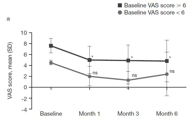
Figure 1.
Figure Painintensity
1. Pain intensityand
andfunctionality
functionalityby
byvisit:
visit:(a)
(a)VAS
VASscore
score (pain);
(pain); and
and (b)
(b) QuickDASH
QuickDASHscore
score
(functionality). * Statistically significant difference between baseline and follow-up assessments
(functionality). * Statistically significant difference between baseline and follow-up assessments at at
Months1,1,33 and
Months and 6;
6; Tukey
Tukey post-hoc analysis; pp144 up assessments (p < 0.05 for all) in both groups.
145 Analysis of the simple effects using the post-hoc Tukey test showed no significant differences in
146 QuickDASH scores between the groups at baseline, first, second, or third follow-up assessments
147 (p > 0.05 for all). The post-hoc Tukey test also showed that within the group with baseline VAS scores
148 ≥Toxins
6, the2019,
QuickDASH
11, 46 scores were significantly different between baseline and first follow-up, baseline
5 of 10
149 and second follow-up, and baseline and third follow-up assessments (p < 0.05 for all). However, no
150 significant differences between the baseline QuickDASH scores and the scores at follow-up
151 at the first, second
assessments or third
in the group follow-up
with baselineassessments
VAS scores 0.05; < 62b).
Figure (p > 0.05 for all;
152 Figure 2a).
153
154 Painintensity
Figure2.2.Pain
Figure intensityand
and functionality
functionality by by visit
visit and
and baseline
baselineVAS VASscore:
score:(a)
(a)VAS
VASscore
score(pain); and
(pain); (b)
and
155 QuickDASH score (functionality). * Statistically significant difference between baseline
(b) QuickDASH score (functionality). *Statistically significant difference between baseline and follow- and follow-up
156 assessments
up assessmentsat at
Months
Months 1, 31,and 6; Tukey
3, and 6; Tukeypost-hoc analysis;
post-hoc p 0.05).
We stratified
the third the population according to sex, and performed a subanalysis of VAS scores using
163 a 2-way Wemixed ANOVA inthe
also investigated which the between-subject
differences in QuickDASH factor was
scores sex (2 levels)
between and theA
the 2 groups. within-subject
further 2-way
164 factor
mixedwas pain level
ANOVA, (VAS
where thescores) at the assessment
between-subject visits
factor was the(baseline and 3 follow-up
group according assessments,
to the baseline ie,
VAS score
165 4(2levels). Main significant effects were found for sex and VAS score (p < 0.05), indicating
levels) and the within-subject factor was the QuickDASH score at all assessment visits (4 levels), significant
166 differences
showed main in VAS scoreseffects
significant acrossfortheboth
4 time points
groups in bothto
according groups. The
baseline VASpost-hoc
score andTukey test revealed
QuickDASH score
167 (p < 0.05), differences
significant between
indicating that bothbaseline and first,significant
groups showed second, and third follow-up
differences assessments
in QuickDASH (p 0.05 for all).effects
the simple However,
using significant
the post-hoc differences
Tukey testwere observed
showed between differences
no significant male and
in QuickDASH scores between the groups at baseline, first, second or third follow-up assessments
(p > 0.05 for all). The post-hoc Tukey test also showed that within the group with baseline VAS scores
≥ 6, the QuickDASH scores were significantly different between baseline and first follow-up, baseline
and second follow-up and baseline and third follow-up assessments (p < 0.05 for all). However, no
significant differences between the baseline QuickDASH scores and the scores at follow-up assessments
in the group with baseline VAS scores < 6 were shown (p > 0.05; Figure 2b).
2.4.2. Patients Grouped by Sex
We stratified the population according to sex and performed a sub analysis of VAS scores using a
2-way mixed ANOVA in which the between-subject factor was sex (2 levels) and the within-subject
factor was pain level (VAS scores) at the assessment visits (baseline and 3 follow-up assessments, i.e.,
4 levels). Main significant effects were found for sex and VAS score (p < 0.05), indicating significant
differences in VAS scores across the 4 time points in both groups. The post-hoc Tukey test revealed
significant differences between baseline and first, second and third follow-up assessments (p < 0.05 for
all) in both males and females.
The analysis of simple effects using a post-hoc Tukey test revealed no significant differences in VAS
scores between male and female patients at baseline and at the first and second follow-up assessments178 mixed ANOVA, where the between-subject factor was sex (2 levels) and the within-subject factor was
179 the QuickDASH score at assessment visits (4 levels), showed main significant effects for sex and
180 functionality of the upper limb (p < 0.05), indicating significant differences in both groups across the
181 4 time points. The Tukey post-hoc test revealed significant differences between baseline and first
182 follow-up, baseline and second follow-up, and baseline and third follow-up assessments (p < 0.05 for
Toxins 2019, 11, 46 6 of 10
183 all) in both males and females.
184 An analysis of the simple effects using the post-hoc Tukey test showed no significant differences
185 > 0.05 for all).
in(pQuickDASH scores between
However, male anddifferences
significant female patients
were at baselinebetween
observed and at the first,
male andsecond,
femaleorpatients
third
186 follow-up assessments
at the third follow-up(passessment (p 0.05 for all). post-hoc
0.05). Tukey test
The post-hoc showed
Tukey that within
test also showedthe male
that group
there were
187 the QuickDASH
significant scores for
differences were statistically
VAS significant
scores in the onlybetween
male group betweenbaseline
baselineandand third
first follow-up
follow-up, (p <
baseline
188 0.05).
and The post-hoc
second Tukeyand
follow-up testbaseline
also showed that within
and third follow-up (p < 0.05
the female group the QuickDASH
for all), scores were
while in the female group
189 significantly
VAS scores were different betweendifferent
significantly baselinebetween
and second follow-up
baseline and first(pfollow-up
< 0.05) and
andbaseline
baseline and third
and second
190 follow-up
follow-up(passessments only (pToxins 2019, 11, 46 7 of 10
with increased holding and loading carried out with that arm, leaving the more delicate handling to
the dominant arm. The mean progression time between the initial symptoms until patients sought
medical care was 20 months. All patients had received previous treatments. The most frequently
affected muscles were the extensor digitorum longus (87.5%), the extensor carpi radialis brevis (83.3%),
the extensor digiti minimi (41.7%) and the extensor carpi ulnaris (29.2%). Patients with >1 affected
muscle received treatment in each of those muscles. This is in contrast to most other published studies,
where patients only had a single muscle treated [10,11,14].
BoNT-A doses reported for treatment of lateral epicondylitis in other published studies are
variable and may be reported as dose per muscle or total dose [7,14–18]; however, doses appear similar
to those used in the present study. BoNT doses in the present study were administered in relation to
muscle size and each patient received BoNT only in symptomatic muscles. Treated muscles did not
display significant atrophy.
The average duration of effect of the BoNT was around 3 months and weakness in the third finger
was the main adverse effect, which is in agreement with previous literature [12]. Therefore, in patients
for whom this side effect would impact on their ability to work, consideration should be taken before
commencing treatment [14].
As a general outcome of the treatment, pain and upper limb function improved at the first
follow-up assessment (1 month post-treatment) and this effect was maintained beyond the paralyzing
effect of the toxin. Consistent with other studies [12], we observed a variation in functionality 3 months
after the injection that appeared to correspond to the maximum effect and the maximum weakness of
the extensor digitorum longus muscle. Weakness of this muscle may cause functional impairment to
the patient, despite pain relief.
When data were stratified according to patients’ baseline VAS pain score (≥6 orToxins 2019, 11, 46 8 of 10
pain refractory to conservative therapy, thus being potential candidates for surgery. As such, these
patients would be predicted to benefit most from BoNT treatment. Further studies are required to
investigate the effectiveness of BoNT in other populations of patients with epicondylitis.
5. Materials and Methods
5.1. Study Design
This was an experimental study of adult patients with epicondylitis, refractory to other treatments,
who attended the Musculoskeletal Unit Rehabilitation Department, Hospital Universitario Virgen del
Rocío between February 2014 and February 2015. The selected sample size was dependent on the
number of patients attending the clinic.
As BoNT is not approved for the treatment of lateral epicondylitis, the Pharmacy Commission
of the hospital was provided with the necessary Guideline for the Introduction of New Drugs in the
Formulary form to obtain approval for this off-label use of BoNT.
The study was approved by the Ethics Committee of the Hospital Universitario Virgen del Rocío
and was conducted according to the Declaration of Helsinki (1964), approval code: 2014PI/055, approval
date: 3 December 2014. Signed, informed consent was obtained from all patients.
5.2. Patients
Adult patients aged ≥ 18 years were eligible for inclusion in the study if they had lateral
epicondylitis resistant or refractory to other treatments (including analgesics such as anaesthetic
injections, anti-inflammatory drugs such as corticoids and physiotherapy), baseline pain intensity > 4
on a VAS ranging from 0 (best) to 10 (worst) [22]. and baseline functional impairment > 30 points on
the QuickDASH scale ranging from 0 (best) to 100 (worst) [23].
Major exclusion criteria were any previous adverse reaction or allergy to treatment with BoNT,
infectious disease with high fever and general discomfort, severe coagulation disorders, pregnancy
or breastfeeding, medical history of concomitant diseases (such as renal or hepatic diseases) or
neuromuscular diseases (such as myasthenia gravis or amyotrophic sclerosis) and previous treatment
with aminoglycosides or other drugs that may interfere with neuromuscular junctions.
5.3. Study Treatment
Ultrasound-guided injections of incobotulinumtoxinA (reconstituted in 1 mL of normal saline)
were administered at the predefined doses per muscle summarized in Table 2. Injections were given
using insulin syringes in 1 site per muscle, with the exception of the extensor digitorum longus, in
which the dose was split between 2 injection sites 4 cm apart. The target muscles for treatment were the
extensor carpi ulnaris, the extensor digiti minimi, the extensor digitorum longus and the extensor carpi
radialis brevis, selected on the basis of clinical symptoms, including the presence of pain during resisted
movements. Patients with more than 1 affected muscle received treatment in each of these muscles.
The anatomical localization of muscles was determined by palpation from the epicondyle
while the patient performed specific, requested movements, followed by ultrasound to ensure the
correct placement of BoNT. Injections were performed through the long axis of the ultrasound probe.
The affected muscles were preferentially injected in their motor end plates in order to control muscular
hyperactivity as well as nociceptive neurotransmission. To avoid procedural variability, all injections
were performed by the same experienced healthcare professional. Patients were assessed for treatment
efficacy at clinical appointments at 1 month, 3 months and 6 months post-treatment.
Patients were permitted to take paracetamol (1 g every 8 h) for 48 h after incobotulinumtoxinA
injection if they experienced pain or discomfort associated with the injection. Non-steroidal
anti-inflammatory drugs and corticosteroids were not allowed during the study period. Patients were
asked to avoid activities that required repetitive supination or strong compression and to interrupt any
physiotherapeutic treatment.Toxins 2019, 11, 46 9 of 10
5.4. Assessed Variables
The study captured information on the participating patients, including sociodemographic
data and data related to disease progression, physical examination, pain (VAS; range 0 [best] to 10
[worst] [22]) and functionality (QuickDASH functional scale for upper limb; range 0 [best] to 100
[worst] [23]). Assessments were performed at the baseline treatment visit and at the assessment visits
1, 3 and 6 months post-treatment. Data on incobotulinumtoxinA doses administered, muscles treated
and any reported adverse events were also collected.
5.5. Statistical Analysis
Data were analysed using descriptive statistics, employing absolute and relative frequencies for
qualitative variables. Quantitative variables were presented as mean ± SD or median, interquartile
range (P50 [P25–P75]), depending on whether or not the data were normally distributed (after applying
the Shapiro-Wilk test [n < 50]). After evaluating the normality of data distribution, an ANOVA for
repeated measurements, with estimations of the effect size and comparisons of the main effects, was
performed to compare measurements at the different time points. As a secondary analysis, we stratified
the population according to sex and baseline VAS score (≥6 orToxins 2019, 11, 46 10 of 10
7. Galvin, R.; Callaghan, C.; Chan, W.-S.; Dimitrov, B.D.; Fahey, T. Injection of botulinum toxin for treatment of
chronic lateral epicondylitis: Systematic review and meta-analysis. Semin. Arthritis Rheum. 2011, 40, 585–587.
[CrossRef]
8. Jabbari, B.; Machado, D. Treatment of refractory pain with botulinum toxins—An evidence-based review.
Pain Med. 2011, 12, 1594–1606. [CrossRef]
9. Krogh, T.P.; Bartels, E.M.; Ellingsen, T.; Stengaard-Pedersen, K.; Buchbinder, R.; Fredberg, U.; Bliddal, H.;
Christensen, R. Comparative effectiveness of injection therapies in lateral epicondylitis: A systematic
review and network meta-analysis of randomized controlled trials. Am. J. Sports Med. 2013, 41, 1435–1446.
[CrossRef]
10. Vicenzino, B.; Coombes, B.K. A single botulinum toxin injection at a precise anatomic point on the forearm
reduces pain at rest, compared to placebo injection in patients with chronic refractory lateral epicondylitis.
Évid.-Based Med. 2010, 15, 149–150. [CrossRef]
11. Placzek, R.; Drescher, W.; Deuretzbacher, G.; Hempfing, A.; Meiss, A.L. Treatment of chronic radial
epicondylitis with botulinum toxin a. A double-blind, placebo-controlled, randomized multicenter study.
J. Bone Joint Surg. Am. 2007, 89, 255–260. [CrossRef] [PubMed]
12. Wong, S.M.; Hui, A.C.; Tong, P.Y.; Poon, D.W.; Yu, E.; Wong, L.K. Treatment of lateral epicondylitis with
botulinum toxin: A randomized, double-blind, placebo-controlled trial. Ann. Intern. Med. 2005, 143, 793–797.
[CrossRef]
13. Climent, J.M.; Fenollosa, P.; Martín del Rosario, F.M. Rehabilitacion Intervencionista, 1st ed.; Ergon: Madrid,
Spain, 2012; pp. 1–592. ISBN 978-8415351-19-1.
14. Espandar, R.; Heidari, P.; Rasouli, M.R.; Saadat, S.; Farzan, M.; Rostami, M.; Yazdanian, S.; Mortazavi, S.M.
Use of anatomic measurement to guide injection of botulinum toxin for the management of chronic lateral
epicondylitis: A randomized controlled trial. Can. Med. Assoc. J. 2010, 182, 768–773. [CrossRef] [PubMed]
15. Villamayor-Blanco, B.; Misa-Agustiño, M.J.; Jorge-Mora, M.T.; López-Castro, A.; López-Pesado, I.;
Jorge-Barreiro, Y.F.J. Resultados terapéuticos a corto plazo de la infiltración con toxina botulínica ante
el fracaso del tratamiento convencional de 7 pacientes con epicondylitis. Rehabilitación 2008, 42, 122–126.
[CrossRef]
16. Santos, B.; Calderón, E.; Ordóñez, P.; García-Hernández, R.; Torres, L.M. Tratamiento de epicondilitis
refractaria con neurotoxina botulínica tipo a libre de complejo proteínico. Rev. Soc. Esp. Dolor 2013, 20, 8–10.
[CrossRef]
17. Kalichman, L.; Bannuru, R.R.; Severin, M.; Harvey, W. Injection of botulinum toxin for treatment of chronic
lateral epicondylitis: Systematic review and meta-analysis. Semin. Arthritis Rheum. 2011, 40, 532–538. [CrossRef]
[PubMed]
18. Oskarsson, E.; Piehl Aulin, K.; Gustafsson, B.E.; Pettersson, K. Improved intramuscular blood flow and
normalized metabolism in lateral epicondylitis after botulinum toxin treatment. Scand. J. Med. Sci. Sports
2009, 19, 323–328. [CrossRef]
19. Fernández-de-Las-Peñas, C.; Ortega-Santiago, R.; Ambite-Quesada, S.; Jiménez-García, R.; Arroyo-Morales, M.;
Cleland, J.A. Specific mechanical pain hypersensitivity over peripheral nerve trunks in women with either
unilateral epicondylalgia or carpal tunnel syndrome. J. Orthop. Sports Phys. Ther. 2010, 40, 751–760. [CrossRef]
20. Ahmad, Z.; Siddiqui, N.; Malik, S.S.; Abdus-Samee, M.; Tytherleigh-Strong, G.; Rushton, N. Lateral
epicondylitis: A review of pathology and management. Bone Joint J. 2013, 95-b, 1158–1164. [CrossRef]
21. Fillingim, R.B.; King, C.D.; Ribeiro-Dasilva, M.C.; Rahim-Williams, B.; Riley, J.L., 3rd. Sex, gender and pain:
A review of recent clinical and experimental findings. J. Pain 2009, 10, 447–485. [CrossRef]
22. Ho, K.; Spence, J.; Murphy, M.F. Review of pain-measurement tools. Ann. Emerg. Med. 1996, 27, 427–432.
[CrossRef]
23. Beaton, D.E.; Wright, J.G.; Katz, J.N.; Upper Extremity Collaborative Group. Development of the quickdash:
Comparison of three item-reduction approaches. J. Bone Joint Surg. Am. 2005, 87, 1038–1046. [CrossRef]
[PubMed]
© 2019 by the authors. Licensee MDPI, Basel, Switzerland. This article is an open access
article distributed under the terms and conditions of the Creative Commons Attribution
(CC BY) license (http://creativecommons.org/licenses/by/4.0/).You can also read