Laser Photocoagulation Combined with Subtenon Injection of Triamcinolone Acetonide for Diabetic Cystoid Macular Edema
←
→
Page content transcription
If your browser does not render page correctly, please read the page content below
Original Article                                                                                                   172

 Laser Photocoagulation Combined with Subtenon Injection of
 Triamcinolone Acetonide for Diabetic Cystoid Macular Edema
    Hsi-Kung Kuo, MD; Pei-Chang Wu, MD; Yi-Hao Chen, MD; Li-Sheng Cheng, MD

        Background: To compare the effects of laser photocoagulation combined with subtenon
                     injection of triamcinolone acetonide with photocoagulation alone, for diabet-
                     ic cystoid macular edema.
        Methods:     This retrospective comparative study analyzed data for 34 eyes with cystoid
                     macular edema treated with photocoagulation alone (n = 17) or combined
                     with triamcinolone (n = 17). All patients were followed for 6 months after
                     the procedure. Laser photocoagulation included focal photocoagulation
                     focused on microaneurysms and light grid photocoagulation spread over the
                     edematous retina. Subtenon injection of triamcinolone (20 mg/0.5 cc) was
                     performed in the superior-temporal conjunctiva.
        Results:     In the photocoagulation only group, the pretreatment mean visual acuity
                     (VA) measured by the logarithm of the minimum angle of resolution
                     (LogMAR) was 1.06 ± 0.49. The number of laser spots was 34 ± 10. The 6
                     -month post- treatment mean LogMAR was 1.13 ± 0.60. In the photocoagu-
                     lation combined with triamcinolone group, the pretreatment mean LogMAR
                     was 1.31 ± 0.49. The number of laser spots was 42 ± 14. The 6 -month
                     post- treatment mean LogMAR was 1.26 ± 0.49. In the photocoagulation
                     only group, 7 eyes had stable vision (improved, stable or loss of < 2 lines)
                     and 8 eyes had vision loss (loss ≥ 2 lines). In the photocoagulation combined
                     with triamcinolone group, 15 eyes had stable vision and 2 eyes had vision
                     loss. Fewer eyes had vision loss in the photocoagulation combined with tri-
                     amcinolone group. (chi-square test, p = 0.024).
        Conclusions: This study, with a follow-up of 6 months, suggests that subtenon injection of
                     triamcinolone combined with macular photocoagulation provides a better
                     chance of stabilizing vision loss in patients with diabetic cystoid macular
                     edema than photocoagulation alone.
                      (Chang Gung Med J 2009;32:172-81)

        Key words: diabetic macular edema, photocoagulation, subtenon injection, triamcinolone ace-
                   tonide, vascular endothelial growth factor (VEGF)

D    iabetic retinopathy is an important public health
     concern and is the leading cause of blindness in
                                                               our working population.(1-3) Diabetic macular edema
                                                               is a primary cause of visual loss in diabetic patients.

From the Department of Ophthalmology, Chang Gung Memorial Hospital-Kaohsiung Medical Center, Chang Gung University
College of Medicine, Kaohsiung, Taiwan.
Received: Dec. 28, 2007; Accepted: May 6, 2008
Correspondence to: Dr. Hsi-Kung Kuo, Department of Ophthalmology, Chang Gung Memorial Hospital. 123, Dapi Rd., Niaosong
Township, Kaohsiung County 833, Taiwan (R.O.C.) Tel.: 886-7-7317123 ext. 2801; Fax: 886-7-7352775; E-mail:
d2767@cgmh.org.tw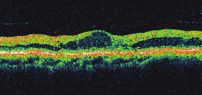
173    Hsi-Kung Kuo, et al
       Laser and TA for diabetic CME

The Early Treatment Diabetic Retinopathy Study              period shorter than 6 months. A total of 113 eyes
(ETDRS) defined macular edema as thickening of              from 76 patients met these criteria. Cystoid macular
the retina and/or hard exudates within 1 disc diame-        edema was defined as an edematous area involving
ter of the center of the macula. In the ETDRS proto-        the fovea, larger than 2/3 of a circle, with a typical
col, microaneurysms were treated with focal photo-          flower-petal picture on fluorescein angiography.
coagulation and areas of nonperfusion or diffuse            Among the 113 eyes, 34 eyes had cystoid macular
leakage associated with macular edema were treated          edema and were included in the analysis.
with grid photocoagulation. Data from the ETDRS
indicate that macular photocoagulation for clinically       Baseline examination
significant macular edema (CSME) effectively                     Initial clinical examination included visual acu-
reduces the risk of moderate visual loss by 50%             ity (VA) with a Landort C chart, intraocular pressure
compared to that in eyes with deferral of treatment.(4-     (IOP) measurement, anterior segment examination,
6)
                                                            and indirect ophthalmoscopy examination with a +
      Although the ETDRS was successful in reduc-           90D conoidal lens (Ocular Instruments, Bellevue,
ing the rates of visual loss due to diabetic macular        WA, U.S.A.). Patients were then assessed with fun-
edema, many patients treated with macular photoco-          dus color photography and fluorescein angiography
agulation do not recover lost vision, especially those      (FA). Optical coherence tomography (OCT) was per-
with diffuse macular edema. In addition, laser photo-       formed in patients treated after 2004. The grading of
coagulation of the macula produces irreversible dam-        the cataract was based on the Lens Opacities
age in the macula.(7) Recently, use of less intense         Classification System III.(17) The IOP was checked
laser burns has been increasingly adopted. New laser        using a pneumotonometer. Patients were counseled
machines and modified photocoagulation techniques           about the prognosis of their condition and treatment
have also been developed to lessen the side effects.(8,9)   with or without subtenon injection of triamcinolone.
Adjunctive and new strategies have been suggested           Patients who gave informed consent for treatment
to improve the treatment results, including intravitre-     were included in the study. Procedures were per-
al triamcinolone for refractory diabetic macular            formed to conform with the tenets of the Declaration
edema.(10-13)                                               of Helsinki.
      Triamcinolone acetonide has been effectively
used in ocular therapeutics for over 50 years. Its use      Treatment and follow-up protocol
for periocular and intraocular treatment of uveitis              The techniques of laser photocoagulation
and retinal vascular disease has increased dramatical-      included focal photocoagulation focused on microa-
ly in recent years.(14-16) Combined subtenon injection      neurysms and light grid photocoagulation spread
of triamcinolone acetonide and macular photocoagu-          over the edematous retina. The procedure was per-
lation might be more effective in reducing edema,           formed with an Ocular Mainster Focal/Grid lens
especially in severe cases. This retrospective study        (Ocular Instruments, Inc, Bellevue, WA, U.S.A.).
compared combined laser photocoagulation with               The laser spot was 200 µm and the laser power was
subtenon injection of triamcinolone acetonide with          adjusted to get the desired effect. Subtenon injection
photocoagulation alone for the treatment of cystoid         of triamcinolone (20 mg/0.5 cc) was performed in
diabetic macular edema.                                     the superior-temporal conjunctiva. Routine post-
                                                            treatment topical medications did not include
                     METHODS                                steroids. The injection was given immediately after
                                                            laser photocoagulation. All patients were given only
     We retrospectively reviewed the records of             one laser photocoagulation treatment with or without
patients who underwent macular photocoagulation             subtenon triamcinolone. Follow-up examinations
for diabetic maculopathy under the care of author           were done at 1, 3 and 6 months postoperatively. The
Kuo at Chang Gung Memorial Hospital from 2000 to            6 month follow-up period was selected because pre-
2005. Patients were excluded if they had undergone          vious studies showed the effects of triamcinolone
panretinal photocoagulation therapy within 3 months         lasted less than 6 months.(12) At each visit, VA, retinal
of macular photocoagulation or had a follow -up             examination and fundus color photography were per-

Chang Gung Med J Vol. 32 No. 2
March-April 2009
Hsi-Kung Kuo, et al   174
                                                                              Laser and TA for diabetic CME

formed. IOP and fluorescein angiography were             nificantly different between groups.
optional examinations.                                        In the photocoagulation only group, the 6 month
                                                         post-treatment LogMAR ranged from 0.1 to 2.0
Statistical analysis                                     (average 1.13 ± 0.60). The change after treatment (6
     The Landort C chart VA was converted to             months post-treatment minus pre- treatment) ranged
LogMAR VA for statistical analysis. A Landort C          from –1.0 to +0.8 (average 0.09 ± 0.56). In the pho-
chart VA of 0.01 was equal to a LogMAR VA of 2.0,        tocoagulation combined with triamcinolone group,
and VA assessed by counting fingers within 1 meter       the post- treatment LogMAR ranged from 0.7 to 2.3
was set as a LogMAR VA of 2.3. For continuous            (average 1.26 ± 0.49). The change after treatment
variables such as VA and number of photocoagula-         ranged from –0.3 to +0.6 (average –0.04 ± 0.23).
tion spots, the Mann-Whitney U test was used to          The change in VA after treatment was not statistical-
determine the statistical significance of between-       ly different between groups (Mann-Whitney test, p =
group differences. The chi-square test was used to       0.245). In the photocoagulation only group, 7 eyes
compare the proportion of eyes with vision loss          had stable vision (VA improved, stable or loss < 2
between groups.                                          lines) and 8 eyes had vision loss (VA loss ≥ 2 lines).
                                                         In the photocoagulation combined with triamci-
                    RESULTS                              nolone group, 15 eyes had stable vision and 2 eyes
                                                         had vision loss (Fig. 1). There were fewer eyes with
Baseline characteristics                                 vision loss in the photocoagulation combined with
     Thirty-four eyes from 26 patients who complet-      triamcinolone group (chi-square test, p = 0.024)
ed the 6 month follow-up were included in this           (Table 2).
analysis (Table 1). The photocoagulation only group
included 17 eyes (10 right eyes, 7 left eyes) from 13    Representative cases
patients (7 men, 6 women). The ages of these                  Case 1: A 54 year-old man had diabetes and reg-
patients ranged from 43-73 years (average, 56 ± 9        ular hemodialysis. He had partial panretinal photoco-
years). Non-proliferative diabetic retinopathy was       agulation for both eyes 1 year previously. He had
diagnosed in 15 eyes and 2 eyes were in post-pan-        diabetic cystoid macular edema in his left eye (Eye
retinal photocoagulation status. There were 17 eyes      No 34, Table 1) (Fig. 2). His VA was 4/200 and the
(8 right eyes, 9 left eyes) from 14 patients (7 men, 7   central foveal thickness measured by OCT was 523
women) in the photocoagulation combined with tri-        µm. He had macular photocoagulation (30 spots)
amcinolone group. The patients’ ages ranged from         only. The macula edema improved and central foveal
45-72 years (average, 59 ± 10 years). Non-prolifera-     thickness decreased to 371 µm 3 months after the
tive diabetic retinopathy was diagnosed in 15 eyes       treatment. The VA remained 4/200.
and proliferative diabetic retinopathy in 2 eyes.             Case 2: A 45 year-old man had non-proliferative
                                                         diabetic retinopathy and maculopathy (OU). FA
Treatment and outcome                                    showed the right eye (Eye No 15, Table 1) had
     In the photocoagulation only group, the pre-        severe blood-retinal barrier breakdown and a flower-
treatment LogMAR ranged from 0.2 to 1.7 (average         petal picture at the macula. OCT also showed cystoid
1.06 ± 0.49). The number of laser spots ranged from      macular edema (Fig. 3). He had macular grid photo-
18 to 50 (average 34 ± 10). In the photocoagulation      coagulation (28 spots) and subtenon injection of tri-
combined with triamcinolone group, the pre- treat-       amcinolone acetonide. His pre-treatment VA was
ment LogMAR ranged from 0.7 to 2.3 (average 1.31         1/20 and the central foveal thickness measured by
± 0.49). The number of laser spots ranged from 20        OCT was 618 µm. Six weeks later, the VA improved
to 76 (average 42 ± 14). Although the average pre-       to 4/20 and central foveal thickness decreased to 198
treatment VA was worse and there were more laser         µm. Three months after treatment, the macula edema
spots in the photocoagulation combined with triamci-     was almost gone and he had panretinal photocoagu-
nolone group, the pre-treatment VA (Mann-Whitney         lation for severe non-proliferative diabetic retinopa-
test, p = 0.160) and number of photocoagulation          thy.
spots (Mann-Whitney test, p = 0.099) were not sig-

                                                                                    Chang Gung Med J Vol. 32 No. 2
                                                                                                March-April 2009
175    Hsi-Kung Kuo, et al
       Laser and TA for diabetic CME

Table 1. Patient Data
 Eye     Patient     Age          Sex         Lesion          DR         TA        Pre-VA        Pre-VA        6M-VA         6M-VA
 no        no       (years)      (M/F)       (OD/OS)         status    (yes/no)   (C chart)    (LogMAR)       (C chart)    (LogMAR)
   1        1           60          F          OD           NPDR         Yes         0.05          1.3          0.04           1.4
   2        2           62          F          OS           NPDR         Yes         0.05          1.3          0.07           1.2
   3        3           52          F          OD           NPDR         Yes         0.2           0.7           0.2           0.7
   4        3                                  OS           NPDR         Yes         0.1           1.0          0.2            0.7
   5        4           62          F          OD           NPDR         Yes         0.03          1.5          0.04           1.4
   6        5           60         M           OD           NPDR         Yes         0.1           1.0          0.08           1.1
   7        6           60         M           OD           NPDR         Yes         0.04          1.4          0.05           1.3
   8        7           58         M            OS          NPDR         Yes         0.01          2.0          0.02           1.7
   9        8           72         M            OS          NPDR         Yes          CF           2.3          0.01           2.0
  10        9           52         M            OS          NPDR         Yes         0.02          1.7           CF            2.3
  11       10           58          F          OD            PDR         Yes         0.04          1.4          0.06           1.2
  12       10                                   OS           PDR         Yes         0.01          2.0          0.02           1.7
  13       11           65          F           OS          NPDR         Yes         0.08          1.1           0.1           1.0
  14       12           58         M           OD           NPDR         Yes         0.2           0.7           0.2           0.7
  15       13           45         M           OD           NPDR         Yes         0.05          1.3           0.2           0.7
  16       13                                   OS          NPDR         Yes         0.2           0.7           0.2           0.7
  17       14           67          F           OS          NPDR         Yes         0.04          1.4          0.02           1.7
  18       15           56          F          OD          S/P PRP        No         0.03          1.5          0.01           2.0
  19       16           47         M           OD           NPDR          No         0.07          1.2          0.02           1.7
  20       17           44         F           OD           NPDR          No          0.2          0.7           0.2           0.7
  21       18           51         M           OD           NPDR          No         0.3           0.5           0.3           0.5
  22       18                                  OS           NPDR          No         0.1           1.0           0.2           0.7
  23        2           62          F          OD           NPDR          No         0.1           1.0          0.02           1.7
  24       19           56         M           OS           NPDR          No         0.7           0.2           0.9           0.1
  25       20           56         M           OD           NPDR          No         0.2           0.7          0.02           1.7
  26       20                                   OS          NPDR          No         0.3           0.5          0.06           1.3
  27       21           71          F           OS          NPDR          No         0.02          1.7          0.01           2.0
  28       22           63         F           OD          S/P PRP        No         0.02          1.7          0.05           1.3
  29       23           43         M           OS           NPDR          No         0.3           0.5           0.4           0.4
  30       24           73         M           OD           NPDR          No         0.2           0.7           0.2           0.7
  31       25           57          F          OD           NPDR          No         0.08          1.1          0.03           1.5
  32       25                                  OS           NPDR          No         0.06          1.2          0.03           1.5
  33       26           54         M           OD           NPDR          No         0.02          1.7           0.2           0.7
  34       26                                  OS           NPDR          No         0.02          1.7           0.2           0.7
Abbreviations: DR: diabetic retinopathy; NPDR: nonproliferative diabetic retinopathy; PDR: proliferative diabetic retinopathy; TA: sub-
tenon injection of triamcinolne acetonide; Pre-VA: pre-treatment visual acuity; 6M-VA: 6 month post-treatment visual acuity; LogMAR:
logarithm of the minimum angle of resolution.

Complications                                                          cataract progression during the follow-up period.
     The most common reported complications of                         There were 12 phakic eyes and 5 pseudophakic eyes
subtenon injection of triamcinolone include cataract                   in the combined group. No cataract surgery was
and elevated IOP. There were 16 phakic eyes in the                     required for these 12 phakic eyes during the follow-
photocoagulation only group and no apparent                            up period, although 1 eye had cataract (posterior sub-

Chang Gung Med J Vol. 32 No. 2
March-April 2009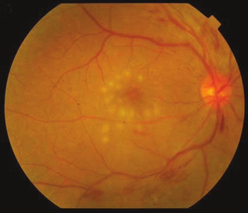
Hsi-Kung Kuo, et al   176
                                                                                                                                              Laser and TA for diabetic CME

                          1.0 Landolt C
                                          LogMAR

                                                                                                                         A
                                          0.0
                                                         subtenon triamcinolone
                                                         control
                          0.32

                                          0.5
6 months post-treatment
                          0.1

                                          1.0
                          0.03

                                          1.5
                          0.01

                                          2.0

                                          2.5
                                                   2.5    2.0           1.5          1.0      0.5       0.0 LogMAR
                                                          0.01         0.03          0.1      0.32      1.0 Landolt C
                                                                              pre-treatment

      Fig. 1 Initial and final vision after macular photocoagulation
      with or without subtenon injection of triamcinolone for dif-                                                       B
      fuse diabetic macular edema. The area beneath the dashed
      line indicates vision loss ≥ 2 lines. The area above and on the
      dashed line indicates stable vision (improved, no change or
      loss of 1 line).

        Table 2. Statistical Summary
                                                          Photo only          Photo + TA
        Pre- treatment                                   1.06 ± 0.49      1.31 ± 0.49         Mann-Whitney test,
        LogMAR                                                                                    p = 0.160              C
        Post-treatment                                   1.13 ± 0.60      1.26 ± 0.49
        LogMAR
        Change in VA                                     0.09 ± 0.56      –0.04 ± 0.23        Mann-Whitney test,
                                                                                                  p = 0.245
        Stable vision                                      9 (53%)             15 (88%)         chi-square test,
                                                                                                   p = 0.024
        Vision loss                                        8 (47%)             2 (12%)
                                                                                                                        Fig. 2 The left eye (Eye No 34, Table 1) of a 54 year-old
        Abbreviations: Photo only: photocoagulation only group; Photo +                                                 man with diabetic macular edema. (A) Pre-treatment fundus
        TA: photocoagulation combined with triamcinolone group; VA: visual                                              color photography. (B) Pre-treatment OCT. The OCT shows
        acuity; Stable VA: VA improved, stable or loss < 2 lines; Vision loss: VA                                       cystoid macular edema. The central foveal thickness is 523
        loss ≥ 2 lines.                                                                                                 µm. (C) Three months after macular photocoagulation, the
                                                                                                                        edema has improved and central foveal thickness has
                                                                                                                        decreased to 371 µm. The VA has remained 4/200.
      capsular opacity) progression. Complete pretreat-
      ment and posttreatment IOP data were available for
      only 6 eyes in the photocoagulation combined with                                                                 visual impairment in diabetic patients. Diffuse dia-
      triamcinolone group, and all values were under 21                                                                 betic macular edema is a more complex therapeutic
      mm Hg.                                                                                                            problem than focal macular edema. The pathogenesis
                                                                                                                        of diffuse diabetic macular edema is not fully under-
                                                              DISCUSSION                                                stood. Proposed mechanisms include leakage from a
                                                                                                                        generally dilated capillary bed, tractional force at the
                                          Diabetic macular edema is the major cause of                                  vitreomacular interface and dysfunction of the retinal

                                                                                                                                                    Chang Gung Med J Vol. 32 No. 2
                                                                                                                                                                March-April 2009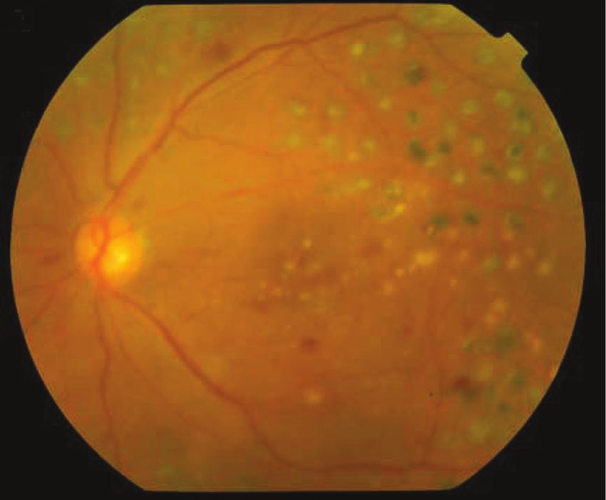
177 Hsi-Kung Kuo, et al
Laser and TA for diabetic CME
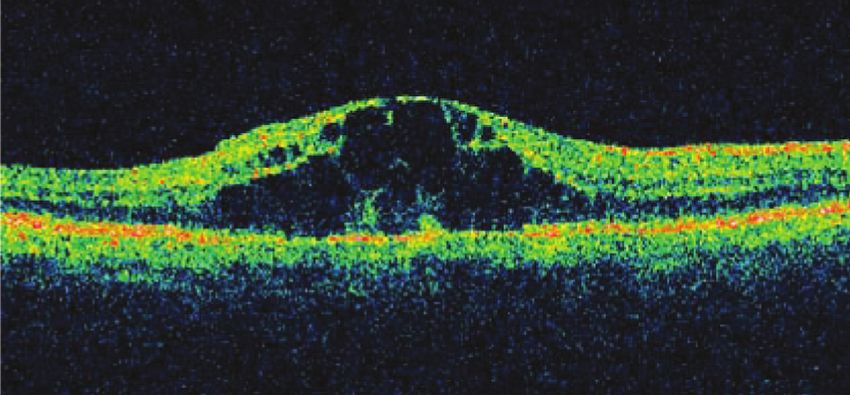
A B
C
D
E
Fig. 3 The right eye (Eye No 15, Table 1) of a 45 year-old man with diabetic maculopathy. (A,B,C) Pre-treatment fundus color
photography, FA, and OCT. The FA shows a flower-petal picture and OCT shows cystoid macular edema. Visual acuity is 1/20. The
central foveal thickness is 618 µm. Six weeks after macular grid photocoagulation (D) and subtenon injection of triamcinolone ace-
tonide, OCT shows the cystoid macular edema has dramatically improved (E). The central foveal thickness has decreased to 198
µm and the VA has improved to 4/20.
pigment epithelium barrier and transport functions.(18) retinopathy, in addition to stimulating new vessel
Previous studies in patients with diabetic macular growth during the proliferative stage of the disease,
edema have reported favorable anatomical and func- VEGF acts as a permeability factor. Anti-VEGF
tional results after vitrectomy and removal of the drugs, including pegaptanib, ranibizumab and beva-
posterior hyaloid (with or without the internal limit- cizumab, have been suggested as promising agents
ing membrane) and tractional force associated with a for the treatment of diabetic retinopathy.(13,22,23)
thickened premacular hyaloid. (11,19,20) Vascular Data from the ETDRS indicate that macular
endothelial growth factor (VEGF) is a potent photocoagulation for CSME effectively reduces the
endothelial-selective angiogenic factor. In diabetic risk of moderate visual loss. Photocoagulation might
Chang Gung Med J Vol. 32 No. 2
March-April 2009
Hsi-Kung Kuo, et al   178
                                                                                     Laser and TA for diabetic CME

resolve macular edema via the opening of new path-            ther suggested the effectiveness of pretreatment of
ways in the retinal pigment epithelium barrier for            the posterior subtenon with triamcinolone. (27) The
fluid transportation between the retina and chorio-           present study was limited by its retrospective design
capillaries.(18,24) However, photocoagulation might           but did include all patients treated since 2000. The
also destroy the photoreceptors, cause blood retinal          results indicate that combined laser photocoagulation
barrier (BRB) breakdown and induce focal inflam-              and triamcinolone is more effective than photocoag-
mation. These processes might lead to the release of          ulation alone in preventing visual loss from cystoid
inflammatory cytokines, such as interleukin-6 and             diabetic macular edema. There was a lack of com-
interleukin-8, which lead to macular edema. (25)              plete IOP data, a short follow-up period and a small
Among the known steroid effects that could amelio-            case number in this study. Further prospective study
rate BRB breakdown are a reduction of                         with a longer follow up is needed to establish the
prostaglandin synthesis, inhibition of cellular prolif-       findings of this study.
eration, blockage of macrophage recruitment and
infiltration of polymorphonuclear leukocytes.(26,27)          Conclusion
Corticosteroids also inhibit the mitogen-dependent                 Our data, with a follow-up of 6 months, suggest
transcription of VEGF.(28)                                    that subtenon triamcinolone combined with macular
      Subtenon injection of corticosteroids might sup-        photocoagulation better stabilizes the vision loss of
press the photocoagulation-induced inflammation,              patients with diabetic cystoid macular edema than
inhibit VEGF and lead to a better outcome.                    photocoagulation alone.
      Triamcinolone is an intermediate-acting corti-
costeroid with marked anti-inflammatory action. It
                                                                                REFERENCES
has been used clinically for macular edema sec-
ondary to retinal vascular occlusion and uveitis.(14-16,29-
                                                              1. Diabetes Control and Complications Trial Research
32)
    The effect of intravitreal triamcinolone for diabetic        Group. The effect of intensive diabetes treatment on the
macular edema unresponsive to laser photocoagula-                progression of diabetic retinopathy in insulin-dependent
tion was investigated in several studies. Generally, it          diabetes mellitus. Arch Ophthalmol 1995;113:36-51.
reduced macular thickening and improved vision for            2. United Kingdom Prospective Diabetes Study Group.
3 months; however, a recurrence of symptoms was                  Intensive blood-glucose control with sulphonylureas or
noted after 6 months.(12,33-36) Subtenon injection of tri-       insulin compared with conventional treatment and risk of
amcinolone for diabetic macular edema refractive to              complications in patients with type 2 diabetes. Lancet
                                                                 1998;352:837-53.
macular photocoagulation was also reported to                 3. Kuo HK, Hsieh HH, Liu RT. Screening for diabetic
improve vision, although the effect was less than                retinopathy by one-field, non-mydriatic, 45° digital pho-
with intravitreal injection in most reports.(37-40) The          tography is inadequate. Ophthalmologica 2005;219:192-
intravitreal concentration of triamcinolone is higher            296.
with the intravitreal route than the subtenon route.          4. Early Treatment Diabetic Retinopathy Research Group.
However, triamcinolone might enter the chorioretina              Photocoagulation for diabetic macular edema, Report
through the sclera via the subtenon route.(41) The sub-          number 1. Arch Ophthalmol 1985;103:1796-806.
                                                              5. Early Treatment Diabetic Retinopathy Research Group.
tenon route also has the advantages of avoiding com-
                                                                 Treatment techniques and clinical guidelines for photoco-
plications associated with the procedure for intravit-           agulation of diabetic maculae edema, Report number 2.
real injection.                                                  Ophthalmology 1987;94:761-74.
      The present study suggests that combined laser          6. Early Treatment Diabetic Retinopathy Research Group.
photocoagulation and triamcinolone might be a bet-               Focal photocoagulation treatment of diabetic macular
ter option than photocoagulation alone for patients              edema, Report number 19. Arch Ophthalmol 1995;113:
with cystoid diabetic macular edema. Verma et al.                1144-55.
                                                              7. Morgan CM, Schatz H. Atrophic creep of the retinal pig-
used a prospective controlled trial to evaluate the
                                                                 ment epithelium after focal macular photocoagulation.
adjunctive role of posterior subtenon triamcinolone              Ophthalmology 1989;96:96-103.
in grid photocoagulation in 2004.(29) They concluded          8. Luttrull JK, Musch DC, Mainster MA. Subthreshold
that posterior subtenon triamcinolone is a useful and            diode micropulse photocoagulation for the treatment of
safe adjunct. Recently, a study by Shimura et al. fur-           clinically significant diabetic maculae oedema. Br J

                                                                                           Chang Gung Med J Vol. 32 No. 2
                                                                                                       March-April 2009
179 Hsi-Kung Kuo, et al
Laser and TA for diabetic CME
Ophthalmol 2005;89:74-80. Strauss R, Gandorfer A, Ulbig M, Kampik A. Intravitreal
9. Bandello F, Polito A, Del Borrello M, Zemella N, Isola bevacizumab therapy for persistent diffuse macular
M. “Light” versus “classic” laser treatment for clinically edema. Retina 2006;26:999-1005.
significant diabetic macular oedema. Br J Ophthalmol 24. Wallow IH. Repair of the pigment epithelial barrier fol-
2005;89:864-70. lowing photocoagulation. Arch Ophthalmol 1984;102:
10. Bhavsar A. Diabetic retinopathy: the latest in current 126-35.
management. Retina 2006;26:S71-9. 25. Er H, Doganay S, Turkoz Y, Cekmen M, Daglioglu MC,
11. Pendergast SD, Hassan TS, Williams GA, Cox MS, Gunduz A, Evereklioglu C, Isci N. The levels of
Margherio RR, Ferrone PJ, Garretson BR, Trese MT. cytokines and nitric oxide in rabbit vitreous humor after
Vitrectomy for diffuse diabetic macular edema associated retinal laser photocoagulation. Ophthalmic Surg Lasers
with a taut premacular posterior hyaloid. Am J 2000;31:479-83.
Ophthalmol 2000;130:178-86. 26. Wilson CA, Berkowitz BA, Sato Y, Ando N, Handa JT, de
12. Martidis A, Duker JS, Greenberg PB, Rogers AH, Juan E. Treatment with intravitreal steroid reduces blood-
Puliafito CA, Reichel E, Baumal C. Intravitreal triamci- retinal barrier breakdown due to retinal photocoagulation.
nolone for refractory diabetic macular edema. Arch Ophthalmol 1992;110:1155-9.
Ophthalmology 2002;109:920-7. 27. Shimura M, Nakazawa T, Yasuda K, Shiono T, Nishida K.
13. Macugen Diabetic Retinopathy Study Group. A phase II Pretreatment of posterior subtenon injection of triamci-
randomized double-masked trial of pegaptanib, an anti- nolone acetonide has beneficial effects for grid pattern
vascular endothelial growth factor aptamer, for diabetic photocoagulation against diffuse diabetic macular oede-
macular edema. Ophthalmology 2005;112:1747-57. ma. Br J Ophthalmol 2007;91:449-54.
14. Jermak CM, Dellacroce JT, Heffez J, Peyman GA. 28. Nauck M, Karakiulakis G, Andre PP, Papakonstantinou E,
Triamcinolone acetonide in ocular therapeutics. Surv Poth M. Corticosteroids inhibit expression of the vascular
Ophthalmol 2007;52:503-22. endothelial growth factor in human vascular smooth mus-
15. Rao NA, Forster DJ. Nonspecific therapy of uveitis. In: cle cells. Eur J Pharmacol 1998;34:309-15.
Podos SM, Yanoff M, eds. Texbook of Ophthalmology, 29. Verma LK, Vivek MB, Kumar A, Tewari HK, Venkatesh
Vol. 2 The uvea. New York: Gower Medical Publishing, P. A prospective controlled trial to evaluate the adjunctive
1992:3.1-3.5. role of posterior subtenon triamcinolone in the treatment
16. Kuo HK, Lai IC, Fang PC, Teng MC. Ocular complica- of diffuse diabetic maculae edema. J Ocul Pharmacol
tions after a sub-Tenon injection of triamcinolone ace- Ther 2004;20:277-84.
tonide for uveitis. Chang Gung Med J 2005;28:85-9. 30. Greenberg PB, Martidis A, Rogers AH, Duker JS, Reichel
17. Chylack LT Jr, Wolfe JK, Singer DM, Leske MC, E. Intravitreal triamcinolone acetonide for macular oede-
Bullimore MA, Bailey IL, Friend J, McCarthy D, Wu SY. ma due to central retinal vein occlusion. Br J Ophthalmol
The Lens Opacities Classification System III. The 2002;86:247-8.
Longitudinal Study of Cataract Study Group. Arch 31. Ip MS, Kumar KS. Intravitreal triamcinolone acetonide as
Ophthalmol 1993;111:831-6. treatment for macular edema from central retinal vein
18. Bresnick GH. Diabetic maculopathy. A critical review occlusion. Arch Ophthalmol 2002;120:1217-9.
highlighting diffuse macular edema. Ophthalmology 32. Chen SD, Lochhead J, Patel CK, Frith P. Intravitreal tri-
1983;90:1301-17. amcinolone acetonide for ischaemic macular oedema
19. Gandorfer A, Messmer EM, Ulbig MW, Kampik A. caused by branch retinal vein occlusion. Br J Ophthalmol
Resolution of diabetic macular edema after surgical 2004;88:154-5.
removal of the posterior hyaloid and the inner limiting 33. Massin P, Audren F, Haouchine B, Erginary A, Bergmann
membrane. Retina 2000;20:126-33. JF, Benosman R, Caulin C, Gaudric A. Intravitreal triam-
20. Kimura T, Kiryu J, Nishiwaki H, Oh H, Suzuma K, cinolone acetonide for diabetic diffuse macular edema:
Watanabe D, Kurimoto M, Takagi H. Efficacy of surgical preliminary results of a prospective controlled trial.
removal of the internal limiting membrane in diabetic cys- Ophthalmology 2004;111:218-24.
toid macular edema. Retina 2005;25:454-61. 34. Sutter FP, Simpson JM, Gillies MC. Intravitreal triamci-
21. Shah SP, Patel M, Thomas D, Aldington S, Laidlaw DAH. nolone for diabetic diffuse macular edema that persists
Factors predicting outcome of vitrectomy for diabetic after laser treatment. Ophthalmology 2004;111:2044-9.
maculae oedema: results of a prospective study. Br J 35. Patflli F, Fasolino G, Radice P, Russo S, Zumbo G, Di
Ophthalmol 2006;90:33-6. Tizio FM, Frisone G, Marchi S. Time course of changes
22. Chun DW, Heier JS, Topping TM, Duker JS, Bankert JM. in retinal thickness and visual acuity after intravitreal tri-
A pilot study of multiple intravitreal injections of amcinolone acetonide for diffuse diabetic macular edema
ranibizumab in patients with center-involving clinically with and without previous macular laser treatment. Retina
significant diabetic macular edema. Ophthalmology 2005;25:840-5.
2006;113:1706-12. 36. Er H, Yilmaz H. Intravitreal cortisone injection for refrac-
23. Haritoglou C, Kook D, Neubauer A, Wolf A, Priglinger S, tory diffuse diabetic macular edema. Ophthalmologica
Chang Gung Med J Vol. 32 No. 2
March-April 2009Hsi-Kung Kuo, et al 180
Laser and TA for diabetic CME
2005;219:394-400. tion of triamcinolone acetonide for diffuse diabetic macu-
37. Ohguro N, Okada AA, Tano Y. Trans-Tenon’s retrobulbar lar edema. Ophthalmol 2005;112:1557-63.
triamcinolone infusion for diffuse diabetic macular 40. Bonini-Filbo MA, Jorge R, Barbosa JC, Calucci D,
edema. Graefes Arch Clin Exp Ophthalmol 2004;242: Cardillo JA, Costa RA. Intravitreal injection versus sub-
444-5. Tenon infusion of triamcinolone acetonide for refractory
38. Bakri S, Kaiser P. Posterior subtenon triamcinolone ace- diabetic macular edema: a randomized clinical trial.
tonide for refractory diabetic macular edema. Am J Invest Ophthalmol Vis Sci 2005;46:3845-9.
Ophthalmol 2005;139:290-4. 41. Inoue M, Takeda K, Morita K, Yamada M, Tanigawara Y,
39. Cardillo JA, Melo LS, Costa RA, Skaf M, Belfort R, Oguchi Y. Vitreous concentrations of triamcinolone ace-
Souza-Filho AA, Farah ME, Kupperman BD. Comparison tonide in human eyes after intravitreal or subtenon injec-
of intravitreal versus posterior sub-Tenon capsule injec- tion. Am J Ophthalmol 2004;138:1046-8.
Chang Gung Med J Vol. 32 No. 2
March-April 2009181
雷射光凝固術合併結膜下注射 Triamcinolone acetonide
對糖尿病囊狀黃斑水腫的治療
郭錫恭 吳佩昌 陳怡粧 鄭力升
背 景翩 比較雷射光凝固術合併結膜下注射 Triamcinolone acetonide 和單獨使用雷射光凝固術
對糖尿病囊狀黃斑水腫的療效。
方 法翩 這個回溯性比較研究分析 34 眼糖尿病囊狀黃斑水腫的治療病例紃其中 17 眼只接受
雷射光凝固術紃17 眼則合併使用 Triamcinolone。所有病人術後追蹤達 6 個月。雷射
光凝固術包括對微小動脈瘤予以局部性光凝固及對水腫視網膜予以輕度柵狀光凝
固。結膜下注射 Triamcinolone (20 mg/0.5 cc) 則位於上方耳側結膜。
結 果翩 在只接受雷射光凝固術組紃術前 LogMAR 視力是 1.06 ± 0.49 紃雷射點是 34 ± 10
發紃術後 6 個月 LogMAR 視力是 1.13 ± 0.60。在合併治療組紃術前 LogMAR 視力是
1.31 ± 0.49紃雷射點是 42 ± 14 發紃術後 6 個月 LogMAR 視力是 1.26 ± 0.49。在只
接受雷射光凝固術組紃7 眼有穩定視力 (進步紃不變或減退少於 2 行)紃8 眼有視力減
退 (減退 > 2 行)。在合併治療組紃15 眼有穩定視力紃2 眼有視力減退。合併治療組在
統計學上有意義的紃較少眼有視力減退 (p = 0.024)。
結 論翩 這個追蹤期 6 個月的研究顯示紃雷射光凝固術合併結膜下注射 Triamcinolone ace-
tonide 比只接受雷射光凝術對於糖尿病囊狀黃斑水腫的病患紃有較好機會可以穩定視
力減退。
(長庚醫誌 2009;32:172-81)
關鍵詞翩 糖尿病黃斑水腫、光凝固術、結膜下注射、triamcinolone acetonide、血管內皮生長因
子 (VEGF)
長庚紀念醫院 高雄院區 眼科系耿長庚大學 醫學院
受文日期芨民國96年12月28日耿接受刊載芨民國97年5月6日
通訊作者芨郭錫恭醫師芚長庚紀念醫院 眼科系。高雄縣833鳥松鄉大埤路123號。Tel.: (07) 7317123轉2801;
Fax: (07) 7352775; E-mail: d2767@cgmh.org.twYou can also read