COVID-19 and EBV Co-Infection in a Child - Scientific ...
←
→
Page content transcription
If your browser does not render page correctly, please read the page content below
Journal of Biosciences and Medicines, 2021, 9, 20-27
                                                                                                     https://www.scirp.org/journal/jbm
                                                                                                               ISSN Online: 2327-509X
                                                                                                                 ISSN Print: 2327-5081

COVID-19 and EBV Co-Infection in a Child

Elda Skenderi, Admir Sulovari, Gjeorgjina Kuli-Lito, Nilsa Shahini, Griselda Toci, Ada Pema

University Hospital Center “Mother Teresa”, Tirana, Albania

How to cite this paper: Skenderi, E., Sulo-         Abstract
vari, A., Kuli-Lito, G., Shahini, N., Toci, G.
and Pema, A. (2021) COVID-19 and EBV                Coronavirus disease 2019 (COVID-19) is caused by severe acute respiratory
Co-Infection in a Child. Journal of Biosciences     syndrome coronavirus 2 (SARS-CoV-2), reported first in December 2019 in
and Medicines, 9, 20-27.                            Wuhan, China. The virus soon spread all over the world and the World
https://doi.org/10.4236/jbm.2021.95003
                                                    Health Organization (WHO) declared a global pandemic on March 11, 2020.
Received: April 13, 2021                            At the beginning of the outbreak, infections were more common in adults
Accepted: May 16, 2021                              then in children; however, in the following months, the number of pediatric
Published: May 19, 2021                             infection cases increased significantly. The disease in children is less severe,
                                                    but occasionally it may be complicated with Multisystem Inflammatory Syn-
Copyright © 2021 by author(s) and
Scientific Research Publishing Inc.                 drome. Some of the symptoms and signs may be overlapped with other infec-
This work is licensed under the Creative            tious diseases of the childhood and confound the appropriate diagnosis. A
Commons Attribution International                   high index of suspicion must be maintained in children while making a diagno-
License (CC BY 4.0).                                sis. We report the case of a 5 years old presented with COVOD-19 and EBV
http://creativecommons.org/licenses/by/4.0/
                                                    co-infection.
                 Open Access

                                                    Keywords
                                                    COVID-19, EBV, Pandemic, Infection, Children

                                                  1. Introduction
                                                  Coronavirus disease 2019 (COVID-19) is caused by severe acute respiratory
                                                  syndrome coronavirus 2 (SARS-CoV-2), reported first in December 2019 in
                                                  Wuhan, Hubei province of China. The virus soon spread through all the world
                                                  and the World Health Organization (WHO) declared a global pandemic on
                                                  March 11, 2020. Since then, in March 15, 2021, confirmed COVID-19 infections
                                                  numbered over 119 million people worldwide and have resulted in over 2.6 mil-
                                                  lion deaths. The first case of COVID-19 in children was confirmed in Shenzhen,
                                                  China on January 20, 2020. At the beginning of the outbreak, COVID-19 infec-
                                                  tions were more common in adults then in children; however, in the following
                                                  months, the number of pediatric infection cases increased significantly. Most
                                                  cases in children are mild and treatment consists in supportive care; only a small

DOI: 10.4236/jbm.2021.95003 May 19, 2021                               20                         Journal of Biosciences and Medicines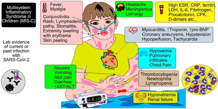
E. Skenderi et al.

                              number need hospitalization and mortality rate is low < 0.1% of diagnosed
                              children [1] [2] [3] [4]. As we are discovering each day more about SARS-CoV-2
                              infection, differences between adults and pediatric disease are probably due to
                              changes within both immune function and the angiotensin-converting enzyme
                              (ACE) 2 receptor, used by the virus to enter type II pneumocytes in the lung.
                              The immune system of children is highly prepared to novel pathogens, due to
                              high levels of innate IgM antibodies with broad reactivity, in addition to the
                              production of the anti-inflammatory interleukin (IL)-10 by neonatal B cells [5].
                              Other probable explanations are alternations in T cell populations in adults due
                              to continuous antigen stimulation and thymic involution, varied levels of ACE-2
                              expression in children, and the simultaneous presence of other viruses in the
                              respiratory mucosa of children, competing with SARS-CoV-2 [6]. Besides, all
                              this children have fewer comorbidities and a stronger pulmonary regenerative
                              potential than adults.

                              2. Case Report
                              A 5 years old male admitted to the University Hospital Center of Tirana with a
                              history of 2 week fever. He was treated with oral antibiotics by a local clinic for
                              acute tonsillitis, but fever persisted and cough, diarrhea and fatigue become dis-
                              turbing in the following days. No family contact with COVID-19 positive indi-
                              viduals was reported. On physical examination he appeared ill. The pharynx was
                              injected with swollen tonsils without exudates. The child was tachypneic, respi-
                              ratory frequency was 40 breaths/min, and oxygen saturation 94%, fine rales were
                              found bilaterally in auscultation. Gastrointestinal manifestations consisted of
                              abdominal pain and diarrhea. The abdomen was soft, not distended, bowel
                              sounds were present. Nor rash on the skin, neither swollen hands or feet, were
                              observed.
                                Laboratory investigations on admission revealed a blood cell count WBC
                              11,400 cells/mm3 (54.7% neutrophils and 34.1% lymphocytes), RBC 4,270,000
                              cells/mm3, Hemoglobin level 11.5 g/dL, Hematocrit value 33.1%, Platelet count
                              1,072,000 cells/mm3, Erythrocyte sedimentation rate 28 mm/h (
E. Skenderi et al.
COVID-19 were negative. As the child did not fulfill all the criteria to meet the
diagnosis of “Multisystem Inflammatory Syndrome in Children”, but the symp-
toms persisted and some of the inflammatory parameters were increased, there
was suspected that another infectious agent could have complicated the scenery.
The EBV panel result indicated acute primary infection IgM antibodies against
viral capsid antigen (VCA) were positive, whereas VCA-IgG antibodies were
negative.
Thrombocytosis was assumed to be of reactive origin due to excessive in-
flammation generated by the combination of two infectious agents COVID-19
and EBV. Once the combined diagnosis was confirmed therapeutic approach
was symptomatic. Fever subsided gradually, lymphadenitis was reduced and the
child appeared playful. Thrombocytosis subsided after a couple of weeks too
(Table 1).
3. Discussion
As SARS-CoV-2 disease is emerging and the world is in the middle of the pan-
demic, it is not easy for the pediatrician, to differ between COVID-19 infection
and other potential viruses of childhood. Almost all medical resources are di-
rected towards COVID-19 infections and still it’s challenging to make a diagno-
sis or predicts its complications in children.
It is already known that fewer cases of COVID-19 disease have been diag-
nosed in children than in adults. The majority of the pediatric cases have been
mild, but severe illness has been reported in 2.5% of pediatric cases in China,
according to the World Health Organization [7]. The most reported signs and
symptoms in children are cough, pharyngeal erythema and fever. Other less
common signs and symptoms include diarrhea, fatigue, rhinorrhea, vomiting
and nasal congestion. A small percent presents with severe disease, dyspnea,
persistent high fever, lethargy, increased levels of enzymes [4]. Since May 2020,
several highly endemic countries reported a high incidence of multisystem in-
flammatory syndrome (MIS) in children [8] [9] [10] [11]. All include fever, ele-
vated inflammatory markers, and organ dysfunction not attributed to another
infectious cause. The median interval from COVID-19 symptom onset to MIS
onset is 25 days [12]. The higher rate of positive serologic tests compared with
nasopharyngeal reverse transcription-polymerase chain reaction (RT-PCR) is
Table 1. Clinical outcome of the patient.
Hositalization time 0 week 1 week 2 weeks 6 weeks
WBC 11,400 cell/mm3 15,100 cell/mm3 9200 cell/mm3 8600 cell/mm3
Lymphocytes 34.1% 51.3% 56.2% 31.4%
CRP 1.16 0.97 0.6 0.05
Thrombocytes 1,072,000 cell/mm3 821,000 cell/mm3 683,000 cell/mm3 464,000 cell/mm3
D-dimer 300 mg/dL 201 mg/dL 190 mg/dL 158 mg/dL
DOI: 10.4236/jbm.2021.95003 22 Journal of Biosciences and MedicinesE. Skenderi et al.
suggestive of a late complication of the disease [8] [12] [13] [14] [15]. Besides
fever, the most common presentations of MIS are gastrointestinal (diarrhea,
vomiting, abdominal pain), cardiovascular, mucocutaneous (rash, mucus mem-
brane changes, conjunctival injection), respiratory (including sore throat),
headache, and limb and periorbital edema [11] [12] [13]. Associated laboratory
findings are elevated inflammation markers (neutrophilia, C-reactive protein,
ferritin, erythrocyte sedimentation rate), thrombocytopenia, lymphopenia, ele-
vated troponin and N-terminal pro-B-type natriuretic peptide (NT-proBNP),
hypertriglyceridemia, and elevated D-dimer and fibrinogen (Table 2, Figure 1)
[16]. Some patients meet the criteria for macrophage activation syndrome (MAS).
As the presenting child had a negative reverse transcriptase protein chain reac-
tion (RT-PCR) for COVID-19 but a positive serology for COVID-19 (increased
IgM levels, which are higher during weeks 2 - 3 of illness), prolonged fever > 10
days, cough, diarrhea, fatigue, cervical lymphadenopathy, pharyngeal erythema
Table 2. CDC case definition for multisystem inflammatory syndrome in children (MIS-C).
1) An individual aged < 21 years with
2) Clinical criteria:
_ A minimum 24-h history of subjective or objective fever _ 38.0 _C AND
_ Severe illness necessitating hospitalization AND
_ Two or more organ systems affected (i.e., cardiac, renal, respiratory, hematologic,
gastrointestinal, dermatologic, neurological)
3) Laboratory evidence of inflammation
_ One or more of the following: an elevated CRP, ESR, fibrinogen, procalcitonin, D-dimer,
ferritin, LDH, or IL-6; elevated neutrophils or reduced; low albumin
4) Laboratory or epidemiologic evidence of SARS-CoV-2 infection
_ Positive SARS-CoV-2 testing by RT-PCR, serology, or antigen OR
_ COVID-19 exposure within 4 weeks prior to onset of symptoms
5) No alternative diagnosis
Abbreviations: CDC, Centers for Disease Control; CRP, C-reactive protein; ESR, erythrocyte
sedimentation rate; LDH, lactate dehydrogenase; RT-PCR, reverse transcriptase polymerase
chain reaction; SARS-CoV-2, severe acute respiratory syndrome coronavirus-2
Figure 1. Infographic showing CDC criteria for the diagnosis of MIS-C.
DOI: 10.4236/jbm.2021.95003 23 Journal of Biosciences and MedicinesE. Skenderi et al.
and some of the inflammatory proteins increased such as CRP and d-Dimer, in-
itially was presumed to be in front of a MIS-C. However there were present some
confounding elements on laboratory results non consistent with MIS-C such as
lymphocytosis and thrombocytosis. A consistent pattern of laboratory abnor-
malities has not yet been identified in children with confirmed COVID-19,
however, a common laboratory abnormalities among hospitalized patients in-
clude lymphopenias (due to destruction of infected T lymphocyte cells) and
thrombocytopenia in cognition with other coagulation parameters abnormali-
ties.
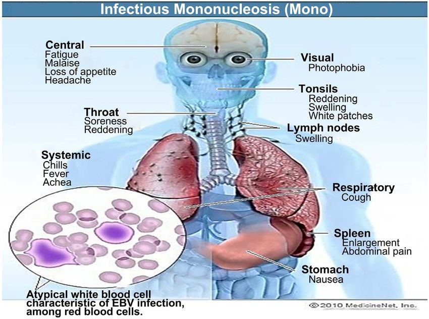
On the other hand Epstein-Barr virus (EBV) which is not a rare agent causing
disease in children manifests overlapping symptoms with COVID-19. EBV is
ubiquitous in nature and infects a large fraction of the world population. In
young children primary infection is usually asymptomatic or produces an acute
illness that is often not recognized as being due to EBV or may be also presented
with the full blown clinic of Infectious Mononucleosis; fever, pharyngitis, lym-
phadenopathy, hepato-splenomegaly and fatigue (Figure 2) [17]. The median
duration of infectious mononucleosis is 16 days, which is much longer than the
duration of most acute viral illnesses, recovery is gradual. A potent innate and
adaptive immune response occurs during primary EBV infection. The innate
immune system is an important first line of defense against viral infections.
Many inflammatory cytokines such as tumor necrosis factor alpha (TNF-α), in-
terleukin-6 (IL-6), IL-1β and IFN-γ are found in the sera of patients infected
with EBV and COVID-19 too. IFN-γ is thought to be important for control of
EBV infection [18] [19]. High levels of IFN-γ likely contribute to the symptoms
experienced during infectious mononucleosis, as this cytokine is known to cause
Figure 2. Infographic showing infectious mononucleosis.
DOI: 10.4236/jbm.2021.95003 24 Journal of Biosciences and MedicinesE. Skenderi et al.
headache, fatigue, and fever [20]. Both CD4 and CD8 T cells make a robust re-
sponse to EBV antigens, the massive lymphocytosis in the blood that characte-
rizes infectious mononucleosis is thought to consist largely of CD8 T cells spe-
cific for EBV lytic antigens [21]. Both agents EBV and COVID-19 modulate the
immune system.
Thrombocytosis is not typical in EBV or COVID-19 infection otherwise they
are companied by mild thrombocytopenia. Thrombocytosis in children are usually
reactive, particularly common during recovery phase of an infection or inflam-
mation and are usually transient and subsides when the primary stimulus ceases.
Reactive thrombocytosis is usually mediated by increased release of numerous
cytokines in response to infections. A wide range of cytokines may participate in
the stimulation of platelet production, IL-3, IL-11, granulocyte-macrophage co-
lony-stimulating factor, erythropoietin but the most imported role is plaid by
thrombopoietin and IL-6 which are initially elevated in response to infections
[22]. In this case thrombocytosis is an exaggerated physiologic response to the
combined infections. Despite the strikingly high platelet count, sometimes ex-
ceeding 1,000,000 cells/mm3, thrombotic and/or hemorrhagic complications are
highly exceptional.
4. Conclusion
COVID-19 has inflicted all the world population. The number of infected child-
ren is progressively increasing. The disease in children is less severe, but some-
times it may be complicated with Multisystem Inflammatory Syndrome. Some of
the symptoms and signs may be overlapped with other infectious diseases of the
childhood, such as EBV and respiratory viruses, and confound the appropriate
diagnosis. So a high index of suspicion must be maintained in children while
making a diagnosis.
Acknowledgements
We thank Prof. Anila Godo for critical reading of the manuscript.
Conflicts of Interest
The authors declare no conflicts of interest regarding the publication of this pa-
per.
References
[1] Dong, Y., Mo, X., Hu, Y., Qi, X., Jiang, F. and Jiang, Z. (2020) Epidemiology of
COVID-19 among Children in China. Pediatrics, 145, e20200702.
https://doi.org/10.1542/peds.2020-0702
[2] Bialek, S., Gierke, R., Hughes, M., McNamara, L., Pilishvili, T. and Skoff, T. (2020)
Coronavirus Disease 2019 in Children—United States, February 12-April 2, 2020.
Morbidity and Mortality Weekly Report, 69, 422-426.
https://www.cdc.gov/mmwr/volumes/69/wr/mm6914e4.htm
https://doi.org/10.15585/mmwr.mm6914e4
DOI: 10.4236/jbm.2021.95003 25 Journal of Biosciences and MedicinesE. Skenderi et al.
[3] Livingston, E. and Bucher, K. (2020) Coronavirus Disease 2019 (COVID-19) in Ita-
ly. JAMA, 323, 1335-1335. https://www.iss.it/infografiche
[4] Lu, X., Zhang, L., Du, H., Zhang, J., Li, Y.Y., Qu, J., et al. (2020) SARS-CoV-2 Infec-
tion in Children. The New England Journal of Medicine, 382, 1663-1665.
https://doi.org/10.1056/NEJMc2005073
[5] Carsetti, R., Quintarelli, C., Quinti, I., Piano Mortari, E., Zumla, A., Ippolito, G., et
al. (2020) The Immune System of Children: The Key to Understanding SARS-CoV-2
Susceptibility? The Lancet Child & Adolescent Health, 4, 414-416.
https://doi.org/10.1016/S2352-4642(20)30135-8
[6] Yuki, K., Fujiogi, M. and Koutsogiannaki, S. (2020) COVID-19 Pathophysiology: A
Review. Clinical Immunology, 215, Article ID: 108427.
https://pubmed.ncbi.nlm.nih.gov/32325252
https://doi.org/10.1016/j.clim.2020.108427
[7] Toubiana, J., Poirault, C., Corsia, A., Bajolle, F., Fourgeaud, J., Angoulvant, F., et al.
(2020) Kawasaki-Like Multisystem Inflammatory Syndrome in Children during the
Covid-19 Pandemic in Paris, France: Prospective Observational Study. BMJ, 369,
m2094. https://doi.org/10.1136/bmj.m2094
[8] Verdoni, L., Mazza, A., Gervasoni, A., Martelli, L., Ruggeri, M., Ciuffreda, M., et al.
(2020) An Outbreak of Severe Kawasaki-Like Disease at the Italian Epicentre of the
SARS-CoV-2 Epidemic: An Observational Cohort Study. The Lancet, 395, 1771-1778.
https://doi.org/10.1016/S0140-6736(20)31103-X
[9] Rauf, A., Vijayan, A., John, S.T., Krishnan, R. and Latheef, A. (2020) Multisystem
Inflammatory Syndrome with Features of Atypical Kawasaki Disease during COVID-19
Pandemic. Indian Journal of Pediatrics, 87, 745-747.
https://doi.org/10.21203/rs.3.rs-29369/v1
[10] Riphagen, S., Gomez, X., Gonzalez-Martinez, C., Wilkinson, N. and Theocharis, P.
(2020) Hyperinflammatory Shock in Children during COVID-19 Pandemic. The Lan-
cet, 395, 1607-1608. https://www.ncbi.nlm.nih.gov/pmc/articles/PMC7204765
https://doi.org/10.1016/S0140-6736(20)31094-1
[11] Godfred-Cato, S., Bryant, B., Leung, J., Oster, M.E., Conklin, L., Abrams, J., et al.
(2020) COVID-19-Associated Multisystem Inflammatory Syndrome in Child-
ren—United States, March-July 2020. MMWR Morbidity and Mortality Weekly
Report, 69, 1074-1080. https://doi.org/10.15585/mmwr.mm6932e2
http://www.cdc.gov/mmwr/volumes/69/wr/mm6932e2.htm?s_cid=mm6932e2_w
[12] Feldstein, L.R., Rose, E.B., Horwitz, S.M., Collins, J.P., Newhams, M.M., Son, M.B.F.,
et al. (2020) Multisystem Inflammatory Syndrome in U.S. Children and Adoles-
cents. The New England Journal of Medicine, 383, 334-346.
[13] Whittaker, E., Bamford, A., Kenny, J., Kaforou, M., Jones, C.E., Shah, P., et al. (2020)
Clinical Characteristics of 58 Children with a Pediatric Inflammatory Multisystem
Syndrome Temporally Associated with SARS-CoV-2. JAMA, 324, 259-269.
https://doi.org/10.1001/jama.2020.10369
[14] WHO (2020) Report of the WHO-China Joint Mission on Coronavirus Disease
2019 (COVID-19). World Health Organization, Geneva.
https://www.who.int/publications-detail/report-of-the-who-china-joint-mission-on
-coronavirus-disease-2019-(covid-19)
[15] Nakra, N.A., Blumberg, D.A., Herrera-Guerra, A. and Lakshminrusimha, S. (2020)
Multi-System Inflammatory Syndrome in Children (MIS-C) Following SARS-CoV-2
Infection: Review of Clinical Presentation, Hypothetical Pathogenesis, and Pro-
posed Management. Child (Basel, Switzerland), 7, 69.
DOI: 10.4236/jbm.2021.95003 26 Journal of Biosciences and MedicinesE. Skenderi et al.
http://www.ncbi.nlm.nih.gov/pubmed/32630212
https://doi.org/10.3390/children7070069
[16] Natasha, N.A., Dean, B.A., Angel, H.-G. and Satyan, L. (2020) Multi-System In-
flammatory Syndrome in Children (MIS-C) Following SARS-CoV-2 Infection: Re-
view of Clinical Presentation, Hypothetical Pathogenesis, and Proposed Manage-
ment. https://www.mdpi.com/journal/children
https://doi.org/10.3390/children7070069
[17] Melissa, S.C. and William, S.C. (2020) Infectious Mononucleosis.
http://medicinenet.com/infectious_mononucleosis/article.htm
[18] Lee, K.S., Groshong, S.D., Cool, C.D., Kleinschmidt-DeMasters, B.K. and van Dyk,
L.F. (2009) Murine Gammaherpesvirus 68 Infection of IFNgamma Unresponsive
Mice: A Small Animal Model for Gammaherpesvirus-Associated B-Cell Lympho-
proliferative Disease. Cancer Research, 69, 5481-5489.
https://doi.org/10.1158/0008-5472.CAN-09-0291
[19] Weck, K.E., et al. (1997) Murine Gamma-Herpesvirus 68 Causes Severe Large-Vessel
Arteritis in Mice Lacking Interferon-Gamma Responsiveness: A New Model for
Virus-Induced Vascular Disease. Nature Medicine, 3, 1346-1353.
https://doi.org/10.1038/nm1297-1346
[20] Schiller, J.H., et al. (1990) Biological and Clinical Effects of the Combination of Be-
ta- and Gamma-Interferons Administered as a 5-Day Continuous Infusion. Cancer
Research, 50, 4588-4594.
[21] Hislop, A.D., Taylor, G.S., Sauce, D. and Rickinson, A.B. (2007) Cellular Responses
to Viral Infection in Humans: Lessons from Epstein-Barr Virus. Annual Review of
Immunology, 25, 587-617.
https://doi.org/10.1146/annurev.immunol.25.022106.141553
[22] Zheng, S.Y., Xiao, Q.Y., Xie, X.H., et al. (2016) Association between Secondary
Thrombocytosis and Viral Respiratory Tract Infections in Children. Scientific Re-
ports, 6, Article No. 22964. https://doi.org/10.1038/srep22964
DOI: 10.4236/jbm.2021.95003 27 Journal of Biosciences and MedicinesYou can also read