Osteomyelitis in Children with Sickle Cell Disease: A Challenging Diagnosis: Case Report from Cameroon - Scientific ...
←
→
Page content transcription
If your browser does not render page correctly, please read the page content below
Open Journal of Pediatrics, 2021, 11, 208-214
                                                                                            https://www.scirp.org/journal/ojped
                                                                                                         ISSN Online: 2160-8776
                                                                                                           ISSN Print: 2160-8741

Osteomyelitis in Children with Sickle Cell
Disease: A Challenging Diagnosis:
Case Report from Cameroon

Djike Puepi Fokam Yolande1,2*, Kukwah Anthony Tufong2,3, Tagakou Mboula Jules2,4,
Andang Paul Mayah5, Eposse Ekoube Charlotte6,7, Diomede Noukeu Njinkui8,
Dominique Enyama8, Helene Kamo Selangai9, Verla Vincent Siysi1,2
1
 Department of Internal Medicine and Paediatrics, Faculty of Health Science, University of Buea, Buea, Cameroon
2
 Buea Regional Hospital, Buea, South West, Cameroon
3
 Department of Microbiology and Parasitology, Faculty of Sciences, University of Buea, Buea, Cameroon
4
 Department of Clinical Sciences, Faculty of Health Sciences, University of Bamenda, Bamenda, Cameroon
5
 Faculty of Health Sciences, University of Buea, Buea, Cameroon
6
 Faculty of Medicine and Pharmaceutical Sciences, University of Douala, Douala, Cameroon
7
 Treatment Centre of Sickle cell Disease, Laquintinie Hospital, Douala, Cameroon
8
 Faculty of Medicine and Pharmaceutical Sciences, University of Dschang, Dschang, Cameroon
9
 Faculty of Medicine and Biomedical sciences, University of Ngaoundere, Ngaoundere, Cameroon

How to cite this paper: Yolande, D.P.F.,      Abstract
Tufong, K.A., Jules, T.M., Mayah, A.P.,
Charlotte, E.E., Njinkui, D.N., Enyama, D.,   Introduction: Sickle Cell Disease (SCD) is the most prevalent genetic disease
Selangai, H.K. and Siysi, V.V. (2021) Os-     in the world predominantly in the African population with Sickle Cell Anae-
teomyelitis in Children with Sickle Cell
                                              mia (SCA) being its dominant form. One of the most frequent complications
Disease: A Challenging Diagnosis: Case Re-
port from Cameroon. Open Journal of Pedia-    of SCD is osteomyelitis. SCA is due to a point mutation in the beta globin
trics, 11, 208-214.                           chain of haemoglobin. This is responsible for the sickled shape of RBCs under
https://doi.org/10.4236/ojped.2021.112020     low oxygen tension conditions leading to obstruction in the microcirculation.
                                              This leads to vaso-occlusive crises (VOC) which has a similar clinical presen-
Received: March 18, 2021
Accepted: May 28, 2021                        tation to that of osteomyelitis, another complication of SCD. Case Presenta-
Published: May 31, 2021                       tion: We present the case of a three-year-old girl with SCA who presented
                                              with an inability to bear weight in a febrile context. A diagnosis of VOC was
Copyright © 2021 by author(s) and
                                              initially made, which was later on changed to both a left chronic tibial and
Scientific Research Publishing Inc.
This work is licensed under the Creative      right distal femoral osteomyelitis following a series of biological, and imaging
Commons Attribution International             investigations. Surgical debridement and drainage were performed, resulting
License (CC BY 4.0).                          9 weeks later in the involution of fever and leg pain. Conclusion: Osteomye-
http://creativecommons.org/licenses/by/4.0/
                                              litis when associated with SCD is a dreadful and deathly disease in low in-
                Open Access
                                              come countries as it also presents like VOC therefore higher suspicion index
                                              is recommended. It is therefore important to take this into consideration at
                                              an early stage in patients with homozygous sickle cell disease so as to rapidly

DOI: 10.4236/ojped.2021.112020         May 31, 2021             208                                    Open Journal of Pediatrics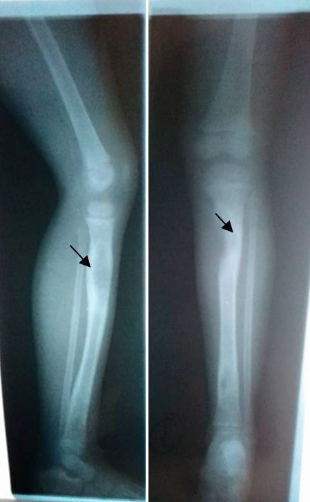
D. P. F. Yolande et al.

                                   initiate multidisciplinary care. Appropriate investigations, appropriate anti-
                                   biotic therapy, and timely surgical intervention would help to greatly reduce
                                   morbidity and mortality.

                                   Keywords
                                   Chronic Osteomyelitis, Sickle Cell Disease, Child

                                 1. Introduction
                                 Sickle Cell Disease (SCD) is the most prevalent genetic disease in the world with
                                 a higher predominance in populations of African descent. This haemoglobino-
                                 pathy is a major public health problem in Africa and particularly in Cameroon,
                                 where between 20% to 25% of the population carries the sickle cell trait [1].
                                 Sickle Cell Anaemia (SCA) is the most severe form of SCD and it is common in
                                 geographical areas where malaria is widespread [2]. In SCD, haemoglobin preci-
                                 pitates as insoluble crystals which lead to an abnormal shape and size of Red
                                 Blood Cells (RBCs) with subsequent phagocytosis of the affected corpuscles.
                                 Under low oxygen concentration, RBCs that contain an abnormal form of Hae-
                                 moglobin (Hb) become deformed (or sickle-shaped) and rigid. This impedes
                                 their ability to pass through microcirculation, with frequent clotting and throm-
                                 bosis. The consequence of the obstruction is the production of ischemia and in-
                                 farction. Tissue infarctions, generally referred to as vaso-occlusive crisis (VOC)
                                 present as pain or swelling. Infarcts can affect several organs of the patient in-
                                 cluding brain, lungs, bones and spleen, and are responsible for most clinical ma-
                                 nifestations. Previous research has shown higher susceptibility of SCA children
                                 towards a rare form of osteomyelitis, i.e. salmonella osteomyelitis [3]. Staphylo-
                                 coccus aureus is the pathogen most commonly involved. Medullary bone infarc-
                                 tion and necrosis create an apt condition for bacterial growth and spreading. In
                                 these patients, osteomyelitis is commonly seen in the tibia, diaphysis of femur
                                 and humerus, along with the infection of the vertebrae. The infection is haema-
                                 togenous in nature due to its delayed onset. Immunodeficiency caused by splenic
                                 dysfunction, tissue infarction, and excess iron content leads to an increased risk
                                 of osteomyelitis [4]. In SCA, osteomyelitis diagnosis can be a major problem for
                                 healthcare providers, where unsuccessful identification of the disease may end
                                 up with severe bone damage and progression of infection to life-threatening sta-
                                 tus. Patients will suffer pain, swelling, fever and increased flow of inflammatory
                                 markers (CRP/ESR) in blood serum. This shows parallel characteristics with that
                                 of painful bone crises, which makes differentiation with bone infarction a diffi-
                                 cult task. Radiographic findings of osteopenia, sclerosis, and periosteal inflam-
                                 mation are seen in both stages of infection and infarction. Therefore, the radio-
                                 graphic features are nonspecific and primarily normal [4] [5]. We report the case
                                 of a three-year-old female sickle cell patient with chronic bone pain, which was
                                 later diagnosed as chronic osteomyelitis and cared for.

DOI: 10.4236/ojped.2021.112020                       209                                   Open Journal of Pediatrics
D. P. F. Yolande et al.
2. Case Presentation
A three-year-old girl, known sickler compliant on daily folic acid supplements
and prophylactic amoxicillin antibiotics, and up-to-date on her immunisation
history. She was also seen to have adequate childhood development. She was
transferred from the paediatric unit to the orthopaedic unit for better manage-
ment after presenting with an inability to bear weight of 9 weeks duration.
The child’s parents reported a history of intermittent left leg pain increasing
in intensity in a context of fever with no signs of inflammation. She was ma-
naged for a VOC in the paediatric unit with oral cloxacillin 150 mg/kg thrice
daily for 10 days and daily alcoholic dressings done on the leg, which later on
subsided the pain. Three weeks later, the pain reoccurred. There was a fever and
signs of inflammation. An X-ray of the left leg done on day 21 of progress was
unremarkable, blood workup revealed C-Reactive Protein (CRP) at 96 mg/l and
a Full Blood Count (FBC) showed leucocytosis at 21,400 cells/µl. There was also
a severe microcytic anaemia with haemoglobin at 7 g/dl, and severe thrombocy-
topenia at 19,000 cells/µL. A blood culture was done which was sterile. A urine
culture was done which was positive for Escherichia coli sensitive to Ciproflox-
acin, Meropenem, Imipenem, Chloramphenicol, Ofloxacin. Ultrasonography of
the urinary tract was normal. The patient was placed on parenteral Imipenem 60
mg/kg in four doses for ten days combined with continuous alcoholic dressing of
the leg. Pain of the leg was thought to be a VOC. The persistence of symptoms
prompted a review from a sickle cell specialist who suspected an osteomyelitis
and placed the patient on parenteral Lincomycin 30 mg/kg in two divided doses
daily and oral Cefixime 8 mg/kg bid daily. For pain Tramadol 1 mg/kg thrice
daily was given alongside Paracetamol syrup 60 mg/kg in four doses. A second
X-ray of the affected leg was requested and done on day 28 of evolution coupled
with blood cultures to be done after a therapeutic window of 48 hours.
The X-ray was normal, blood culture was negative, and urine culture was pos-
itive for Kluyvera sp. sensitive to Imipenem, Meropenem, Doxycycline, Nitrofu-
rantoin and Quinolones. At this point all parenteral antibiotics were stopped and
the patient was placed on oral Nitrofurantoin 50 mg daily alongside all other as-
pects of management (antalgics and alcohol dressing). Fever subsided by the 10th
day of treatment, but the pain in the leg, swelling and inability to bear weight
persisted and prompted a second orthopaedic review. On orthopaedic examina-
tion, signs of inflammation were found on the left proximal leg. Patient was
subjected to a third, X-ray of both legs which revealed a sequestrum surrounded
by involucrum on the left proximal tibia with periosteal reaction on the distal
right femur (Figure 1).
A pre-operative workup was done preceding a sequestrectomy and drainage
of the proximal left tibia, and a drain put in place. The result of the pus culture
obtained on the second day post-surgery was positive for Salmonella sp. and
sensitive to Imipenem, Ceftriaxone, Chloramphenicol, Ofloxacin and Gentamy-
cin. Her treatment was Ceftriaxone 75 mg/kg/day and Gentamycin 3 mg/kg/day.
DOI: 10.4236/ojped.2021.112020 210 Open Journal of PediatricsD. P. F. Yolande et al.
Results of the bone biopsy histopathologic analysis were in favour of a “subacute
osteitis”.
On the second day post-op, the patient became paler and more dyspnoeic;
FBC revealed a haemoglobin level drop to 6.8 g/dl. She was thus transfused
350cc of whole blood.
Control CRP on the sixth day post-op was at 24 mg/L.
Post-op evolution was favourable as the patient was discharged on the 7th day
post-op, relayed on oral thiamphenicol 75 mg/kg in two doses daily for six
weeks, oral paracetamol 60 mg/kg in four divided doses daily to alternate with
oral ibuprofen tid daily in case of pains, alongside daily wound dressing.
Patient came for follow, six weeks later; there was no pain or swelling on the
right leg, no fever, and she was now able to bear weight. Blood workup showed
leucocytosis at 17,100 cells/µl of lymphocytic and monocytic predominance.
There was also a normocytic anaemia with haemoglobin at 8.2 g/dl and throm-
bocytosis at 784,000/µl. CRP was atD. P. F. Yolande et al.
3. Discussion
We presented a three-year-old, known sickler with a history of chronic long bone
pain initially thought to be VOC which later turned out to be an osteomyelitis.
Osteomyelitis often occurs in the long bones (the case of our patient in whom
the left proximal tibia was affected) in which there is slow vascular flow within
the looped vessels of the metaphysic. This combined with microtrauma (ische-
mia following VOC), is believed to encourage seeding of infection during bacte-
raemia. Inflammation follows thus, and if purulent material develops, the pres-
sure effect of the subsequent abscess will lead progressively to bone destruction
[6]. In osteomyelitis, the primary site of infection is the bone and bone marrow.
Any part of the bone may be involved but there is preferential targeting of me-
taphyseal regions of long bones adjacent to joints [7], the case in our patient was
typical of this. Like this case, the usual means of infection is by the haemato-
genous spread through the bone marrow to the cortex, although direct introduc-
tion may complicate trauma or orthopaedic surgery, or may arise from radiation
therapy [7] [8]. When pus accumulates, intramedullary pressure increases, re-
sulting in vascular collapse, venous stasis, and further ischemia. Pus accumulat-
ing underneath the periosteum elevates it from the cortex, thus further reduces
the vascular supply. As this continues to accumulate, the periosteum is breached,
followed by the development of mucosal or cutaneous abscesses and fistulae [8].
In cases of untreated or chronic infection, this new bone or involucrum may
surround the dead bone, the sequestrum, leading to a “bone-within-bone” ap-
pearance. Infection-controlled surgery and being bed-ridden for seven days af-
firms hospitalization duration according to the classification system whereas pa-
tients with advanced osteomyelitis (type B4) are noted for extended hospitaliza-
tion (months) and high risk of repeated infections and surgery [6].
Mindful of the inadequate diagnostic imaging modalities like bone scintigra-
phy such as in the present case, diagnosing osteomyelitis in children with SCD
can be extremely difficult as they often present with fever and a painful, swollen,
tender limb with limited range of motion. These signs and symptoms are similar
to those found in patients with a VOC. There was no definitive features on his-
tory, physical examination, laboratory testing, or radiological study that could
reliably differentiate between osteomyelitis and VOC in our patient, with the
possible exception of a positive bacterial culture from the bone [9]. However, a
bone biopsy or aspiration is often not performed because it is an invasive pro-
cedure and should be done before starting antibiotics to maximize the chances of
obtaining a positive culture result [9]. Furthermore, positive bone biopsy cul-
tures such as in this case are reported in only 30% to 86% of cases [10], implying
that there is still a high false-negative rate. Negative cultures from the peripheral
blood such as in this case are even less specific in supporting a diagnosis of os-
teomyelitis, and they are reported to demonstrate an organism in 30% to 76% of
cases [10]. Thus, even though the patient was confirmed to have subacute os-
teomyelitis, there are many cases in which no definitive diagnosis can be made
DOI: 10.4236/ojped.2021.112020 212 Open Journal of PediatricsD. P. F. Yolande et al.
as osteomyelitis may show similar clinical and para clinical presentation to VOC
in the acute stage. The patient, in tandem with pain in the bone and fever, was
initially treated empirically for possible osteo-articular infection or sepsis with
broad spectrum antibiotics. These antibiotics were discontinued 48 hours later
as the blood culture returned negative. However, in the case of the patient, fever
and pain were persistent despite negative blood culture. Clinicians are often
faced with the dilemma of whether to treat for osteomyelitis or not [9]; we nev-
ertheless continued antibiotics on this basis and that of a positive urine culture.
Failure to treat an unconfirmed case of osteomyelitis can have serious conse-
quences, including weight loss, growth retardation chronic bone damage, per-
sistent sepsis [3], chronic limb deformity and stiffness as well as social stigmati-
zation [9]. However, unnecessary treatment for what is mistakenly thought to be
osteomyelitis can have significant psychosocial, financial and healthcare re-
source implications and can contribute to antibiotic resistance. Patients may also
unnecessarily experience adverse effects of the antibiotics and are at risk of com-
plications from central or peripheral intravenous lines that may be inserted to
facilitate administration of antibiotics [10].
It is important to highlight that osteomyelitis is much less common than
VOC. Studies suggest that VOC is 50 times more common than osteomyelitis in
a patient with SCD [11] [12]; as in the onset of symptoms of this patient, she was
managed for painful bone crisis which later turned out to be an osteomyelitis.
Therefore, even if a patient with SCD presents with fever and bony pain and has
risk factors for osteomyelitis, this should be considered in the context of the rar-
ity of osteomyelitis compared with that of VOC. The presence of some or all of
the risk factors should nevertheless increase the index of suspicion for osteomye-
litis and make the physician more inclined to request imaging or a definitive
bone aspirate for culture.
4. Conclusion
Differentiating VOC from osteomyelitis is a medically bent tree to straighten as
there is no clear-cut clinical difference at an initial stage. These areas biomarkers
and/or blood workup and imaging show similar abnormalities in the acute stag-
es. Osteomyelitis, a difficult and dreadful disease of childhood when associated
with sickle cell disease and its complications can be severe and even deathly, es-
pecially in low income countries. Therefore, there should always be a high index
of suspicion of osteomyelitis in all sickle cell patients who present with a painful
bone crisis. The clinician should never hesitate to seek more advanced imaging
techniques or perform repeated radiographies in order to make an early diagno-
sis and initiate timely adequate treatment.
Conflicts of Interest
The authors declare no conflicts of interest regarding the publication of this pa-
per.
DOI: 10.4236/ojped.2021.112020 213 Open Journal of PediatricsD. P. F. Yolande et al.
References
[1] Sap Ngo Um, S., et al. (2019) A Cross Sectional Study of Growth of Children with
Sickle Cell Disease, Aged 2 to 5 Years in Yaoundé, Cameroon. The Pan African Medi-
cal Journal, 34, 13. https://doi.org/10.11604/pamj.2019.34.85.16432
[2] Ankit, M., et al. (2020) StatPearls-Sickle Cell Anemia.
https://www.ncbi.nlm.nih.gov/books/NBK482164/#_ncbi_dlg_citbx_NBK482164
[3] Fontalis, A., et al. (2019) The Challenge of Differentiating Vaso-Occlusive Crises
from Osteomyelitis in Children with Sickle Cell Disease and Bone Pain: A 15-Year
Retrospective Review. Journal of Children’s Orthopaedics, 13, 33-39.
https://doi.org/10.1302/1863-2548.12.180094
[4] AlDallal, S.M. (2017) Osteomyelitis: A Manifestation of Sickle Cell Anaemia. Clini-
cal and Medical Investigation, 2,1-3. https://doi.org/10.15761/CMI.1000132
[5] Mark, W., et al. (1998) Etiology of Osteomyelitis Complicating Sickle Cell Disease.
Pediatrics, 101, 296-297. https://doi.org/10.1542/peds.101.2.296
[6] Walker, B.R., Colledge, N.R., Ralston, S.H. and Penman, I.D. (2014) Davidson’s
Principles and Practice of Medicine. Churchil Livingstone Elsevier, London, 1095.
[7] Kamakshi, S.S., Naik, V., Vittal, K. and Shriyanka, R. (2016) Chronic Suppurative
Osteomyelitis: A Case Report. Journal of Advanced Clinical and Research Insights,
3, 220-223. https://doi.org/10.15713/ins.jcri.143
[8] Williams, N.S., Bulstrode, C.J.K. and O’Connel, R. (2013) Bailey and Love’s Short
Practice of Surgery. CRC Press, New York, 571.
[9] Berger, E., Saunders, N., Wang, L. and Friedman, J.N. (2009) Sickle Cell Disease:
Differentiating Osteomyelitis from Vaso-Occlusive Crisis. Archives of Pediatrics
and Adolescent Medicine, 163, 251-255.
https://doi.org/10.1001/archpediatrics.2008.545
[10] Keeley, K. and Buchanan, G.R. (1982) Acute Infarction of Long Bones in Children
with Sickle Cell Anemia. The Journal of Pediatrics, 101, 170-175.
https://doi.org/10.1016/S0022-3476(82)80111-X
[11] Buchanan, G.R. (1996) Differentiation of Bone Infarct from Infection in a Child in
Sickle Cell Disease. The Pediatric Infectious Disease Journal, 15, 724-725.
https://doi.org/10.1097/00006454-199608000-00028
[12] Rudolph, C.D. (2003) Rudolph’s Pediatrics. McGraw-Hill, New York, 904-906.
DOI: 10.4236/ojped.2021.112020 214 Open Journal of PediatricsYou can also read