Case Report: A Case of Acute Cellular Rejection Due to Atopic Dermatitis Exacerbation 3 Years After Heart Transplantation - Frontiers
←
→
Page content transcription
If your browser does not render page correctly, please read the page content below
CASE REPORT
                                                                                                                                        published: 22 February 2021
                                                                                                                                   doi: 10.3389/fimmu.2021.630051

                                          Case Report: A Case of Acute
                                          Cellular Rejection Due to Atopic
                                          Dermatitis Exacerbation 3 Years
                                          After Heart Transplantation
                                          Nobutaka Kakuda 1, Eisuke Amiya 1,2*, Masaru Hatano 1,2, Hisataka Maki 3, Chie Bujo 1,
                                          Masaki Tsuji 1, Koichi Narita 1, Kanna Fujita 1, Junichi Ishida 1, Minoru Ono 4
                                          and Issei Komuro 1
                                          1Department of Cardiovascular Medicine, Graduate School of Medicine, The University of Tokyo, Tokyo, Japan,
                                          2Department of Therapeutic Strategy for Heart Failure, The University of Tokyo, Tokyo, Japan, 3 Department of
                                          Cardiovascular Medicine, Saitama Medical Center, Jichi Medical University, Saitama, Japan, 4 Department of Cardiac
                                          Surgery, Graduate School of Medicine, The University of Tokyo, Tokyo, Japan

                           Edited by:
         Niels Olsen Saraiva Camara,      Background: Little evidence has been presented about the association between
        University of São Paulo, Brazil
                                          previous atopic/allergic disease and graft rejection after solid organ transplantation.
                      Reviewed by:
                           Luc Colas,     Thus, we present a case wherein acute cellular rejection (ACR) after heart
INSERM U1087 L’unité de recherche        transplantation (HTx) was noted along with exacerbation of atopic disease.
      de l’institut du thorax, France
                     Luiza Guilherme,     Case Summary: A 32-year-old man was admitted at our hospital for regular monitoring
     University of São Paulo, Brazil      of graft rejection. He had undergone heart transplant 3 years prior due to dilated
                 *Correspondence:         cardiomyopathy. Echocardiogram revealed good biventricular function, and no
                        Eisuke Amiya
               amiyae-tky@umin.ac.jp      abnormal findings were found in blood sampling tests. However, biopsy showed
                                          moderate ACR [Grade 2R(ISHLT 2004)/3A(ISHLT 1990)], which required twice-
                   Specialty section:     repeated steroid pulses with intensified immunosuppression. Meanwhile, his atopic
         This article was submitted to
    Alloimmunity and Transplantation,
                                          dermatitis, which was diagnosed before having heart failure, was getting worse for the
               a section of the journal   past 6 months. The exacerbation of atopic dermatitis was presumed to be related to the
              Frontiers in Immunology
                                          development of the intractable cellular rejection.
      Received: 17 November 2020
        Accepted: 06 January 2021         Discussion: This case suggested the association of atopic disease and graft rejection
       Published: 22 February 2021        after HTx. We examined 76 patients from a cohort of previous studies who underwent HTx
                             Citation:    at our hospital, which suggested that patients with atopic/allergic disorders such as atopic
      Kakuda N, Amiya E, Hatano M,
   Maki H, Bujo C, Tsuji M, Narita K,
                                          dermatitis and asthma tended to have a significantly higher frequency of moderate
        Fujita K, Ishida J, Ono M and     rejection than non-allergic patients. (p = 0.012; Fisher’s exact test). Our case also
Komuro I (2021) Case Report: A Case       suggests that exacerbation of atopic dermatitis might cause graft rejection of the
   of Acute Cellular Rejection Due to
    Atopic Dermatitis Exacerbation 3      transplanted organ, so that it is important to carefully evaluate the risk of graft rejection
   Years After Heart Transplantation.     if there is a previous history of atopic/allergic disease.
         Front. Immunol. 12:630051.
   doi: 10.3389/fimmu.2021.630051         Keywords: atopic dermatitis, regulatory T cell, acute cellular rejection, heart transplantation, late rejection of graft

Frontiers in Immunology | www.frontiersin.org                                    1                                     February 2021 | Volume 12 | Article 630051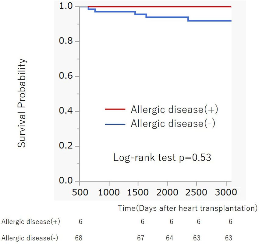
Kakuda et al. Case Report: Can Allergies Cause Graft Rejection?
INTRODUCTION the arms and legs. A blood test on admission revealed mild
anemia. The blood counts of the other two strains were within
Heart transplantation (HTx) is a radical treatment that saves the the normal range, and the eosinophil count was normal. Liver
lives of those with end-stage heart failure. Although the surgical and kidney function tests were normal. Chest X-ray showed
procedures and perioperative management methods have been normal heart shadow and no pleural effusion. Electrocardiogram
established, control of acute and chronic rejection and the (ECG) revealed sinus rhythm of 73 bpm with no ST-T changes.
harmful effects of immunosuppressants remain as the biggest Transthoracic echocardiography showed normal left and right
challenges. Previous studies have reported that through the ventricular function, and left ventricle ejection fraction was
response of the T-cells, the frequency of graft rejection estimated at 73% (Teichholz). Echocardiography parameters of
increases in the presence of atopic/allergic diseases (1, 2). The intraventricular and posterior left ventricle wall diameters were 6
pathophysiology of many atopic/allergic diseases is associated and 8 mm, respectively. The immunosuppressive drugs taken by
with a type 2 T helpler cell (Th2)-based inflammatory response the patient at the time of hospitalization were MMF 1,750 mg/
that involves the production of cytokines such as interleukin day and cyclosporine 150 mg/day. Cyclosporine serum level (the
(IL)-4, IL-5, and IL-13 (3). The untoward effect of Th2-based trough value) was 145 ng/ml, which was within the optimum
inflammation and cytokines is presumed to increase graft range. Right heart catheterization showed that the mean right
rejection by making CD4 + effector T-cells resistant to atrial pressure, mean pulmonary artery wedge pressure, mean
regulatory T-cells (Tregs) (4). Several studies have also pulmonary artery pressure, and cardiac index (Fick) were 5, 11,
reported the critical role Tregs play in immune tolerance and 16 mmHg, and 3.63 L/min/m2, respectively, suggesting that
immunosuppression (5, 6). In this report, we present a case of intracardiac pressure was within the normal range and cardiac
graft rejection reaction that correlated with aggravation of atopic output was maintained. Coronary artery angiography showed no
dermatitis 3 years after HTx. significant stenosis in the coronary arteries. On the other hand,
EMB showed infiltration of multiple inflammatory cells with
myocardial damage, which corresponded ACR with grade 2R
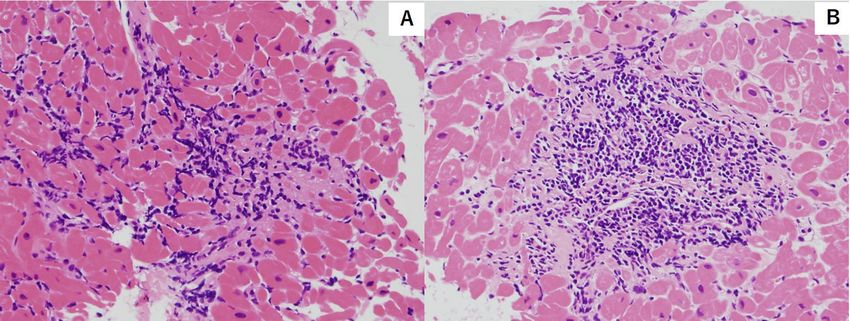
CASE DESCRIPTION (ISHLT 2004)/grade (3A ISHLT 1990) (Figure 1). To counteract
the graft rejection, he was treated with a 3-day course of parenteral
We present the case of a 32-year-old man who underwent methylprednisolone (1,000 mg/day) and then oral prednisolone 25
orthotropic HTx at our hospital due to dilated cardiomyopathy mg/day, which was gradually reduced every two days. In addition,
(DCM). The patient was diagnosed with atopic dermatitis during the dose of cyclosporine was increased from 150 mg/day to 160 mg/
childhood which necessitated the use of topical drugs. At 24 day. However, repeat EMB after 1 week also revealed similar
years old, he noted dyspnea on exertion. Shortly after, he was findings of ACR with a grade of 2R (ISHLT 2004)/3A (ISHLT
diagnosed with DCM and subsequently developed severe drug- 1990). The patient was given steroid pulse therapy for 3 days, and
resistant heart failure. At 25 years old, a left ventricular assist MMF was shifted to everolimus 1 mg/day to intensify
device was implanted as bridge therapy for heart failure. At 29 immunosuppression. Repeat EMB after 1 week showed the
years old, orthotropic HTx was performed at our hospital. The improvement of graft rejection (Grade 0). He was discharged
patient was treated with standard immunosuppression therapy without any overt complications. In addition, atopic dermatitis
consisting of cyclosporine, mycophenolate mofetil (MMF), and also improved by intensifying immunosuppressive drugs with
prednisolone. We performed endomyocardial biopsy according topical drugs. During subsequent follow-ups, no adverse or
to our institutional protocol, which consists of examinations at clinical events were observed. One year after, 4 years after HTx,
the 1st, 2nd, 3rd, 4th, 6th, 8th, 10th, 12th, 18th, and 24th post- significant ACR was not noted [grade 1R (ISHLT 2004)/1A (ISHLT
procedural weeks and the 1st, and 2nd post-procedural years. 1990)] and there was no exacerbation of atopic dermatitis.
During the patient’s follow-ups, there were three events of mild
acute cellular rejection (ACR) (grade 1R ISHLT2004/grade 2
ISHLT 1990) within two years, whereas in all other examinations DISCUSSION
there were no findings of ACR. In addition, anti-HLA antibodies
were not detected during any of the follow-ups. There were also Previous studies have reported cases in which concomitant atopic/
no signs of heart failure such as dyspnea and edema, and the allergic disorders had an increased incidence of graft rejection.
immunosuppressive treatment course was stable. The dose of Previous investigations using mice have demonstrated that classic
prednisolone was gradually reduced according to the policy of allergic disease such as airway hyperresponsiveness and allergic
our institution and it was turned off about one year after HTx. conjunctivitis, exacerbates corneal allograft rejection (1, 4). Nguyen
After 3 years, the patient was hospitalized to undergo a regular et al. have indicated that the frequency of corneal graft rejection
endomyocardial biopsy (EMB). On admission, his vital signs following normal-risk keratoplasty was significantly increased in
were normal, with no leg edema and no jugular dilation. Of note patients with atopic dermatitis (2). Seung et al. have shown that
is that his atopic dermatitis worsened from the previous winter acute rejection after renal transplantation is more common and
season and six months before hospitalization. This was severe in patients with atopy (7). However, no case reports exist
evidenced by erythema and scaling on the neck, precordium, about the association of atopic/allergic disease and graft rejection
and back, and erythema with scabs and exudate were found on after HTx, and in our knowledge, this is the first report of its kind.
Frontiers in Immunology | www.frontiersin.org 2 February 2021 | Volume 12 | Article 630051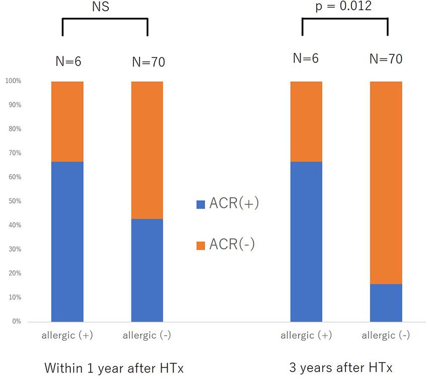
Kakuda et al.                                                                                                                         Case Report: Can Allergies Cause Graft Rejection?

  FIGURE 1 | Hematoxylin and eosin stain of endocardial biopsy sample featuring myocyte injury with multiple lymphocytic infiltration. [Grade2R(ISHLT 2004)/3A
  (ISHLT 1990)]. (A) First endocardial biopsy (B) Second endocardial biopsy.

    Several hypotheses have been proposed to explain the                                           transplantation (10–12). Acute graft rejection was reported to
association between atopic/allergic disease and graft rejection.                                   be associated with the local productions of IL-4 and IL-5
Atopic dermatitis and allergic airway inflammation are Th2-                                         together with eosinophil infiltration (10). Several studies have
dominant allergic diseases. Th2 produces cytokines such as IL-4,                                   reported on the impact of eosinophil on the development of
which induces immunoglobulin E (IgE) production by acting on                                       graft rejection (11, 12). Eosinophil was reported to correlate
B-cells, while mast cells release cytokines such as IL-6 and tumor                                 with the severity of atopic dermatitis (13), and thus, might add
growth factor-beta (TGF-b) in response to the involvement of                                       some hints for the explanation of the association between atopy
the IgE receptor (FcϵRI) complex on the cell surface (3, 8). The                                   and graft rejection. However, the present case did not represent
cytokines induced by Th2 are reported to suppress the effect of                                    eosinophilia, so the correct explanation of the association
Tregs, leading to the enhancement of CD4+ effector T-cells. In                                     between atopic dermatitis and graft rejection in this case had
studies with mice, it has been reported that IL-4 exacerbates                                      not been clarified.
corneal allograft rejection by making CD4+ effector T-cells                                           For further validation of the association between previous
resistant to Tregs (9). Tregs have been reported to play                                           history of atopic/allergic disease and the risk of ACR, we
important roles in the suppression of graft rejection following                                    examined 76 patients from a cohort of previous study who
organ transplant (3). From these findings, there is a                                               underwent heart transplantation at our hospital between
possibility that the risk of rejection may increase via such T-                                    August 2007 and May 2017 (14). Six patients (7.9%) had a
cell responses in patients with atopic/allergic diseases. On the                                   history of atopic/allergic diseases such as bronchial asthma (n =
other hand, several reports have described the association                                         4) and atopic dermatitis (n = 3), one patient had both atopic
between eosinophil and graft rejection after heart and lung                                        dermatitis and bronchial asthma (Table 1). The basic

TABLE 1 | Basic characteristics.

                                                All                                With allergic disease                          Without allergic disease                            P value
                                             (n = 76)                                      (n = 6)                                        (n = 70)

Age, years                                40 (29–53)                                   44.5 (27.5–53.0)                                 39.5 (28.8–53.2)                                0.80
Male                                       53 (69.7)                                       6 (100)                                         47 (67.1)                                    0.09
BMI, kg/m2                              20.0 (17.1–23.1)                               23.8 (20.9–27.2)                                 19.8 (17.4–23.1)                               0.0075
WBC, ml                               5,600 (4,200–6,850)                           5,400 (3,950–10,200)                              5,600 (4,200–6,750)                               0.79
Eosinophils,/ml                          11.2 (0–51.7)                                 17.3 (6.3–61.5)                                   10.8 (0–51.9)                                  0.65
Hb, g/dl                                11.4 (10.4–12.8)                               12.1 (10.9–13.8)                                 11.4 (10.2–12.8)                                0.20
Plt, ×104/ml                            21.8 (19.0–24.7)                               22.0 (15.9–23.8)                                 21.8 (19.1–24.8)                                0.54
eGFR,                                   51.6 (39.2–70.1)                               45.9 (40.4–62.6)                                 52.4 (38.9–71.0)                                0.59
ml/min/1.73m2
CRP, mg/dl                              0.06 (0.02–0.2)                              0.035 (0.018–0.18)                                0.06 (0.02–0.25)                                 0.37
BNP, pg/ml                             79.2 (44.0–116.5)                              64.7 (44.8–88.8)                                 81.5 (43.7–121.9)                                0.45

All variables presented as median (interquartile range) or n (%).
Age was the age at the time of the heart transplantation. Blood test data was data one year after the heart transplantation. P-values were calculated by Fisher’s exact test, t-test or Wilcoxon
rank-sum test comparing those with allergic disease and without allergic disease.
BMI, body mass index; WBC, white blood cell; Hb, hemoglobin; Plt, platelet; eGFR, estimated glomerular filtration rate; CRP, C-reactive protein; BNP, B-type natriuretic peptide.

Frontiers in Immunology | www.frontiersin.org                                                  3                                            February 2021 | Volume 12 | Article 630051
Kakuda et al. Case Report: Can Allergies Cause Graft Rejection?
characteristics are presented in Table 1. The percentage of
atopic/allergic disease was slightly low possibly owing to the
selection of candidates for HTx. During the chronic phase after
HTx (1–3 years), patients with atopic/allergic disease tended to have
a significantly higher frequency of moderate rejection [(Grade 2R
(ISHLT 2004)/3A (ISHLT 1990) or higher)] than patients without
atopic/allergic disease [(p = 0.012; Fisher’s exact test), Odds ratio
(95% CI) 10.73 (1.75 to 65.90)] (Figure 2). On the other hand, there
was no significant difference in the frequency of moderate rejection
[p = 0.40, odds ratio (95% CI) 2.67 (0.46 to 15.53)] less than 1 year
after HTx. Based on the above, the risk of graft rejection, especially
during the chronic phase, increases in atopic/allergic diseases. The
survival curve was not significantly different between these two
groups (Figure 3).
Another lingering question is that despite immunosuppressive
therapy, why did the patient develop exacerbation of atopic
dermatitis? Both cyclosporin and MMF have been reported to
be highly effective for atopic dermatitis. However, some studies
have reported paradoxical development of atopic dermatitis after
solid organ transplantation (15). Ozdemir et al. demonstrated
newly-developed allergies after HTx (16). In addition, some
studies have reported the development of allergies after receiving
immunosuppressive therapy, but there had been no report about FIGURE 3 | Kaplan-Meier survival curve of patients with and without
the mechanism of action, which should be elucidated more previous allergic disease after heart transplantation. There was no significant
difference in survival curves between two groups (Log-rank p = 0.53).
robustly in the future.
In fact, at our institution, prednisolone is turned off within 1 year
after HTx (17). If there is concern about exacerbation of rejection However, triggers for late-onset ACR have not been identified.
due to exacerbation of atopic/allergic disease as in this case, a Future studies may focus on finding out the specific causes for
regimen such as continuing a small amount of steroid, which will be late-onset ACR, which can ultimately lead to improvements in
more effective for atopic/allergic disease, might be better. the treatment for post-HTx patients.
Generally speaking, late-onset ACR has been known to have In a limitation of this study, the chronological association
more adverse clinical outcomes as compared to early-onset ACR. between the graft rejection and the exacerbation of atopic
dermatitis in this case was difficult to presume. However, it is
more likely that the state of atopic dermatitis affected the state of
graft rejection because the impact derived from atopic dermatitis
on the systemic immune response might be greater than the
impact derived from graft rejection (18). In addition, more
concise evaluation of the state of atopic dermatitis might help
the correct clarification of the association between atopic
dermatitis and graft rejection.
Similar to this case, no reports of graft rejection due to
exacerbation of atopic dermatitis have yet to be reported.
Patients with a history of allergic disorders such as atopic
dermatitis may be at an increased risk of developing
immunological rejection after transplantation, requiring a more
intensive immunosuppressive regimen and careful follow-up.
CONCLUSIONS
ACR following exacerbation of atopic dermatitis 3 years post-
HTx is rare and has never been reported. This case highlights the
importance of considering all factors that may contribute to graft
FIGURE 2 | Difference in the development of acute cellular rejection within rejection, regardless of diagnostic findings. However, it is also
one year after heart transplantation (HTx) and three years after HTx between necessary to determine what processes are involved in
patients with and without previous allergic disease. NS, not significant.
this relationship.
Frontiers in Immunology | www.frontiersin.org 4 February 2021 | Volume 12 | Article 630051Kakuda et al. Case Report: Can Allergies Cause Graft Rejection?
DATA AVAILABILITY STATEMENT AUTHOR CONTRIBUTIONS
The raw data supporting the conclusions of this article will be NK: Data collection, statistical analysis, and writing of the manuscript.
made available by the authors, without undue reservation. EA: Conception of the idea, data collection, critical feedback on the
manuscript, and writing of the manuscript. CB, MT, JI, MH, KN, KF,
and HM: Data collection and critical feedback on the manuscript.
MH, MO, and IK: Critical feedback on the manuscript. All authors
contributed to the article and approved the submitted version.
ETHICS STATEMENT
The studies involving human participants were reviewed and FUNDING
approved by the institutional review board at the University of
Tokyo (approval number: 2,650). The patients/participants This work was supported by the Ministry of Education, Culture,
provided their written informed consent to participate in Sports, Science and Technology of Japan through Grant-in-Aid
this study. 17K09488 (to EA).
IFN-gamma. Eur J Immunol (2000) 30(5):1290–6. doi: 10.1002/(SICI)1521-
REFERENCES 4141(200005)30:53.0.CO;2-H
1. Niederkorn JY, Chen PW, Mellon J, Stevens C, Mayhew E. Allergic airway 13. Dhar S, Malakar R, Chattopadhyay S, Dhar S, Banerjee R, Ghosh A.
hyperreactivity increases the risk for corneal allograft rejection. Am J Transpl Correlation of the severity of atopic dermatitis with absolute eosinophil
(2009) 9(5):1017–26. doi: 10.1111/j.1600-6143.2009.02603.x counts in peripheral blood and serum IgE levels. Indian J Dermatol
2. Nguyen NX, Martus P, Seitz B, Cursiefen C. Atopic dermatitis as a risk factor Venereol Leprol (2005) 71(4):246–9. doi: 10.4103/0378-6323.16615
for graft rejection following normal-risk keratoplasty. Graefes Arch Clin Exp 14. Bujo C, Amiya E, Hatano M, Tsuji M, Maki H, Ishida J, et al. Association
Ophthalmol (2009) 247(4):573–4. doi: 10.1007/s00417-008-0959-4 between infectious event and de novo malignancy after heart transplantation.
3. Miura K, Inoue K, Ogura A, Kaminuma O. Role of CD4+ T cells in Airway Heart Vessels (2020). doi: 10.1007/s00380-020-01715-9. (in press).
Diseases: learning from Murine Models. Int J Mol Sci (2020) 21(20):7480. 15. Marcus N, Amir AZ, Grunebaum E, Dipchand A, Hebert D, Ng VL, et al. De
doi: 10.3390/ijms21207480 novo allergy and immune-mediated disorders following solid-organ
4. Niederkorn JY, Chen PW, Mellon J, Stevens C, Mayhew E. Allergic transplantation-prevalence, natural history, and risk factors. J Pediatr
conjunctivitis exacerbates corneal allograft rejection by activating Th1 and (2018) 196:154–160.e2. doi: 10.1016/j.jpeds.2017.11.026
Th2 alloimmune responses. J Immunol (2010) 184(11):6076–83. doi: 10.4049/ 16. Ozdemir O, Arrey-Mensah A, Sorensen RU. Development of multiple food
jimmunol.0902300 allergies in children taking tacrolimus after heart and liver transplantation.
5. Joffre O, Santolaria T, Calise D, Al Saati T, Hudrisier D, Romagnoli P, et al. Pediatr Transpl (2006) 10(3):380–3. doi: 10.1111/j.1399-3046.2005.00474.x
Prevention of acute and chronic allograft rejection with CD4+CD25 17. Kittleson MM, Kobashigawa JA. Cardiac Transplantation Current Outcomes
+FOxp3+ regulatory T lymphocytes. Nat Med (2008) 14(1):88–92. and Contemporary Controversies. JACC Heart Fail (2017) 5: (12):857–68.
doi: 10.1038/nm1688 doi: 10.1016/j.jchf.2017.08.021
6. Dijke IE, Korevaar SS, Caliskan K, Balk AHMM, Maat APWM, Weimar W, 18. Miraglia del Giudice M, Decimo F, Leonardi S, Maioello N, Amelio R, Capasso
et al. Inadequate immune regulatory function of CD4+CD25bright+FoxP3+ A, et al. Immune dysregulation in atopic dermatitis. Allergy Asthma Proc
T cells in heart transplant patients who experience acute cellular rejection. (2006) 27(6):451–5. doi: 10.2500/aap.2006.27.2887
Transplantation (2009) 87(8):1191–200. doi: 10.1097/TP.0b013e31819ec2fb
7. Seung LM, Lorincz AL. Incidence of acute renal transplant rejection in atopic Conflict of Interest: EA and MH belong to the Department of Therapeutic
individuals. Arch Dermatol (1994) 130(5):584–8. doi: 10.1001/archderm.130.5.584 Strategy for Heart Failure, Graduate School of Medicine, University of Tokyo,
8. Foley John F. Ceramide keeps mast cells in check. Sci Signal (2012) 252:302. which is endowed by Actelion Pharmaceuticals Japan Ltd., Otsuka
doi: 10.1126/scisignal.2003810 Pharmaceutical, NIPRO CORPORATION, Terumo Corp., Senko Medical
9. Reyes NJ, Chen PW, Niederkorn JY. Allergic conjunctivitis renders CD4+ T Instrument Mfg., Century Medical Inc., Kinetic Concepts Inc., and St. Jude
cells resistant to T regulatory cells and exacerbates corneal allograft rejection. Medical.
Am J Transpl (2013) 13(5):1181–92. doi: 10.1111/ajt.12198 The remaining authors declare that the research was conducted in the absence of
10. Goldman M, Le Moine A, Braun M, Flamand V, Abramowicz D. A role for any commercial or financial relationships that could be construed as a potential
eosinophils in transplant rejection. Trends Immunol (2001) 22(5):247–51. conflict of interest.
doi: 10.1016/s1471-4906(01)01893-2
11. Kaes J, Van der Borght E, Vanstapel A, Van Herck A, Sacreas A, Heigl T, et al. Copyright © 2021 Kakuda, Amiya, Hatano, Maki, Bujo, Tsuji, Narita, Fujita, Ishida,
Group The Leuven Lung Transplant. Peripheral Blood Eosinophilia Is Ono and Komuro. This is an open-access article distributed under the terms of the
Associated with Poor Outcome Post-Lung Transplantation. Cells (2020) 9 Creative Commons Attribution License (CC BY). The use, distribution or
(11):2516. doi: 10.3390/cells9112516 reproduction in other forums is permitted, provided the original author(s) and the
12. Braun MY, Desalle F, Le Moine A, Pretolani M, Matthys P, Kiss R, et al. IL-5 copyright owner(s) are credited and that the original publication in this journal is
and eosinophils mediate the rejection of fully histoincompatible vascularized cited, in accordance with accepted academic practice. No use, distribution or
cardiac allografts: regulatory role of alloreactive CD8(+) T lymphocytes and reproduction is permitted which does not comply with these terms.
Frontiers in Immunology | www.frontiersin.org 5 February 2021 | Volume 12 | Article 630051You can also read