Intralesional Cryotherapy Versus Intralesional Corticosteroid and 5-Fluorouracil in the Treatment of Hypertrophic Scars and Keloids: A Clinical ...
←
→
Page content transcription
If your browser does not render page correctly, please read the page content below
http://www.cjmb.org

    Open Access                                                                                            Original Article
                              Crescent Journal of Medical and Biological Sciences
                              Vol. 5, No. 3, July 2018, 215–221
                              eISSN 2148-9696

Intralesional Cryotherapy Versus Intralesional Corticosteroid
and 5-Fluorouracil in the Treatment of Hypertrophic Scars
and Keloids: A Clinical Trial
Hamideh Azimi Alamdari¹, Ghazaleh Davarnia1*, Hamideh Herizchi Ghadim¹, Asal Sadri1

    Abstract
    Objectives: Keloids are raised fibrous scars that extend beyond the boundaries of the original wound and usually reappear
    after surgical excision. Hypertrophic scars are similar lesions but are limited to wound edges and may regress over time.
    The aim of the present study was to provide a comparative assessment of intralesional cryotherapy with 2 methods of
    intralesional injection of triamcinolone and 5-fluorouracil.
    Materials and Methods: The study was conducted on Iranian Azeri patients with hypertrophic and keloid scars in Sina
    hospital (Tabriz-Iran) from August 2016 to May 2017. Twenty-one scars were assigned to each group. The first group
    received intralesional cryotherapy and the second and third groups were treated with intralesional triamcinolone acetonide
    (40 mg/mL) 0.2 mL/cm2 and 5-fluorouracil (50 mg/mL) 0.2 mL/cm2, respectively. Dimensions of the scars (including surface
    area, height and volume) were measured before and after the study. Therapies were repeated every 4 weeks and would be
    reapplied for 6 sessions depending on the presence of scar tissue.
    Results: A significant decline was found in surface area, height and volume of the scars with cryotherapy after the first
    session compared to other 2 methods. The decline in surface area, height and volume after the sixth session was 52.9%,
    61.37%, and 78.06% respectively with steroid injection and 32.16%, 58.07%, and 60.67% with 5-FU injection. Results
    also showed that in terms of surface area, height and volume of the disease, there was a significant difference between
    steroid and 5-FU groups in the sixth session, with the results being more favorable in the former group. Side effects were
    permanent hypopigmentation, telangiectasia and atrophy in the steroid group; surface wound, hyperpigmentation and
    increased pain in the 5-FU group; and temporary hypopigmentation in the cryotherapy group.
    Conclusions: Results showed that intralesional cryotherapy accelerates keloid healing and has fewer side effects than other
    treatments.
    Keywords: Keloid, Cryotherapy, Steroid, 5-Fluorouracil, Hypertrophic scar

Introduction                                                           inflammation (first 3-10 days), 2) proliferation (next 10-
Keloid and hypertrophic scars are benign fibroproliferative            14 days), 3) maturation or remodeling (3 weeks to several
dermal lesions developed due to overproduction of                      years later). Keloids and hypertrophic scars have similar
collagen at the site of an earlier skin wound. Common                  treatments, but the latter has better prognosis (3).
causes include surgery, vaccination, burns, ear piercing                 Keloid is a rather painful mass with clear edges, rubbery
and acne (1).                                                          texture and shiny surface and is mostly telangiectatic.
  Keloids are raised fibrous scars that extend beyond                  Common affected places are anterior chest, shoulders,
the boundaries of the original wound and while not                     earlobes, cheeks, and the skin on joints. Patients complain
regressing, they usually relapse after surgical excision.              about pain, itching, and hyperesthesia of the lesions and
The name comes from the Greek cheloides meaning crab’s                 these symptoms are because of stretched scar tissue and
claw (2).                                                              cause discomfort for the patients. The risk of developing
  Hypertrophic scars are similar lesions but are limited               keloids is probable in all individuals (except for albinos),
to wound edges and may regress over time. They usually                 but the highest incidence rate is related to dark-skinned
appear one month after the injury while keloids may take               people (4).
3 months or years to develop. Both lesions are caused                    Treatment of scars is an old challenge and a single
by body’s abnormal response to dermal damage and are                   effective treatment has not yet been proven for it.
related to excessive collagen deposition in three stages: 1)           Although many therapeutic protocols show varying

Received 14 December 2017, Accepted 20 May 2018, Available online 7 June 2018
1
 Department of Dermatology, Sina Hospital, Tabriz University of Medical Sciences, Tabriz, Iran.
*Corresponding Author: Ghazaleh Davarnia, MD; Tel/Fax: +98 041335406612, Email: ghd_md@yahoo.com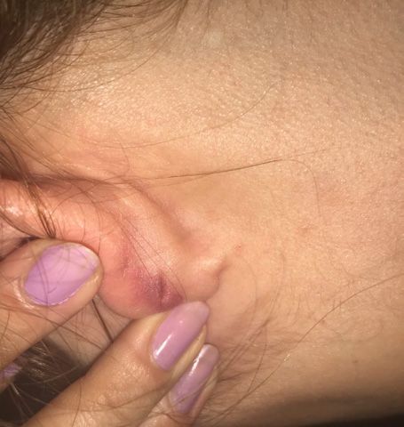
Azimi Alamdari et al

degrees of effectiveness, there is little systematic research                  was 21 (sample size included 33 patients with 63 scars)
on this issue (5). Therapeutic modalities for hypertrophic                     that were distributed in the three groups.
scars and keloids include pressure therapies, silicone gel                       The first group received intralesional cryotherapy
dressing, intralesional injection (corticosteroid, 5-FU,                       alone. The second and third groups were treated with
methotrexate, bleomycin), radiotherapy, cryotherapy and                        intralesional steroid and 5-FU respectively. Type of the
laser (1). Intralesional steroid injection has been used                       scars (hypertrophic or keloid) was identified prior to
since 1960 to treat hypertrophic and keloid scars. Steroid                     the treatments and their dimensions were measured and
reduces collagen and glycosaminoglycan synthesis,                              recorded along with physical appearance such as color,
inflammatory processes, and fibroblast proliferation and                       erythema, telangiectasia and accompanying symptoms.
raises hypoxia in the scar tissue (6). Its side effects include                  Intralesional cryotherapy was performed using a special
atrophy, hypopigmentation and telangiectasia (1). In recent                    needle (designed by an engineer from Sarma Darman
decades, intralesional 5-FU, either alone or in combination                    Co., Tehran, Iran) and freezing by liquid nitrogen into
with steroid, has been suggested for treatment (7). It has                     the scar tissue (Figure 1). Type of the steroid used in this
been shown that 5-FU reduces fibroblast proliferation                          study was triamcinolone acetonide (Triamhexal) 40 mg/
in the tissue. The only side effects reported are painful                      mL and 0.2 mL/cm2 of it was injected intralesionally using
injection, hypopigmentation, purpura, and sometimes                            insulin syringe (gauge 27). Then, 0.2 mL/cm2 of 5-FU
wound at the site (1). Intralesional steroid and 5-FU                          250 mg/5 mL (EBEWE) was injected intralesionally. The
have played a successful role in decreasing pain, itching                      maximum dose of triamcinolone and 5-FU injected in
and volume of scar; however, several therapy sessions are                      each session was 30 mg/mL and 100 mg/mL, respectively.
required and the rate of relapse is high (8).                                  The treatments were repeated every 4 weeks and would be
   Intralesional cryotherapy is a new therapeutic method                       reapplied for further 6 sessions depending on the presence
that freezes the scar tissue from inside. Intralesional probe                  of scar tissue. Lesions were imaged before and after the
allows the destruction of scar by forming intracellular                        treatment. Dimensions (including height) of the scars were
ice crystals and cellular anoxia; in fact, cold damages the                    measured by means of Vernier scale. Moreover, scar size
endothelial cell connections and causes blood stasis and                       and physical appearance were examined in each session
vascular injury and necrosis (9). Numerous studies have                        and recorded in the checklist. Prior to the treatment in the
confirmed the effectiveness of intralesional cryotherapy                       5-FU group, blood cell count and liver function tests were
as a monotherapy for scars. Given the intralesional nature                     performed and repeated after the first and sixth sessions.
of the treatment, complications such as hypopigmentation                         Collected data were analyzed in SPSS 17.0 using
(compared to normal surface cryotherapy) are much less                         descriptive statistics (mean, standard deviation and
and the patients experience a shorter recovery period (4).                     frequency, means comparison test, one-way ANOVA and
Intralesional cryotherapy reduces the volume and relapse                       chi-square test). P
Azimi Alamdari et al

   One patient with one scar was excluded from the study                     A                                       B
in the 5-FU group because of increased pain after the third
session. In total, of the 63 scars, 56 were itching, 2 scars
caused pain and others showed no symptom.
   Considering the changes in scar color compared to
surrounding skin, 39 scars had mild erythema, 20 showed
marked redness, and 4 had the same color as the skin.
   Comparison of the means of height, surface area and
volume of scars in the steroid group showed that the effect            Figure 3. (A) keloid resulted from Acne vulgaris on shoulder before
of therapies was manifested for scar height in short term              treatment. (B) same keloid after 6 session treatment by intralesional
and for surface area and volume in long term. The χ2 values            5-FU. Hyperpigmentation due to 5-FU is notable.
of the measured qualities of surface area and height of
scare during the therapy sessions, age, gender and itching
showed that no statistically significant difference existed            Explicit change in scar volume was noticed after the
between observations measurement and calculations in                   first session in the cryotherapy group and after the sixth
the steroid group (Figure 2).                                          session in the steroid and 5-FU groups (Tables 1 and 2).
   Results showed that in the 5-FU group, the effect of                   The biggest reduction in scar height after the first
therapies on scar height and volume became clear after the             therapy session was observed in the cryotherapy group
sixth session and that no significant difference was found             (Figures 4-6). After the sixth session of steroid and 5-FU
between therapy sessions in the surface area. The χ2 values            therapy, the smallest size of surface area and height was
of the measured qualities of surface area and height of                found in the steroid group indicating the greater effect
scar during the therapy sessions, age and gender showed                of steroid on the reduction of scar height and surface
that the difference between observations measurement                   area compared to 5-FU. Results showed that the effect of
and calculations was statistically insignificant (P > 0.05)            steroid therapy on the reduction of scar volume after the
(Figure 3).                                                            sixth session was greater than the effect of 5FU.
   Results in the cryotherapy group showed that the                       Side effects in the steroid group included permanent
qualities of surface area, height and volume of scar, original         hypopigmentation: 4 cases, telangiectasia: 4 cases, and
size and the size after the first session were significantly           atrophy: 3 cases. In the 5-FU group, side effects were
different (P > 0.05). The χ2 values of the measured qualities          surface wound: 4 cases, hyperpigmentation: 16 cases, and
during the therapy sessions, age and gender showed that                increased pain: 1 case (excluded from the study after 3
the difference between observations measurement and                    months) (Figure 7). Side effects in the cryotherapy group
calculations was statistically insignificant.                          included 3 cases of temporary scar hypopigmentation that
   Results of the study showed that the three methods of               disappeared in the follow-up 6 months after the treatment.
cryotherapy, 5-FU and steroid had a significant difference             No infection, liver enzymes disorder or hematologic
at P < 0.01 in terms of the original size of scars and the size        disorder was observed (Table 3).
after the first therapy session.
   Comparison of the means of scar height and surface                  Discussion
area in the 3 groups showed that the original size of scars            Keloids and hypertrophic scars are rather prevalent
in the cryotherapy group was larger than that in the other             diseases that generally develop in response to the injuries
2 groups. However, after the first therapy session, the                in which damage to the skin is associated with proliferation
size in this group was smaller than that in other groups;              of dense fibrous tissues developed after dermal lesion
therefore, results showed a remarkable reduction in scar               recovery. Hypertrophic scars can occur in all races, but
surface area in the cryotherapy than the other 2 groups.               keloids are more common in specific races, especially in
                                                                       the individuals who have more pigmented skin (10). Some
                                                                       reports indicate the effect of hormones on the growth and
        A                     B                                        development of these diseases. After 3-4 weeks, the tissue
                                                                       inflammation becomes bigger and thicker. The injury
                                                                       usually becomes tough and turns pink to red and grows
                                                                       for months or years. The surface of keloids is smoother
                                                                       and rounder and continues to grow beyond the original
                                                                       lesion. Various techniques have been used to remove this
                                                                       lesion, but results have not been completely satisfactory
                                                                       (11). Therefore, the purpose of this study was to provide
                                                                       a comparative assessment of three different therapeutic
Figure 2. (A) Hypertrophic scar before treatment. (B) flattened        methods, namely steroid, 5-FU, and cryotherapy in the
scar after 3 session intralesional Triamcinolone therapy.              keloid treatment.
Hypopigmentation is one of the steriod complication.

                                                Crescent Journal of Medical and Biological Sciences, Vol. 5, No. 3, July 2018         217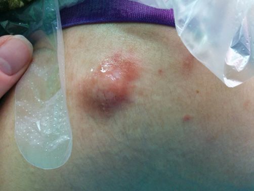
Azimi Alamdari et al

Table 1. The Effect of Applied Methods on Height, Surface Area and Volume of Scar at Different Stages of the Study
                                                             Cryotherapy                           5-FU                          Steroid
 Scar height                Original size                        3.482a                           2.0450a                        1/8600a
                        After the 1st session                    0.783b                           1.9300a                        1/5525b
                        After the 6th session                       -                            1.5015b                         0/7855c
                         Significance                            0.0001                            0.004                         0.0001
 Scar surface area          Original size                      129.1545a                         98.5050a                       44/1995a
                        After the 1st session                  23.7805b                          97.6750a                       43/3820a
                        After the 6th session                       -                            69.8300a                       20/8240b
                        Significance                            0.00001                            0/556                         0.0001
 Scar volume                Original size                       460.58 a                          221.41a                        103/87a
                        After the 1st session                    18.76b                           213.30a                        91/15ab
                        After the 6th session                       -                             85.08b                          22/78b
                        Significance                             0.0001                           0.0001                         0.0001
* Letters in each column of the qualities scar surface area, height and volume show statistically significant difference.

Table 2. Comparison of the Qualities of Surface Area, Height and Volume of the Scar in the 3 Therapy Groups at Different Stages

                                   Original Size                                After the 1st Session                     After the 6th Session
Therapy            Surface area                         Volume        Surface Area                    Volume        Surface       Height       Volume
                                     Height (mm)                                     Height (mm)
                      (mm2)                              (mm3)           (mm2)                         (mm3)       Area (mm2)      (mm)         (mm3)
Cryotherapy           129.15 a
                                       3.4820a         460.58a         23.7805c           0.7830c       18.76c         -             -            -
Steroid              44.1995c          1.8600b         103.87c         43.3820ab          1.5525ab     91.15b        20.82b        0.79b        22.78b
5-FU                 98.5050b          2.0450b         221.41b         97.6750a           1.9300a      213.30a       66.83a        1.46a        85.08c
* Letters in each column of the qualities scar surface area, height and volume show statistically significant difference.

Table 3. Side effects Associated With Different Therapies

                                                                                 Side Effects
                     Atrophy          Telangiectasia       Increased Pain          Hyperpigmentation         Hypopigmentation        Surface Wound
5-FU                     -                   -                   5%                        80%                        -                    20%
Steroid                15%                 20%                    -                          -                       20%                    -
Cryotherapy              -                   -                    -                          -                   Temporary15%               -

   Intralesional 5-FU injection acts by restraining the                            triamcinolone group than in the 5-FU group. Side effects
proliferation of fibroblasts and activity of antimetabolites.                      in the 5-FU group including itching, pain, dryness, limited
It also has a preventive effect on type 1 collagen gene                            mobility, and aesthetic problems were more than those in
expression stimulated by TGF-β in the human fibroblasts                            the triamcinolone group. No relapse was observed in the
(12). 5-FU interrupts DNA and RNA synthesis at different                           scars (11).
levels by preventing thymidylate synthase and production                              Researcher found that atrophy occurred only in
of toxic metabolites (13). Intralesional triamcinolone                             8% of the cases following 5-FU injection, no case of
injection also prevents protein synthesis and fibroblast                           telangiectasia was reported and hyperpigmentation and
migration. Steroids prevent collagen synthesis and have                            hypopigmentation were found in 4% of the cases. On
anti-inflammatory effects. Atrophy is a side effect of                             the other hand, in the triamcinolone group, atrophy was
steroids which are used in the keloid treatment (14).                              observed in 12% of the cases, hypopigmentation in 8%,
   One research in 2004 showed that the average recovery                           hyperpigmentation in 4%, and telangiectasia in 4% of the
in cryotherapy and intralesional triamcinolone injection                           cases (11). Some studies report that unless other easier
was 67.22 ± 3.5 (15). Another study showed that the                                therapies are effective, steroid (TAC, triamcinolone) is
therapeutic response to this type of treatment varied from                         the first line therapy for keloid treatment and the second
50% to 100% and that the chance of lesion relapse was up                           line therapy for HTSs with a response rate of 50% to 100%
to 50% (2).                                                                        and relapse rate of 9% to 50% (16). Roques and Teot also
   Results of a study by Sharma et al on 5-FU and                                  showed that intralesional steroid injection caused pain and
triamcinolone acetonide intralesional injection revealed                           that 63% of the patients who received therapy reported
that following the therapies, 96% and 72% of the cases                             post-therapy complications including hypopigmentation,
recovered in triamcinolone acetonide group and 5-FU                                subcutaneous atrophy and skin fat, telangiectasia, and
group, respectively. The recovery was also better in the                           ineffectiveness (17).

218       Crescent Journal of Medical and Biological Sciences, Vol. 5, No. 3, July 2018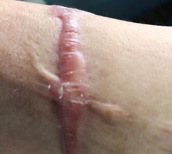
Azimi Alamdari et al
A B C
Figure 4. (A) keloid on back region before treatment. (B) Reduction
near 50% in size after 1 session intralesional cryotherapy. (C)
complete remission of keloid after second cryosurgery.
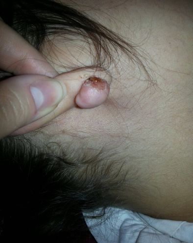
Figure 7. Superficial ulcer is a complication of 5FU injection.
A B
rate at 47.4% (19). Another study in China also showed
that the success rate of 5-FU therapy was 62.5% while it
was 92% in 5-FU therapy combined with glucocorticoid
(22).
In 2012, Prabhu et al in a randomized controlled trial
compared the efficacy of intralesional triamcinolone and
5-FU and showed that the treatment with triamcinolone
(71.23%) was significantly better that the treatment with
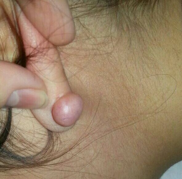
Figure 5. (A) Ear lobe keloid induced after piercing. (B) After one
session cryotherapy the keloid is flattened.
5-FU (57.48%) (P = 0.04). Pain reduction was 18% in
the 5-FU group and 24% in the TAC group, though the
difference was not significant. Although side effects in the
5-FU group were 5 times greater than the side effects in
A B the TAC group, their difference was not significant (5).
Nevertheless, keloid size reduction in the TAC group was
significantly better than that in the 5-FU group (5).
Results of a research in 2017 showed that TAC/5-FU
combination instead of TAC alone led to a significant
improvement in itching, pain, dryness, mobility limitation,
and aesthetic complications (23).
Research has also shown that cryotherapy with liquid
nitrogen combined with corticosteroid has been highly
successful in treating keloid scars (24). Findings of Har-
Figure 6. (A) keloid on forearm before treatment by cryosurgery. Shai et al showed that intralesional cryotherapy is capable
(B) Notable reduction in keloid size after one session cryosurgery. of concentrating cold temperature inside the wound and
thus less affects the external skin tissue. In one study
In one study by Khan et al in 2014, 150 patients were using CryoProbe, a 67.4% size reduction was observed 6
divided into 2 groups of TAC therapy alone and TAC/5- months after the cryotherapy. The redness also decreased
FU combination therapy. Results showed that in the and none of the patients developed hypopigmentation or
TAC alone group, recovery was 68% and in the TAC/5- relapse (25). Results of a review study in 2015 showed that
FU group recovery was 84%. In the former group, 24% in 8 studies conducted, scar volume had decreased from
of the cases showed side effects and in the latter group, 51% to 63%, but scars had not been removed completely in
side effects were reported in 8% of the cases. Results of any of the cases. In addition, relapse rate ranged from 0%
the same study showed that TAC/5-FU combination had to 24%. Post-treatment hypopigmentation was reported
better outcomes for keloid treatment (18). in some studies. Pain and itching had been significantly
Nanda and Reddy treated the keloid scars of 28 patients decreased in most of the studies (8). Researchers have
by intralesional injection of 50 mg/mL 5-FU weekly for 12 reported that intralesional cryotherapy leads to reductions
weeks. Results showed that 78.5% of the patients reported in lesion volume, hypopigmentation and other dermal
a recovery rate (in terms of keloid size, height and related complications (26). Two different studies showed that
symptoms) above 50% and none of the patients reported no permanent or specific hypopigmentation developed
relapse 24 weeks after the treatment (19). Similar results after cryotherapy in white patients (25,26). However,
were reported for small keloids (20). Results of another other studies reported hypopigmentation in most of the
study showed that recovery rate was above 50% in 85% of patients including those with lighter Fitzpatrick skin
the patients (21). One study reported the one-year relapse types. Although hypopigmentation was improved over
Crescent Journal of Medical and Biological Sciences, Vol. 5, No. 3, July 2018 219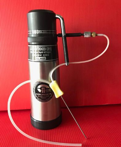
Azimi Alamdari et al

time in both studies, it remained permanent in 31% to                          fewer complications. Intralesional cryotherapy is effective
37% of the cases (27,28). Research also showed that skin                       in reducing the volume of scars in fewer sessions and can
redness decreased by 52%-83% after therapies (8).                              be used as the primary treatment for scar followed by
  Findings of a review study revealed that pain decreased                      other modalities such as steroids to achieve good results.
by 52.5% (35%-78%) after treatments and itching
decreased 43.6% (28%-61%) but did not completely                               Conflict of Interests
disappear (8).                                                                 Authors declare that they have no conflict of interests.
  In most of the studies, post-treatment lesion volume
decreased by more than 50% (8). However, researchers                           Ethical Issues
reported that lesions did not disappear after 10 sessions of                   This study was approved by Ministry of Health and
intralesional cryotherapy (29).                                                Medical Education, Iran, and confirmed (identifier:
  Significant reduction in scar volume, deformity,                             IRCT20100314003566N9) by the Research and Ethics
hardness, redness, itching, dryness and aesthetic                              Committee of Tabriz University of Medical Sciences, Iran.
complications through cryotherapy has been reported
by researchers (30). In addition, hypopigmentation is                          Financial Support
the most common side effect of contact cryotherapy                             None.
(91.7) which is less observed in intralesional cryotherapy
because of increased viability of melanocytes (26,31).                         Acknowledgments
Treatment of the patients with intralesional cryotherapy                       The authors would like to thank Engineer Framarz Jenabi
changes the structure of scar, and collagen fibers are                         for designing the probe.
arranged in parallel rows and form the natural structure
of dermis (26).                                                                References
  Researchers have suggested that numerous cryotherapy                         1.  Mutalik S. Treatment of keloids and hypertrophic scars.
sessions have functional effects on keloids and                                    Indian J Dermatol Venereol Leprol. 2005;71(1):3-8.
hypertrophic scars and prevent relapse (32,33). Results                            doi:10.4103/0378-6323.13777
                                                                               2. Dunsky K, Brissett A. Keloids and Hypertrophic Scars.
also showed that cryotherapy is more effective (85%) than
                                                                                   In: Sataloff RT, ed. Sataloff ’s Comprehensive Textbook of
intralesional steroid injection (34). Findings of another                          Otolaryngology: Head & Neck Surgery: Facial Plastic and
study also showed that the combination of cryotherapy                              Reconstructive Surgery. Jp Medical Ltd; 2015.
and intralesional steroid injection is much more effective                     3. Thareja S, Kundu RV. Keloids and Hypertrophic
than cryotherapy or steroid therapies alone (35).                                  Scarring. In: Neelam A. Vashi NA, Maibach HI, eds.
  Researchers treated 25 keloid lesions with intralesional                         Dermatoanthropology of Ethnic Skin and Hair. Springer;
cryotherapy and silicone gel and 7 keloids with cryotherapy.                       2017:233-255.
Results showed that keloid volume decreased significantly                      4. Goldenberg G, Luber AJ. Use of intralesional cryosurgery
                                                                                   as an innovative therapy for keloid scars and a review of
in all of the cases. In the combinatory method, hardness
                                                                                   current treatments. J Clin Aesthet Dermatol. 2013;6(7):23-
and patients’ pain and discomfort were more favorable                              26.
while redness and itching in cryotherapy were less than                        5. Prabhu A, Sreekar H, Powar R, Uppin V. A randomized
those in the combination therapy (36).                                             controlled trial comparing the efficacy of intralesional
  Results of the present study in reducing the surface                             5-fluorouracil versus triamcinolone acetonide in
area, height and volume of keloids with different therapy                          the treatment of keloids. J Sci Soc. 2012;39(1):19-25.
methods are consistent with the literature. Most of the                            doi:10.4103/0974-5009.96466
previous studies have highlighted the better effectiveness                     6. Wong TS, Li JZ, Chen S, Chan JY, Gao W. The Efficacy of
of steroid in healing the keloid and its lesser side effects                       Triamcinolone Acetonide in Keloid Treatment: A Systematic
                                                                                   Review and Meta-analysis. Front Med (Lausanne).
compared to 5-FU method. The present findings also
                                                                                   2016;3:71. doi:10.3389/fmed.2016.00071
indicated better effectiveness and fewer side effects of                       7. Rabello FB, Souza CD, Farina Junior JA. Update on
steroid in comparison with 5-FU.                                                   hypertrophic scar treatment. Clinics (Sao Paulo).
  Previous studies have confirmed better effectiveness of                          2014;69(8):565-573.
cryotherapy compared to steroid therapy. Present results                       8. van Leeuwen MC, Bulstra AE, Ket JC, Ritt MJ, van
also show that cryotherapy have better outcomes than                               Leeuwen PA, Niessen FB. Intralesional Cryotherapy for the
steroid and 5-FU which is in line with previous findings.                          Treatment of Keloid Scars: Evaluating Effectiveness. Plast
Although different complications have been reported for                            Reconstr Surg Glob Open. 2015;3(6):e437. doi:10.1097/
                                                                                   gox.0000000000000348
cryotherapy, similar to previous studies, only temporary
                                                                               9. Har-Shai Y, Mettanes I, Zilberstein Y, Genin O, Spector I, Pines
hypopigmentation was observed in this study in 15% of                              M. Keloid histopathology after intralesional cryosurgery
the cases which is consistent with previous studies.                               treatment. J Eur Acad Dermatol Venereol. 2011;25(9):1027-
  Results of this study showed that cryotherapy is more                            1036. doi:10.1111/j.1468-3083.2010.03911.x
effective than steroid and 5-FU in treating keloids, and it                    10. Shaffer JJ, Taylor SC, Cook-Bolden F. Keloidal scars: a
provides better outcomes in shorter timeframes, and has                            review with a critical look at therapeutic options. J Am Acad

220    Crescent Journal of Medical and Biological Sciences, Vol. 5, No. 3, July 2018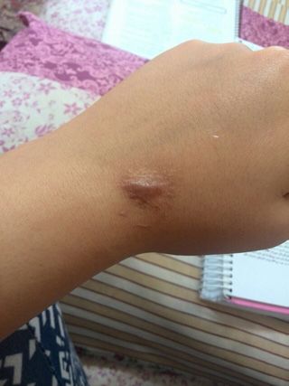
Azimi Alamdari et al

    Dermatol. 2002;46(2 Suppl Understanding):S63-97.                            of cryosurgery in keloids and hypertrophic scars. A
11. Sharma S, Bassi R, Gupta A. Treatment of small keloids                      prospective consecutive trial of case series. Arch Dermatol.
    with intralesional 5-fluorouracil alone vs. intralesional                   1993;129(9):1146-1151.
    triamcinolone acetonide with 5-fluorouracil. J Pak Assoc              25.   Har-Shai Y, Sabo E, Rohde E, Hyams M, Assaf C, Zouboulis
    Dermatol. 2012;22(1):35-40.                                                 CC. Intralesional cryosurgery enhances the involution
12. Wendling J, Marchand A, Mauviel A, Verrecchia F.                            of recalcitrant auricular keloids: a new clinical approach
    5-fluorouracil blocks transforming growth factor-beta-                      supported by experimental studies. Wound Repair Regen.
    induced alpha 2 type I collagen gene (COL1A2) expression in                 2006;14(1):18-27. doi:10.1111/j.1743-6109.2005.00084.x
    human fibroblasts via c-Jun NH2-terminal kinase/activator             26.   Har-Shai Y, Amar M, Sabo E. Intralesional cryotherapy for
    protein-1 activation. Mol Pharmacol. 2003;64(3):707-713.                    enhancing the involution of hypertrophic scars and keloids.
    doi:10.1124/mol.64.3.707                                                    Plast Reconstr Surg. 2003;111(6):1841-1852. doi:10.1097/01.
13. Bulstrode NW, Mudera V, McGrouther DA, Grobbelaar AO,                       prs.0000056868.42679.05
    Cambrey AD. 5-fluorouracil selectively inhibits collagen              27.   van Leeuwen MC, Bulstra AE, van Leeuwen PA, Niessen
    synthesis. Plast Reconstr Surg. 2005;116(1):209-221;                        FB. A new argon gas-based device for the treatment of
    discussion 222-203.                                                         keloid scars with the use of intralesional cryotherapy.
14. Ahuja RB, Chatterjee P. Comparative efficacy of intralesional               J Plast Reconstr Aesthet Surg. 2014;67(12):1703-1710.
    verapamil hydrochloride and triamcinolone acetonide in                      doi:10.1016/j.bjps.2014.08.046
    hypertrophic scars and keloids. Burns. 2014;40(4):583-588.            28.   van Leeuwen MC, van der Wal MB, Bulstra AE, et al.
    doi:10.1016/j.burns.2013.09.029                                             Intralesional cryotherapy for treatment of keloid scars: a
15. Fatemi F, Ahmadpoor K. Evaluation of therapeutic response                   prospective study. Plast Reconstr Surg. 2015;135(2):580-
    of Keloid and Hypertrophic scars to bleomycin tatto and                     589. doi:10.1097/prs.0000000000000911
    to cryotherapy followed by intralesional triamcinolone                29.   Gupta S, Kumar B. Intralesional cryosurgery using lumbar
    injection. J Shahrekord Univ Med Sci. 2004;5(4):5-10.                       puncture and/or hypodermic needles for large, bulky,
16. Ho WS, Ying SY, Chan PC, Chan HH. Use of onion extract,                     recalcitrant keloids. Int J Dermatol. 2001;40(5):349-353.
    heparin, allantoin gel in prevention of scarring in chinese           30.   Har-Shai Y, Brown W, Labbe D, et al. Intralesional
    patients having laser removal of tattoos: a prospective                     cryosurgery for the treatment of hypertrophic scars
    randomized controlled trial. Dermatol Surg. 2006;32(7):891-                 and keloids following aesthetic surgery: the results of a
    896. doi:10.1111/j.1524-4725.2006.32192.x                                   prospective observational study. Int J Low Extrem Wounds.
17. Roques C, Teot L. The use of corticosteroids to treat keloids:              2008;7(3):169-175. doi:10.1177/1534734608322813
    a review. Int J Low Extrem Wounds. 2008;7(3):137-145.                 31.   Har-Shai Y, Dujovny E, Rohde E, Zouboulis CC. Effect of
    doi:10.1177/1534734608320786                                                skin surface temperature on skin pigmentation during
18. Khan MA, Bashir MM, Khan FA. Intralesional                                  contact and intralesional cryosurgery of keloids. J Eur Acad
    triamcinolone alone and in combination with 5-fluorouracil                  Dermatol Venereol. 2007;21(2):191-198. doi:10.1111/j.1468-
    for the treatment of keloid and hypertrophic scars. J Pak                   3083.2006.01890.x
    Med Assoc. 2014;64(9):1003-1007.                                      32.   Rusciani L, Rossi G, Bono R. Use of cryotherapy in the
19. Nanda S, Reddy BS. Intralesional 5-fluorouracil as a treatment              treatment of keloids. J Dermatol Surg Oncol. 1993;19(6):529-
    modality of keloids. Dermatol Surg. 2004;30(1):54-56.                       534.
20. Gupta S, Kalra A. Efficacy and safety of intralesional                33.   Zouboulis CC, Zouridaki E, Rosenberger A, Dalkowski
    5-fluorouracil in the treatment of keloids. Dermatology.                    A. Current developments and uses of cryosurgery in the
    2002;204(2):130-132. doi:10.1159/000051830                                  treatment of keloids and hypertrophic scars. Wound Repair
21. Kontochristopoulos G, Stefanaki C, Panagiotopoulos                          Regen. 2002;10(2):98-102.
    A, et al. Intralesional 5-fluorouracil in the treatment of            34.   Layton AM, Yip J, Cunliffe WJ. A comparison of intralesional
    keloids: an open clinical and histopathologic study. J Am                   triamcinolone and cryosurgery in the treatment of acne
    Acad Dermatol. 2005;52(3 Pt 1):474-479. doi:10.1016/j.                      keloids. Br J Dermatol. 1994;130(4):498-501.
    jaad.2004.09.018                                                      35.   Yosipovitch G, Widijanti Sugeng M, Goon A, Chan YH, Goh
22. Zhang GY, Gao WY, Li X, et al. Effect of camptothecin                       CL. A comparison of the combined effect of cryotherapy
    on collagen synthesis in fibroblasts from patients with                     and corticosteroid injections versus corticosteroids and
    keloid. Ann Plast Surg. 2009;63(1):94-99. doi:10.1097/                      cryotherapy alone on keloids: a controlled study. J Dermatolog
    SAP.0b013e3181872775                                                        Treat. 2001;12(2):87-90. doi:10.1080/095466301317085363
23. Paul A, Kumar N, Moirangthem T, Singh MK, Hussain KA.                 36.   Stromps JP, Dunda S, Eppstein RJ, Babic D, Har-Shai Y,
    Comparative study of intralesional triamcinolone alone and                  Pallua N. Intralesional cryosurgery combined with topical
    in combination with 5-flurourouracil for the treatment of                   silicone gel sheeting for the treatment of refractory keloids.
    keloid and hypertrophic scars. Hellenic Journal of Surgery.                 Dermatol Surg. 2014;40(9):996-1003.
    2017;89(1):13-17. doi:10.1007/s13126-017-0371-9
24. Zouboulis CC, Blume U, Buttner P, Orfanos CE. Outcomes

 Copyright © 2018 The Author(s); This is an open-access article distributed under the terms of the Creative Commons Attribution
 License (http://creativecommons.org/licenses/by/4.0), which permits unrestricted use, distribution, and reproduction in any medium,
 provided the original work is properly cited.

                                                   Crescent Journal of Medical and Biological Sciences, Vol. 5, No. 3, July 2018        221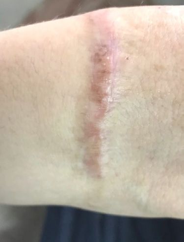


You can also read